. 2025 Jul 30:16:1621222.
doi: 10.3389/fimmu.2025.1621222. eCollection 2025.

Engineered human B cells targeting tumor-associated antigens exhibit antigen presentation and antibody-mediated functions

Affiliations

Engineered human B cells targeting tumor-associated antigens exhibit antigen presentation and antibody-mediated functions

Alexander Boucher et al. Front Immunol. .

Abstract

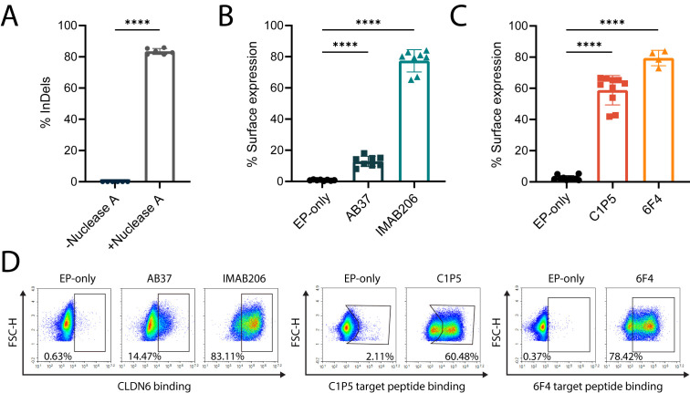
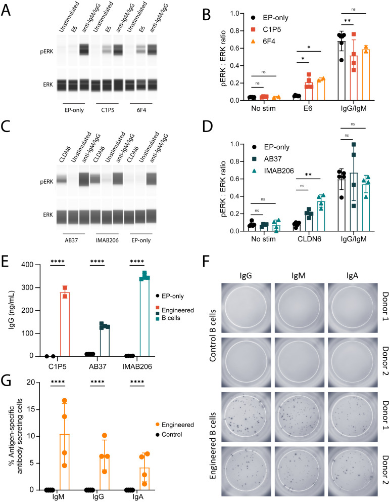
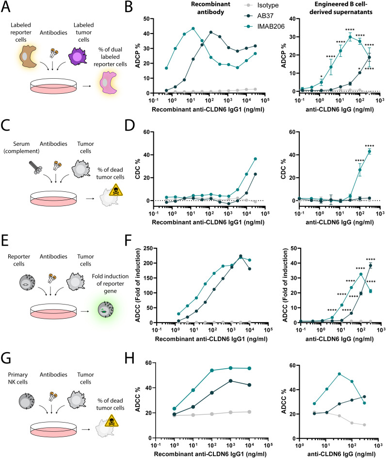
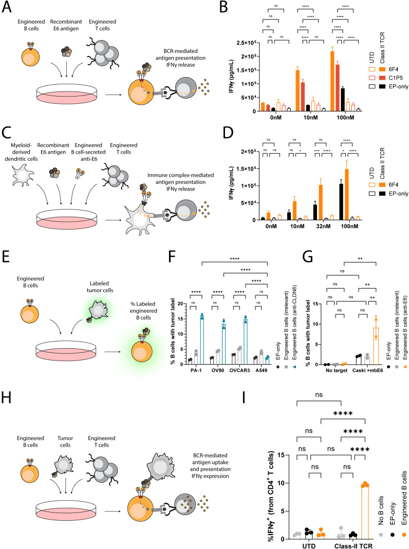
B cell engineering represents a promising therapeutic strategy that recapitulates adaptive immune functions, such as memory retention, antibody secretion and affinity maturation in murine models of viral infection. These mechanisms may be equally beneficial in oncology. Recent studies have linked endogenous anti-tumor B cell immunity to favorable prognosis across multiple malignancies. Here, we present functional validation of human B cells engineered to target tumor-associated membrane and intracellular antigens. We demonstrate that engineered B cells express therapeutically relevant membrane B cell receptors that are secreted as antibodies upon differentiation. Additionally, engineered B cells take up tumor-associated antigens and demonstrate potent antigen presentation capabilities, while their secreted antibodies activate T cell responses via immune complexes and induce tumor-directed cytotoxic responses. B cell engineering to target tumor-associated antigens may thus have utility as a novel modality for solid tumor therapy.

Keywords: B cell; antibody; antigen presentation; cell engineering; genome editing; immune complex; tertiary lymphoid structure (TLS).


Conflict of interest statement

ABo, TW, IK, EK, BK, HS, LS, JM, CP and AMC were employed by ElevateBio. CA, RH, MK, JF, LNK, SH, DBu, HL, DBr, and ADN were employed by Tabby Therapeutics. CR, MVO, and PB were employed by Life EditTherapeutics. The remaining authors declare that the research was conducted in the absence of any commercial or financial relationships that could be construed as a potential conflict of interest.

Figures

Figure 1
Figure 1
Engineering of primary human B cells with antigen-specific BCRs. (A) Percent of InDels in the targeted IgH locus of edited B cells with and without Nuclease A provided with Guide 10. ****=pv<0.0001, two-tailed unpaired t-test. (B) Percentage of surface expression of CLDN6-specific BCRs (AB37 and IMAB206), as measured by flow cytometry, compared to EP-only control B cells. Data are the mean of 6 experiments with 2 donors for IMAB206 and 4 experiments with 2 donors for AB37. (C) Same as (B) for E6-specific BCRs (C1P5 and 6F4). Data are the mean of 3 experiments with 3 donors for C1P5 and 2 experiments with 2 donors for 6F4. For (A-C) mean is indicated and error bars represent SD, each dot represents an independent experiment. For (B, C) ****=pv<0.0001 for one-way ANOVA with Tukey’s multiple comparison test. (D) Representative flow cytometry plots for (B, C) of surface BCR expression of engineered B cells.
Figure 2
Figure 2
Engineered B cells activate BCR signaling cascades and secrete isotype-switched antigen-specific antibodies. (A) Representative immunoblot of B cells engineered to express either C1P5 or 6F4 E6-binding BCRs, stimulated with recombinant E6 protein, anti-IgM/IgG protein control or unstimulated. Protein extracts were analyzed using anti-pERK antibody (top panel) or anti-total ERK antibody (bottom panel). (B) Quantification of mean ratio expression of ERK to pERK as in (A) ns=pv>0.05, *=pv<0.05, **=pv<0.01 for two-way ANOVA with Dunnett’s multiple comparisons test. (C) Same as A for the CLDN6-binding BCRs AB37 or IMAB206. Cell stimulation occurred with recombinant CLDN6 protein. (D) Quantification of experiments as in (C) For (A-D), data representative of 2–3 experiments with 2 donor cells. ns=pv>0.05, **=pv<0.01 for two-way ANOVA with Dunnett’s multiple comparisons test. (E) Quantification of IgG antibodies in the supernatants of engineered B cells expressing either C1P5, AB37 or IMAB206, by ELISA. ****=pv<0.0001 for two-way ANOVA with Šídák’s multiple comparisons test. (F) Representative ELISPOT images of E6-specific IgM, IgG and IgA secreted from B cells engineered to express the anti-E6 6F4 antibody. (G) Quantification of data as in (F) as normalized to the total amount of cells seeded in the wells. ****=pv<0.0001 for two-way ANOVA with Šídák’s multiple comparisons test. For (E-G) pooled data from 2–3 experiments with 2–4 donor cells. For (B, D, E), (G) mean and error bars are indicated, each dot represents an independent experiment.
Figure 3
Figure 3
Antigen-specific IgGs secreted by engineered B cells trigger Fc-mediated functions. (A) Schema depicting the flow cytometry-based ADCP assay. Labeled reporter cells were incubated with labeled tumor cells and either recombinant antibodies or concentrated antibodies from the supernatant of engineered B cells. The frequency of dual labeled reporter cells was analyzed. (B) ADCP response of recombinant IgG1 antibodies (left) and concentrated engineered B cell supernatants (right) for Isotype, AB37, and IMAB206 against CLDN-6-expressing PA-1 cells determined by flow cytometry. *=pv<0.05, ****=pv<0.0001 for two-way ANOVA with Dunnett’s multiple comparisons test to the isotype control. (C) Schema depicting the flow cytometry-based CDC assay. Tumor cells were incubated with sera and either recombinant antibodies or concentrated antibodies from the supernatant of engineered B cells. The frequency of dead tumor cells was analyzed. (D) CDC response of recombinant IgG1 antibodies (left) and concentrated engineered B cell supernatants (right) for Isotype, AB37, and IMAB206 against CLDN-6-expressing PA-1 cells, as determined by flow cytometry. ****=pv<0.0001 for two-way ANOVA with Dunnett’s multiple comparisons test to the isotype control. (E) Schema depicting the ADCC reporter assay. Reporter cells were incubated with tumor cells and either recombinant antibodies or concentrated antibodies from the supernatant of engineered B cells. The fold induction of the luciferase reporter gene in the reporter cells was analyzed. (F) ADCC response of recombinant IgG1 antibodies (left) and concentrated engineered B cell supernatants (right) for Isotype, AB37, and IMAB206 against CLDN-6-expressing PA-1 cells determined by luciferase fold of induction. ****=pv<0.0001 for two-way ANOVA with Dunnett’s multiple comparisons test to the isotype control. (G) Schema depicting the flow cytometry-based ADCC assay. Primary NK cells were incubated with tumor cells and either recombinant antibodies or concentrated antibodies from the supernatant of engineered B cells. The frequency of dead tumor cells was analyzed. (H) ADCC response of recombinant IgG1 antibodies (left) and concentrated engineered B cell supernatants (right) for Isotype, AB37, and IMAB206 against CLDN-6-expressing PA-1 cells, as determined by flow cytometry. For the engineered B cell-derived supernatants, data are representative of technical replicates and two (B) or one (D-F) experiments using two independent donors to generate supernatants or one experiment using one donor to generate supernatants (H). Error bars indicate SD.
Figure 4
Figure 4
Engineered B cells and immune complexes taken up by dendritic cells present E6 antigen to activate E6-specific MHC class II-restricted T cells. (A) Schema depicting the engineered B cell antigen presentation assay. B cells, either non-engineered (EP-only) or engineered to express anti-E6 antibodies (6F4 or C1P5), are loaded with recombinant E6 antigen at multiple concentrations and then incubated with T cells, either engineered (Class II TCR) or non-engineered (UTD) to express an anti-E6 TCR. The concentration of IFNγ in the supernatants is then analyzed by ELISA. (B) ELISA for IFNγ as in (A) Data representative of 2 experiments with 2 donors. Bars and error bars represent mean and SD. ns=pv>0.05, ****=pv<0.0001 for two-way ANOVA with Šídák’s multiple comparisons test. (C) Schema depicting the immune complex-mediated antigen presentation assay. Immune complexes were formed with concentrated antibodies from the supernatants of B cells, either non-engineered or engineered to express anti-E6 antibodies, and recombinant E6 antigen at multiple concentrations. These were then incubated with myeloid-derived dendritic cells and then co-cultured with T cells, either engineered or non-engineered to express an anti-E6 TCR. The concentration of IFNγ in the supernatants is then analyzed by ELISA. (D) ELISA for IFNγ as in (C) Data representative of 2 experiments with 2 donors. Bars and error bars represent mean and SD. ns=pv>0.05, *=pv<0.05, ***=pv<0.001, ****=pv<0.0001 for two-way ANOVA with Šídák’s multiple comparisons test. (E) Schema depicting the flow cytometry-based engineered B cell membrane-bound antigen uptake assay. Engineered or non-engineered B cells are incubated with labeled tumor cells either expressing or not expressing the targeted antigen. The frequency of B cells with the tumor label is then analyzed by flow cytometry. (F) Flow cytometry results as in (E) for B cells engineered to express anti-CLDN6 IMAB206 (blue) or anti-E6 (6F4) antibodies (grey). ns=pv>0.05, ****=pv<0.0001 for two-way ANOVA with Šídák’s multiple comparisons test. (G) Flow cytometry results as in (E) for B cells engineered to express anti-E6 6F4 antibodies (orange), or anti-CLDN6 IMAB206 antibodies (grey). ns=pv>0.05, **=pv<0.01, for two-way ANOVA with Tukey’s multiple comparisons test. (H) Schema depicting a flow cytometry-based engineered B cell antigen presentation assay. B cells, either non-engineered or engineered to express anti-E6 antibodies, were incubated with tumor cells, engineered to express the membrane-bound E6 antigen, and with T cells, either engineered or non-engineered to express an anti-E6 TCR. The frequency of T cells expressing intracellular IFNγ was then analyzed by flow cytometry. (I) Flow cytometry results as in (H) for B cells engineered to express anti-E6 6F4 (orange) antibodies. Data representative of 3 experiments using one donor. Bars and error bars represent mean and SD. ns=pv>0.05, ****=pv<0.0001, for two-way ANOVA with Tukey’s multiple comparisons test.

Similar articles

References

    1. June CH, Sadelain M. Chimeric Antigen Receptor Therapy. N Engl J Me. (2018) 379:64–73. doi: 10.1056/NEJMra1706169, PMID: - DOI - PMC - PubMed
    1. Liu E, Marin D, Banerjee P, Macapinlac HA, Thompson P, Basar R, et al. Use of CAR-transduced natural killer cells in cd19-positive lymphoid tumors. N Engl J Me. (2020) 382:545–53. doi: 10.1056/NEJMoa1910607, PMID: - DOI - PMC - PubMed
    1. Choi BD, Gerstner ER, Frigault MJ, Leick MB, Mount CW, Balaj L, et al. Intraventricular CARv3-TEAM-E T cells in recurrent glioblastoma. N Engl J Me. (2024) 390:1290–8. doi: 10.1056/NEJMoa2314390, PMID: - DOI - PMC - PubMed
    1. Mullard A. CAR T cell therapies raise hopes - and questions - for lupus and autoimmune disease. Nat Rev Drug Discover. (2023) 22:859–61. doi: 10.1038/d41573-023-00166-x, PMID: - DOI - PubMed
    1. Müller F, Taubmann J, Bucci L, Wilhelm A, Bergmann C, Völkl S, et al. CD19 CAR T-Cell therapy in autoimmune disease - A case series with fllow-up. N Engl J Me. (2024) 390:687–99., PMID: - PubMed

LinkOut - more resources