. 2025 Jul 31:1247:217-403.
doi: 10.3897/zookeys.1247.137606. eCollection 2025.

ANNiKEY Linear - diagnoses, descriptions, and a single-access identification key to Annelida family-level taxa

Affiliations

ANNiKEY Linear - diagnoses, descriptions, and a single-access identification key to Annelida family-level taxa

Christopher J Glasby et al. Zookeys. .

Abstract

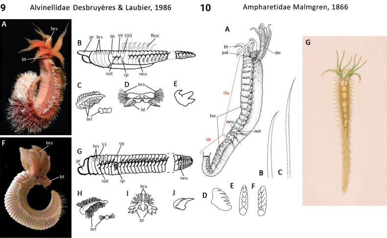
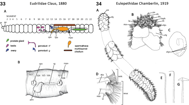
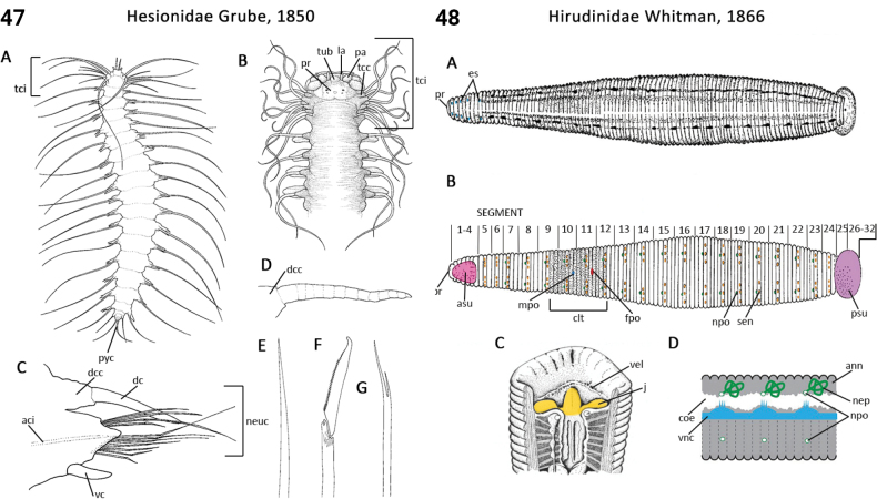
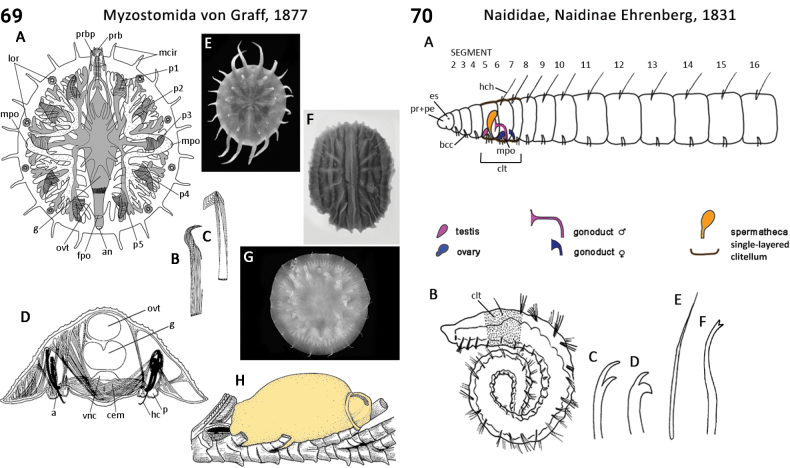
Phylum Annelida are ubiquitous metazoans found in almost every terrestrial and aquatic habitat on Earth. Historically, taxonomic studies on the phylum have been focused largely on its majorgroups, polychaetes, oligochaetes and leeches, so that while family-level keys for each group are available, no single-source identification guide exists to the world's annelid families. Here, the first illustrated linear key to annelid families is provided and family-level descriptions and diagnoses that distinguish individuals of each family from those of other families in the phylum are updated. This information is generated from an annelid DELTA database of 334 characters and 166 mostly family-level taxa. A link is provided to downloadable software (ANNiKEY Interactive) allowing the same data to be interrogated using the open-source DELTA program Intkey, which enables both interactive identification and taxonomic query functionality. For each family-level taxon, a diagnosis, full description, links to taxonomic data at the World Register of Marine Species, illustrations of diagnostic features, and a summary of the recent literature, including a list of published keys to genera and species are provided.

Keywords: ANNiKEY; Annelid; DELTA; computer taxonomy; diagnosis; interactive key; linear key; natural language descriptions; taxonomic verification.


Conflict of interest statement

The authors have declared that no competing interests exist.

Figures

Figures 1, 2.
Figures 1, 2.
Distinguishing features: 1.Aberrantidae: A–E, G, H.Aberrantaenignatica; F, I.A.banyulensis. A. Anterior region, dorsal, prostomial antenna missing; B. Anterior region, lateral; C. Parapodium from chaetiger 11; D. Posterior region, ventral (right pygidial cirrus missing); E, F. Capillary notochaetae; G–I. Lyriform and hooked neurochaetae. Abbreviations: cbr ciliated branchia; gpa grooved palp; ma median antenna; pc pygidial cirrus; pp pygidial papilla; tpe triannulate peristomium. Sources: A–I derivatives of Mackie (2019: fig. 7.2.1.2). 2.Acanthobdellidae: A–C.Acanthobdella sp.: A. General appearance; B. Anterior region; C. Diagram of anatomy, lateral view; D.Acanthobdellapeledina reproductive system, female and male. Abbreviations: an anus antc anterior chaetigers atr atrium eb ejaculatory bulb es eye spot fpo female pore hc hook chaeta int intestine mo mouth mpo male pore nep nephridium ovi ovisac pr prostomium psu posterior sucker S segment spo spermathecal pore tes testis vag vagina vnc ventral nerve cord. Sources: A–C derivatives of fig. 12, Mann (1962), D derivative of fig. 4, Davies and Govedich (2001)
Figures 3, 4.
Figures 3, 4.
Distinguishing features: 3.Acanthodrilidae: A. Schematic image of reproductive organs, dorsal side up; B. Arrangement of pores; C.Diplotremaaustralis, genital field. Abbreviations: fpo female pore gp genital papilla mpo male pore pe peristomium prpo prostate pore pr prostomium prg prostate gland S segment sd sperm duct spo spermathecal pore. Sources: A, B derivatives of fig. 8.4 B, 8.8 A Jamieson (2006), C derivative of fig. 17 Dyne and Jamieson (2004). 4.Acoetidae: A.Eupanthalis sp., animal with details of anterior, middle and posterior segments, dorsal view. B–F.Polyodontesaustraliensis: B. Anterior end, dorsal view; C. Parapodium of chaetiger 26; D. Superior neurochaetae from chaetiger 26; E. Spinose neurochaeta from chaetiger 27; F. Stout aristate neurochaeta from chaetiger 27; G. Anterior section of tube; H.Panthalisoerstedi entire animal, dorsal view. Abbreviations: ely elytron la lateral antenna ma median antenna neuc neurochaetae notc notochaetae omm ommatophore pa palp pbr pinnate branchia pr prostomium pyp pygidial papilla sf silk fibre spg spinning gland vc ventral cirrus. Sources: A–G after fig. 1.68 Beesley et al. (2000), H after Pl. XXVIA, fig. 20, MacIntosh (1900–1922).
Figures 5, 6.
Figures 5, 6.
Distinguishing features: 5.Acrocirridae: A, B, D–G.Macrochaetaaustraliensis; A. Entire animal lateral view; B. Anterior end dorsal view; C.Chauvinelia sp. dorsal view of entire animal, chaetae are not drawn; D. Parapodium of chaetiger 30; E, F. Capillary from parapodium of chaetiger 10; G. Compound falciger from parapodium of chaetiger 35. Abbreviations: bra branchia cap capillary chaetae cf compound falciger intc interramal cirrus neu neuropodium not notopodium pa palp pr prostomium. Sources: A, B, D–G derivatives of fig. 1.111 Beesley et al. (2000), C derivative of fig. 7.3.1.7.8 Martínez et al. (2019). 6.Aeolosomatidae: A.Aeolosoma sp., live animal, dorsal view; B, C.Aeolosomacf.hemprichi, entire animal with three zooids, dorsal view (B) anterior end, dorsal view (C); D, E.Aeolosomapsammophilum, chaetal fascicle (D), sigmoid chaeta (E); F, G.Rheomorphaneizvestnovae dorsal view (F), dorsal view of anterior end (G); H–K.Hystricosomachappuisi, dorsal view (H), ventral view of anterior end (I), chaeta fascicle (J), sigmoid chaeta (K). Abbreviations: asc anterior sensory cilia nuo nuchal organ per peristomium pl pygidial lobe pr prostomium ps pigment spot zoo zoid. Sources: A after Malcolm Storey, Bioimages, The Virtual Field-Guide, UK https://www.bioimages.org.uk/, under cc by nc-sa-3.0; B, C after fig. 1.129 Beesley et al. (2000), D–G redrawn from Bunke (1967), H–K redrawn from Pop (1975).
Figures 7, 8.
Figures 7, 8.
Distinguishing features: 7.Alluroididae: A. Diagram of reproductive organs, dorsal side up. Abbreviations: mpo male pore pe peristomium pr prostomium. Sources: A derivative of fig. 8.4 A Jamieson (2006). 8.Almidae: A. Schematic image of reproductive organs, dorsal side up; B.Glyphidriluskukenthali, anterior end showing genital region, ventral view. Abbreviations: ala alae fpo female pore mpo male pore pap papilla pe peristomium pr prostomium S segment. Sources: A, B derivatives of fig. 8.4B, 8.38A Jamieson (2006).
Figures 9, 10.
Figures 9, 10.
Distinguishing features: 9.Alvinellidae: A.Alvinellapompejana entire animal; B–E.A.pompejana; B. Body shape and position of the uncinigerous tori; C. Gill shape; D. Buccal apparatus with tentacles; E. Shape of uncini; F.Paralvinellasulfincola, entire animal; G–J.P.sulfincola; G. Body shape and position of the uncinigerous tori; H. Gill shape; I. Buccal apparatus with tentacles; J. Shape of uncini. Abbreviations: bra branchia brl branchial lamella brs branchial scar bt buccal tentacles fbac filamentous bacteria neu neuropodium not notopodium pr prostomium S segment sp spine. Sources: A–J derivatives of fig. 4, fig. 10 Jollivet and Hourdez (2021). 10.Ampharetidae: A–E.Amphicteisdalmatica; A. Entire animal lateral view; B, C. Chaetae: B. Palea; C. Capillary notochaeta; D, E. Neurochaetal uncinus from chaetiger 15 lateral (D) and frontal (E) views; F.Auchenoplax sp., neurochaetal uncinus almost frontal view; G.Amphareteacutifrons, entire animal. Abbreviations: ab abdomen bt buccal tentacles neu neuropodium not notopodium pal paleae sbr smooth branchia thx thorax tor torus. Sources: A–F derivatives of fig. 1.112 Beesley et al. (2000), K derivative of MacIntosh (1900–1922).
Figures 11, 12.
Figures 11, 12.
Distinguishing features: 11.Amphinomidae: A–H.Eurythoe sp.; A. Entire animal; B. Anterior end dorsal view; C. Parapodium of chaetiger 13; D–F. Chaetae from parapodium of chaetiger 13: D. Notoacicular spine; E. Fine ‘spurred’ notochaeta; F. Detail of spur as indicated in E. G, H. Chaetae from parapodium of chaetiger 38: G. Furcate neurochaeta; H. ‘Harpoon’ notochaeta. Abbreviations: bra branchia car caruncle dc dorsal cirrus es eye spot la lateral antenna ma median antenna neuc neurochaetae notc notochaetae nuo nuchal organ pa palp pr prostomium vc ventral cirrus. Sources: A–H derivatives of fig. 1.66 Beesley et al. (2000). 12.Aphroditidae: A.Laetmonicemoluccana dorsal view; B–O. Generalised aphroditid: B. Prostomium, first segment dorsal view; C. Parapodium posterior view; D–I. Notochaetal types; J–O. Neurochaetal types; P.Aphroditaaculeata, dorsal view. Abbreviations: dtc dorsal tentacular cirri ely elytron elys elytral scar es eye spot ft facial tubercle ma median antenna nc notopodial capillary neuc neurochaetae nfe notopodial felt nsp notopodial spine pa palp pph palophore pr prostomium tnph tentaculophore vc ventral cirrus vtc ventral tentacular cirri. Sources: A–O derivatives of fig. 1.70 Beesley et al. (2000), P derivative of MacIntosh (1900–1922).
Figures 13, 14.
Figures 13, 14.
Distinguishing features: 13.Apisthobranchidae: A–G.Apistobranchus, various species; A, B. Head and tail ends, ventral view; C. Head, dorsal view; D. Chaetiger 7; E–G. Neurochaetae showing differently worn tips. Abbreviations: ci cilia cip ciliated prostomium dpc dorsal pygidial cirrus intc interramal cirrus nel neuropodial lobe nell neuropodial lamella nol notopodial lobe nuo nuchal organ pa palp pr prostomium py pygidium vpc ventral pygidial cirrus vcb ventral ciliary band. Sources: A, B after fig. 5.1.1 Blake (1996a), C–G after fig. 1.103 Beesley et al. (2000). 14.Arenicolidae: A, E, F.Arenicolabombayensis; B.Arenicolamarina; C.Abarenicolawellsi; D.Arenicoloidesbranchialis; A. Whole body, lateral view, showing three distinct parts, achaetous head, chaetigerous trunk and achaetous tail (tail may be chaetigerous in some species); B. Whole body; C, D. Two types of prostomia: C. Trilobate; D. Transverse; E. Pilose notopodial capillary; F. Neuropodial hook. Abbreviations: as achaetous segment, bra branchia, notc notochaetae, ntp notopodium, nup neuropodium, pe peristomium, pr prostomium, prb proboscis (everted). Sources: A, E, F derivatives of fig. 1.48, Beesley et al. (2000), B pl. LXXXVIII, fig. 5, McIntosh (1915), C, D derivatives of Darbyshire (2020).
Figures 15, 16.
Figures 15, 16.
Distinguishing features: 15.Biwadrilidae: A. Diagram of reproductive organs, dorsal side up. B.Biwadrilusbathybates, ventral view. Abbreviations: clt clitellum, fpo female pore, mpo male pore, pe peristomium, pr prostomium, S segment. Sources: A derivative of fig. 8.4, Jamieson (2006), Non-leech Clitellata, B derivative of fig. 1, Blakemore (2008). 16.Branchiobdellidae: A. Diagram of lateral aspect of a hypothetical branchiobdellid showing anatomic characters; B. Ventral view of Triannulatamagna; C–E.Branchiobdellaparasita: C. Dorsal view; D. Upper jaw; E. Lower jaw. Abbreviations: an anus clt clitellum dap dorsal appendage dr dorsal ridge g gut j jaw llo lateral lobe mo mouth mpo male pore nep nephridium npo nephridial pore ph pharynx prg prostate gland psu posterior sucker spo spermathecal pore spt spermatheca ten tentacle vnl ventral lip. Sources: A–C derivatives of fig. 49, 55, 56 Brinkhurst and Gelder (2001), D, E derivatives of Timm (2009).
Figures 17, 18.
Figures 17, 18.
Distinguishing features: 17.Capilloventridae: A. Diagram showing location of reproductive organs, dorsal side up; B.Capilloventeraustralis, hair and crotchet chaeta, C. Anterior of C.longicapitus with long prostomium; D. Anterior of C.acheronensis with rounded prostomium. Abbreviations: mo mouth pe peristomium pr prostomium. Sources: A derivative of fig. 12.3 Thorp et al. (2019), C, D derivatives of figs 6, 45, 46 Pinder (2013). 18.Capitellidae: A.Notomastustorquatus parts of the entire animal; B.Dasybranchus species posterior segments bearing dorsal branchiae; C.Scyphoproctustowraensis posterior end with anal plate; D.Capitellacapitata’ thoracic segments (7 to 9) showing genital spines lateral view; E, F.Notomastustorquatus: E. Transverse section of body at chaetiger 10 showing arrangement of notopodia and neuropodia; F. Capillary notochaeta from thorax; G, H. Abdominal notochaetae of Mediomastusaustraliensis; G. Frontal view; H. Lateral view; I. Entire animal Capitellacapitata’. Abbreviations: apl anal plate gs genital spine bra branchia notc notochaetae pc pygidial cirrus pr prostomium prb proboscis py pygidium SI segment 1 Sources: A–G after fig. 1.49 Beesley et al. (2000), I after MacIntosh (1900–1922), pl. XCII, fig. 3.
Figures 19, 20.
Figures 19, 20.
Distinguishing features: 19.Chaetopteridae: A–D.Spiochaetopterus sp.: A. Anterior end of worm extended from its tube; B, C. Long notochaetae from chaetiger 6; D. ‘Cup’ chaeta from chaetiger 4; E. Uncinus from chaetiger 7; F.Chaetopterusvariopedatus, entire animal. Abbreviations: anot ailiform notopodia anr anterior region dcg dorsal ciliary groove gec green epithelial cells gpa grooved palp mir mid region mo mouth pe peristomium pls piston-like segment por posterior region pr prostomium. Sources: A–E after fig. 1.104 Beesley et al. (2000), F after MacIntosh (1900–1922), pl. LXXXIX, fig. 3. 20.Chrysopetalidae, Calamyzinae: Ichthyotomussanguinarius: A. Complete juvenile individual 1 mm long; B. Stylet jaws in open position; C. Mid-body parapodium; D. Neuropodial spiniger and falcigers. Abbreviations: dc dorsal cirrus es eye spot j jaw jc jaw carrier no nuchal organ pa palp pr prostomium vc ventral cirrus. Sources: A derivative of fig. 1 Watson (2022), B–D derivatives of text fig. 13, plate 1, fig. 12, plate 6, fig. 3b Eisig (1906).
Figures 21, 22.
Figures 21, 22.
Distinguishing features: 21.Chrysopetalidae, Chrysopetalinae: A.Paleanotus sp. entire animal dorsal view; B–H.Paleaequorsetula: B. Anterior end dorsal view; paleae on chaetigers 1 and 2 removed; C. Anterior end ventral view; D. Parapodium from chaetiger 30; notochaetal paleal groups shown; E. Main palea from the midbody showing internal and external structure; F–H. Compound neurochaetae from chaetiger 34: F. Superior spiniger; G. Mid-superior falciger; H. Mid-inferior falciger. Abbreviations: aci aciculae acn acirrose neuropodium segment dc dorsal cirrus dtc dorsal tentacular cirri la lateral antenna lap lateral paleae ma median antenna map main paleae mep median paleae mof mouth fold neuc neurochaetae notc notochaetae nuf nuchal fold pa palp pal parapodial palea vc ventral cirrus vc2 ventral cirrus, chaetiger 2 vc ventral cirrus, chaetiger 3 vtc ventral tentacular cirri. Sources: A–H after fig. 1.71 Beesley et al. (2000). 22.Chrysopetalidae, Dysponetinae: A.Dysponetuscaecus, anterior end, dorsal view; B, D, E.Dysponetuspygmaeus: B. 7-segmented juvenile ventral view; D. Neurochaetae; E. Notochaetae; C. Notochaetae. Abbreviations: buc buccal cirrus, dc dorsal cirrus, la lateral antenna, ma median antenna, neuc neurochaetae, pa palp, py pygidium, pyc pygidial cirrus. Sources: A derivative of fig. 3 Watson et al. (2014), B derivative of fig. 27.1 Bhaud and Cazaux (1987), C derivative of Handbook of Zoology, Watson (2022), D, E derivatives of Pl. 10, Imajima and Hartman (1964).
Figures 23, 24.
Figures 23, 24.
Distinguishing features: 23.Cirratulidae: A. Entire animal of Cirriformiacf.filigera dorsal view; B. Anterior end of Dodecaceria species dorsal view; C. Posterior end of Chaetozonesetosa; D–I. Chaetae: D. Capillary notochaeta from mid-body chaetiger Cirratulus species; E. Spine from mid-body neuropodia of Caullerielladimorphosetosa; F. Hook from mid-body neuropodia Dodecaceria species; G. Capillary notochaeta from posterior chaetiger Monticellinaaphelocephalus; H. Spine from posterior chaetiger of Chaetozonesetosa; I. Spine from posterior chaetiger of Tharyxlongisetosa; J.Cirriformiatentaculata, entire animal. Abbreviations: bra branchia cap capillary chaetae pa palp pe peristomium pr prostomium py pygidium sp spine. Sources: A–I after fig. 1.113 Beesley et al. (2000), J after McIntosh (1900–1922), pl. XCI, fig. 1. 24.Cossuridae: Cossura sp. A. Anterior end dorsal view; B. Parapodium of chaetiger 18; C. Neurochaeta from parapodium of chaetiger 8; D. Neurochaeta from parapodium of chaetiger 23; E. Posterior abdominal chaeta. Abbreviations: dbf dorsal branchial filament neuc neurochaetae notc notochaetae pe peristomium pr prostomium SI segment 1. Sources: A–E after fig. 1.50 Beesley et al. (2000).
Figures 25, 26.
Figures 25, 26.
Distinguishing features: 25.Criodrilidae: A. Diagram of reproductive organs dorsal side up; B–D.Criodriluslacuum: B. Anterior body of sexually mature specimen in ventral view with glandular pads, beginning of clitellum and a spermatophore; C. Spermathecal chaeta; D. Chaetal bundle, paired simple-pointed crotchets. Abbreviations: fpo female pore sch spermathecal chaeta mpo male pore pe peristomium pr prostomium scc simple crochet chaeta. Sources: A derivative of fig. 8.4, Jamieson (2006), B–D derivatives of fig. 12.2, 12.28, 12.3 Thorp et al. (2019). 26.Ctenodrilidae: A–C.Aphropharynx sp.: A. Lateral view of entire animal; B. Capillary chaeta from parapodium of chaetiger 12; C. Multidentate spine from parapodium of chaetiger 12; D.Ctenodriluscf.serratus, chaetal hooks; E.Raricirrusvariabilis, pectinate chaetiger. Abbreviations: cap capillary chaetae pe peristomium pr prostomium py pygidium sp spine. Sources: A–D after fig. 1.114 Beesley et al. (2000), E derivative of fig. 7.15.5.2 Dean and Blake (2019).
Figures 27, 28.
Figures 27, 28.
Distinguishing features: 27.Dinophilidae: A.Trilobodrilusaxi live specimen, mature female; B.Trilobodrilus species, details of head, dorsal view. Abbreviations: crg ciliary ring, ct ciliary tuft, mo mouth, no nuchal organ, pe peristomium, pr prostomium. Sources: A, B derivatives of fig. 1B, fig. 2A Westheide (2019), respectively. 28.Diurodrilidae: A, B.Diurodrilus sp.: A. Entire adult female, ventral view; B. Entire young female, dorsal view showing ‘cuticular plates’. Abbreviations: crg ciliary ring, ct ciliary tuft, ctp cuticular plate, mo mouth, pe peristomium, pr prostomium, pt pygidial toe. Sources: A, B after fig. 1.64 Beesley et al. (2000).
Figures 29, 30.
Figures 29, 30.
Distinguishing features: 29.Dorvilleidae: A–H.Schistomeringosloveni: A. Entire animal dorsal view; B. Anterior end dorsal view; C, D. Jaw parts: C. Maxillae dorsal view; D. Mandibles ventral view; E. Parapodium from chaetiger 10; F–H. Chaetae from parapodium of chaetiger 10: F. Simple chaeta; G. Furcate chaeta; H. Compound falciger; I.Dorvillearubrovittata entire animal. Abbreviations: aci aciculae bpe biannulate peristomium dc dorsal cirrus es eye spot ibp inferior base plate ifd inferior free denticle j jaw mc marginal cirrus neuc neurochaetae pa palp pr prostomium sbp superior base plate sfd superior free denticle vc ventral cirrus. Sources: A–H after figs 1.58 A–H, Beesley et al. (2000), I after MacIntosh (1900–1922), pl. LV, fig. 1. 30.Dorydrilidae: A. Schematic diagram showing location of reproductive organs, dorsal side up; B.Dorydrilustetrathecus, anterior end; C.Dorydrilusmichaelseni, side view of anterior body with a protruded penis. Abbreviations: fpo female pore mpo male pore pe peristomium pes penis sheath pr prostomium S segment sc sigmoid chaeta. Sources: A derivative of fig. 12.3 Thorp et al. (2019). B, C derivatives of Timm (2009), fig. 135.
Figures 31, 32.
Figures 31, 32.
Distinguishing features: 31.Enchytraeidae: A. Schematic diagram showing location of reproductive organs, dorsal side up; B.Enchytraeusalbidus anterior body of sexually mature specimen with clitellum; C–J. Chaetae types. Abbreviations: clt clitellum mpo male pore pe peristomium pr prostomium scc simple crochet chaeta. Sources: A derivative of fig. 12.3 Thorp (2019), B–J derivatives of Timm (2009) figs 139, 161. 32.Erpobdellidae: A.Dina orientalis, dorsal view; B. Head end, dorsal view showing arrangement of eyespots in Erpobdellaoctoculata; C. Reproductive system of Erpobdellacostata; D. Reproductive system of Motobdellasedonensis. Abbreviations: atr atrium ed ejaculatory duct ep epididymus fpo female pore mpo male pore ova ovary ovd oviduct psu posterior sucker tes testis vd vas deferens. Sources: A after fig. 1 Grosser et al. (2011), B, C after fig. 2 Oceguera-Figueroa et al. 2010, D after fig. 3 Govedich et al. 1998.
Figures 33, 34.
Figures 33, 34.
Distinguishing features: 33.Eudrilidae: A. Diagram of reproductive organs, dorsal side up; B.Stuhlmanniavariabilis showing ventral view and seminal groove. Abbreviations: fpo female pore mpo male pore pe peristomium pen penis pr prostomium S segment sg seminal grove spo spermathecal pore. Sources: A, B derivatives of fig. 8.4 B, 8.10 Jamieson (2006). 34.Eulepethidae: Mexieulepis sp. A. Entire animal dorsal view, anterior, middle and posterior sections; B. Dorsal view of anterior end with first two pairs of elytra removed; C. Elytron from chaetiger 13; D. Parapodium of chaetiger 21 with elytron removed; E, F. Chaetae from parapodium of chaetiger 27: E. Superior notochaetal spine; F. Neurochaetal spine. G. Neuroacicula from parapodium of chaetiger 27. Abbreviations: dc dorsal cirrus dtc dorsal tentacular cirrus ely elytron elyn elytron notch elys elytral scar la lateral antenna ma median antenna neuc neurochaetae notc notochaetae nta notoacicula nua neuroacicula pa palp tnph tentaculophore vc ventral cirrus vtc ventral tentacular cirrus. Sources: A–G derivative of fig. 1.72 Beesley et al. (2000).
Figures 35, 36.
Figures 35, 36.
Distinguishing features: 35.Eunicidae: A–H.Euniceantennata: A. Anterior end, dorsal view; B, C. Jaw parts: B. Maxillae dorsal view; C. Mandibles ventral view; D. Parapodium from chaetiger 26; E–H. Chaetae: E. Simple limbate, view from parapodium of chaetiger 41; H. Subacicular hook from parapodium of chaetiger 29; I.Marphysabelli entire animal. Abbreviations: aci acicula bra branchia cpl cutting plate dc dorsal cirrus la lateral antenna M maxilla ma median antenna MC maxillary carrier paa palpal antenna pc peristomial cirrus pe peristomium pr prostomium sah subacicular hook sha shaft vc ventral cirrus. Sources: A–H derivatives of fig. 1.59 Beesley et al. (2000), I derivative of MacIntosh (1900–1922), pl. LV, fig. 5. 36.Euphrosinidae: A–C. An unidentified euphrosinid species: A. Entire animal dorsolateral view; B. Anterior end dorsal view; C. Anterior end ventral view. D–F.Euphrosinecf.superba: D. Parapodium of chaetiger 19; E, F. Furcate and ringent notochaetae from parapodium of chaetiger 32; G.Euphrosinefoliosa dorsal, ventral surfaces. Abbreviations: bra branchia car caruncle dc dorsal cirrus es eye spot la lateral antenna ma median antenna mo mouth neuc neurochaetae notc notochaetae py pygidium vc ventral cirrus vll ventrolateral lip. Sources: A–F derivatives of fig. 1.67 Beesley et al. (2000), G derivative of MacIntosh (1900–1922; pl. XXIV, fig. 3).
Figures 37, 38.
Figures 37, 38.
Distinguishing features: 37.Fabriciidae: A.Fabricinuda sp. thoracic uncini; B, C.Fabriciola sp.: B. Neurochaetal uncinus; C. Abdominal notochaetal uncinus; D, E. Capillary chaetae Fabricialeidyii; F. Whole body Manayunkiaspeciosa. Abbreviations: ab abdomen brh branchial heart es eye spot neu neuropodium not notopodium pe peristomium rad radiole rc radiolar crown thx thorax vfl ventral filamentous appendage. Sources: A derivative of fig. 1.100, B, C derivatives of fig. 1.99 Beesley et al. (2000), D–F derivatives of Leidy (1884 [1883]; pl. IX, figs 1, 4, 14). 38.Fauveliopsidae: A.Fauveliopsis sp. entire animal ventrolateral view; B. Notopodial spine from chaetiger 2; C. Simple chaeta from posterior end; D, E.Fauveliopsisglabra: D. Parapodium of chaetiger 10; E. Parapodium of chaetiger 21. Abbreviations: cap capillary chaetae ip interramal papilla pe peristomium pr prostomium sp spine. Sources: A–C derivatives of fig. 1.115 of Beesley et al. (2000), D, E derivatives of fig. 7.15.3.7 of Zhadan and Salazar-Vallejo (2019).
Figures 39, 40.
Figures 39, 40.
Distinguishing features: 39.Flabelligeridae: A, B.Diplocirrus sp.: A. Entire animal dorsolateral view; B. Chaetae and papillae of chaetiger 15; C. Anterior end of Therochaeta sp. showing the cephalic cage dorsal view; D–F. Chaetae from chaetiger 20 of Pherusa sp.: D. Barred capillary with details of surface shown in E; F. Spine; G.Pherusaplumosa entire animal. Abbreviations: as1 achaetous segment 1 as2 achaetous segment 2 bra branchia ccc cephalic cage chaetae ccp cephalic cage papillae cph cephalic hood neuc neurochaetae notc notochaetae pap papillae. Sources: A–F after fig. 1.116 of Beesley et al. (2000), G after MacIntosh (1900–1922; pl. LXXXIX, fig. 1). 40.Glossiphonidae: A.Placobdellacostata, dorsal view; B. Ventral view of head of Placobdella sp. showing mouth pore of sucker; C. Anatomy of Glossiphoniacomplanata. Abbreviations: ann annulus es eye spot fpo female pore mpo male pore ova ovary ovd oviduct pap papillae ppo proboscis pore pr prostomium psu posterior sucker S segment tes testis. Sources: A derivative of fig. 1 Sket and Trontelj (2008), B derivative of fig. 4 Mann (1962), C derivative of fig. 14 Mann (1962).
Figures 41, 42.
Figures 41, 42.
Distinguishing features: 41.Glossoscolecidae: A. Schematic image of reproductive organs, dorsal side up; B, C.Glossoscolex (Praedrilus) lutocolus: B. Lateral view; C. Ventral view. Abbreviations: fpo female pore mpo male pore pe peristomium pr prostomium S segment. Sources: A derivative of fig. 8.4 B Jamieson (2006), B, C derivatives of fig. 1a, b Bartz et al. (2012). 42.Glyceridae: A. Anterior end of Glycera species with pharynx everted; inset showing papilla; B. Jaw apparatus; C. Mid-body parapodium of Glycera species; D, E. Chaetae of Glycera species: D. Simple notochaeta; E. Compound neurochaeta. Abbreviations: al aileron bra branchia dc dorsal cerrus j jaw pa palp ph pharynx pcl postchaetal lobe pr prostomium pra prostomial appendages prl prechaetal lobe vc ventral cirrus. Sources: A, C–E derivative of fig. 1.73 Beesley et al. (2000), B derivatives of fig. 2 Böggemann (2022).
Figures 43, 44.
Figures 43, 44.
Distinguishing features: 43.Goniadidae: A. Anterior end of Goniada species with pharynx everted partially; inset showing papilla; B. Details of pharynx of Glycindearmigera showing the micrognaths and macrognaths, terminal anterior view; C. Anterior parapodium of Goniada species; D. Posterior parapodium of Goniada species; E–H. Compound neurochaetae of Goniada species from chaetiger 23 (E) chaetiger 21 (F) chaetiger 27 (G) chaetiger 40 (H). Abbreviations: ac acicular spine dc dorsal cirrus mac macrognath mic micrognath neu neuropodium not notopodium ph pharynx pr prostomium pra prostomial appendage vc ventral cirrus. Sources: A–I derivatives of fig. 1.74 Beesley et al. (2000). 44.Haemadipsidae: A, B.Haemadipsa sp.: A. Anterior end ventral view; B. Posterior end dorsal view; C, D.Idiobdellaseychellensis: C. Ventral view showing position of gonopores and medioventral papillae; D. Lateral view of tail end, lacking caudal auricles; E. Reproductive organs of Idiobdellaseychellensis; F.Haemadipsarjukjuana, dorsal view. Abbreviations: an anus atr atrium buf buccal frill caa caudal auricle ed ejaculatory duct ep epididymus es eye spot fpo female pore mo mouth mpo male pore nep nephridiopore ovi ovary ovd oviduct pap papilla psu posterior sucker S segment sd sperm duct tes testis vag vagina. Sources: A, B derivatives of fig. 21 Mann (1962), C, D derivatives of fig. 2 Borda and Siddall (2010), E derivative of fig. 6 Harding (1913), F derivative of fig. 1 Lai et al. (2011).
Figures 45, 46.
Figures 45, 46.
Distinguishing features: 45.Haplotaxidae: A. Schematic diagram showing location of reproductive organs, dorsal side up; B–E.Haplotaxisgordioides: B. General view; C. Anterior end; D. Ventral chaeta; E. Dorsal chaeta. Abbreviations: clt clitellum, mpo male pore pe peristomium pr prostomium scc simple crochet chaeta. Sources: A–C derivatives of fig. 12.24, 12.3 Thorp et al. (2019), D, E derivatives of Timm (2009), fig. 163. 46.Hartmaniellidae: A–I.Hartmaniella sp.: A. Anterior end in ventral view; B. Anterior end in dorsal view; C. Anterior end in lateral view; D. Parapodium from anterior chaetiger; E. Parapodium from mid-body chaetiger; F. Compound spiniger; G. Acicular spine; H. Acicula; I. Jaw apparatus (schematic). Abbreviations: antc anterior chaetigers bpe biannulate peristomium bra branchia ci cilia dc dorsal cirrus j jaw MI maxilla 1 MII maxilla 2 MIII maxilla 3 MIV maxilla 4 MC maxillary carrier midc midbody chaetigers nea neuroacicula noa notoacicula pr prostomium SI segment 1 vc ventral cirrus. Sources: A, B photo Chris Glasby, C–I after fig. 2 Carrera Parra (2003).
Figures 47, 48.
Figures 47, 48.
Distinguishing features: 47.Hesionidae: A. Hesionid sp. entire animal, dorsal view; B. Anterior end Leocrates sp. dorsal view; C. Median parapodium Nereimyra sp.; D–F. Cirrus and chaetae Nereimyra sp.: D. Details of dorsal cirrus; E. Simple chaeta; F. Compound chaeta; G. Forked chaeta Gyptis sp. from chaetiger 14. Abbreviations: aci acicula dc dorsal cirrus dcc dorsal cirri cirrophore la lateral antenna neuc neurochaetae pa palp pr prostomium pyc pygidial cirri tcc tentacular cirri cirrophore tci tentacular cirri tub tubercle vc ventral cirrus. Sources: A–G derivatives of fig. 1.75 Beesley et al. (2000). 48.Hirudinidae: A–C.Hirudomedicinalis: A. Dorsal view of the medicinal leech; B. Ventral view; C. Dissected anterior end to show jaws; D. General organisation of body segments in leeches. Abbreviations: ann annulus asu anterior sucker coe coelom clt clitellum es eye spot fpo female pore j jaw (coloured) mpo male pore nep nephridium npo nephridial pore pr prostomium psu posterior sucker sen sensillium vel vellum vnc ventral nerve cord. Sources: A–C derivatives of Mann (1962), D derivative of fig. 2, Kuo and Lai (2019).
Figures 49, 50.
Figures 49, 50.
Distinguishing features: 49.Histriobdellidae: Stratiodrilustasmanicus. A. Entire animal ventral view; B. Prionagnath-type jaws with maxillae everted ventral view. Maxillae (blue) showing long carrier and multiple jaw elements; mandibles (orange) with long shaft and unfused cutting plate. Abbreviations: cla clasper cpl cutting plate cpo copulatory organ j jaw la lateral antenna lca locomotory appendage ma median antenna MI maxilla 1 MII maxilla 2 MIII maxilla 3 MIV maxilla 4 pa palp ppc parapodial cirrus. Sources: A, B after fig. 1.65 Beesley et al. (2000). 50.Hormogastridae: A. Schematic image of reproductive organs, dorsal side up; B.Hormogasterrediiinsularis, anterior end showing genital fields; C.Ailoscolexlacteospumosus, anterior end showing genital fields. Abbreviations: fpo female pore mpo male pore pe peristomium pr prostomium S segment spo spermathecal pore tpu tubercular pubertatis. Sources: A, B, C derivatives of figs 8.4B, 8.39B, 8.39A Jamieson (2006).
Figures 51, 52.
Figures 51, 52.
Distinguishing features: 51.Hrabeiellidae: A–C.Hrabeiellaperiglandulata: A. Whole specimen showing characteristic arrangement of epidermal glands; B. Living adult animal; C. Typical arrangement of chaetae segment. Abbreviations: ch chaeta eg epidermal gland gc gland cell mo mouth. Sources: A–C derivative of fig. 7.5.1.1 Purschke (2019a). 52.Iospilidae: A–C.Iospilusphalacroides: A. Entire animal dorsal view; B. Head dorsal view; C. Uniramous parapodium of anterior body; D. Compound chaeta Phalacrophorusuniformis. Abbreviations: bip biramous parapoda dc dorsal cirrus es eye spot nea neuroacicula pr prostomium py pygidium tci tentacular cirri unp uniramous parapodia. Sources: A–D derivatives of fig. 1.76, Beesley et al. (2000).
Figures 53, 54.
Figures 53, 54.
Distinguishing features: 53.Iphionidae: A.Iphionetreadwelli anterior end dorsal view (right palp regenerating); B–F.Iphionemuricata: B. Left middle elytron, with detail of polygonal area and spinous tubercles; C. Right parapodium from segment 2, anterior view; E. Neurochaeta from segment 2; D. Right middle elytrigerous parapodium, anterior view; F. Upper, middle, and lower neurochaeta; G, H. Entire animal (elytra pairs numbered). Abbreviations: aci acicula aprn anterior prostomial notch dtc dorsal tentacular cirrus ely elytron elys elytral scar epa elytra polygonal area etub elytra tubercle la lateral antenna opap occipital papilla pa palp pap papilla tnph tentaculophore vc ventral cirrus vtc ventral tentacular cirri. Sources: A–F derivative of figs 1, 2, 7 Pettibone (1986), G, H photo: Arthur Anker. 54.Kynotidae: A. Schematic image of reproductive organs, dorsal side up; B.Kynotuscingulatus ventral surface of segment 13–16, showing the pores of three pairs of prostates; a fourth pair discharges at the male pores; clasper is shown evaginated through the left male pore (clasper pore). Abbreviations: cla clasper fpo female pore mpo male pore pe peristomium pr prostomium prpo prostate pore S segment. Sources: A, B derivatives of fig. 8.4 B, 8.37 B, Jamieson (2006).
Figures 55, 56.
Figures 55, 56.
Distinguishing features: 55.Lacydoniidae: A. Entire animal Lacydoniabrasiliensis, dorsal view; B, C. Anterior end of Lacydoniaoculata, dorsal view (B), vental view (C); D. Posterior end of Lacydoniaoculata; E. Parapodium of Lacydoniamiranda; F. Notopodial capillary chaeta of Lacydonialaureci; G. Neuropodial spinigerous chaeta of Lacydonialaureci. Abbreviations: dc dorsal cirrus es eye spot eye eye la lateral antenna ma median antenna nea neuroacicula nta notoacicula pa palp pyc pygidial cirrus pyp pygidial papilla ps pigment spot py pygidium vc vental cirrus. Sources: A derivative of fig. 1 Rizzo and Magalhães (2022a), B, C, D derivatives of fig. 1.77 Beesley et al. (2000), E, F, G derivatives of fig. 2, Rizzo and Magalhães (2022a). 56.Longosomatidae: A.Heterospiolongissima posteriorly incomplete; B.Heterospio sp. anterior end dorsal view; C. Detail of head dorsal view, palps missing; D–F. Chaetae: D. Capillary notochaeta; E. Spine of posterior chaetiger with detail of tip; F. Aristate notochaeta; G.Heterospiomediterranea parapodium from elongated segment with circle of spines and capillaries; H.H.longissima posterior end lateral view. Abbreviations: bra branchia neuc neurochaetae notc notochaetae pe peristomium pr prostomium py pygidium S8 segment 8 S9 segment 9. Sources: A, G derivatives of fig. 1 H derivative of fig. 2 Blake and Maciolek (2019a), B–F derivative of fig. 1.105 Beesley et al. (2000).
Figures 57, 58.
Figures 57, 58.
Distinguishing features: 57.Lopadorrhynchidae: A–D.Pelagobiaviguieri; A. Entire animal ventral view; B. Anterior end ventral view; C. Parapodium from chaetiger 2; D. Compound neurochaeta from parapodium of chaetiger 2. Abbreviations: dc dorsal cirrus dtc dorsal tentacular cirrus la lateral antenna pa palp pc pygidial cirrus pr prostomium vc ventral cirrus. Sources: A–D derivative of fig. 1.78 Beesley et al. (2000). 58.Lumbricidae: A. Diagram of reproductive organs, dorsal side up; B.Eiseniellatetraedra, anterior body lateral view; C. Chaeta; D.Lumbricusterrestris external features of worm showing genital pores, seminal groove and clitellum. Abbreviations: clt multilayered clitellum fpo female pore mpo male pore pe peristomium pr prostomium S segment sg seminal grove spo spermathecal pore. Sources: A, D derivatives of fig. 8.4 B, 8.53 A Jamieson (2006), B derivative of fig. 30, Martin and Aït Boughrous (2012), C derivatives of Timm (2009: 189).
Figures 59, 60.
Figures 59, 60.
Distinguishing features: 59.Lumbriculidae: A. Diagram showing typical location of reproductive organs (highly variable), dorsal side up; B, C.Lumbriculusvariegatus: B. Anterior end; C. Bifid crotchet chaeta; D–F.Styloscolextubulatus: D. Anterior body of mature specimen with clitellum; E. Simple crotchet chaeta. Abbreviations: bcc bifid crotchet chaeta clt clitellum mpo male pore pe peristomium pes penis sheath pr prostomium scc simple crochet chaeta tes testis. Sources: A, D, E derivatives of fig. 12.26, 12.3 Thorp et al. (2019), B, C derivatives of Timm (2009: 171). 60.Lumbrineridae: A–G.Lumbrineriscf.latreilli; A. Anterior end dorsal view; B, C. Jaw parts: B. Maxillae dorsal view; C. Mandibles ventral view; D. Parapodium from chaetiger 11; E–G. Chaetae: E. Simple limbate chaeta from parapodium of chaetiger 11; F. Compound hook from parapodium of chaetiger 11; G. Simple hook from parapodium of chaetiger 43; H.L.latreillii entire animal. Abbreviations: aci acicula MI maxilla 1 MII maxilla 2 MIII maxilla 3 MIV maxilla 4 MV maxilla 5 MC maxillary carrier neuc neurochaetae pcl postchaetal lobe pr prostomium. Sources: A–G derivatives of fig. 1.60 Beesley et al. (2000), H derivative of MacIntosh (1900–1922), pl. LIV, fig. 6.
Figures 61, 62.
Figures 61, 62.
Distinguishing features: 61.Lutodrilidae: A. Diagram of reproductive organs, dorsal side up; B.Lutodrilusmultivesiculatus, anterior end with genital region, in ventral view, showing alae. Abbreviations: ala alae fpo female pore mpo male pore pe peristomium pr prostomium S segment spm spermatophore. Sources: A, B derivatives of figs 8.4B, 8.38A Jamieson (2006). 62.Magelonidae: A.Magelonapapillicornis entire animal; B. Anterior end of Magelonadakini dorsal view; C. Thoracic parapodium of Magelonasinbadi; D. Abdominal parapodium of Magelonacrenulifrons; E–G. Notochaetae of Magelona sp. H. Neurochaeta of Magelona sp. Abbreviations: ab abdomen pa palp pr prostomium th thorax. Sources: A derivative of McIntosh (1915), pl. XC, fig. 6, B, E–H derivatives of fig. 1.106 Beesley et al. (2000), C, D derivatives of fig. 4.2.8 Mortimer (2019).
Figures 63, 64.
Figures 63, 64.
Distinguishing features: 63.Maldanidae: A.Micromaldanepamelae, entire animal, lateral view; B. Anterior end of Euclymenetrinalis dorsolateral view; C. Mid-body parapodium of Micromaldanepamelae; D. Double row of uncini of Rhodineloveni; E. Anterior hook of Lumbriclymeneinterstricta; F.Nicomachemaculata, entire animal. Abbreviations: anf anal funnel cap capillary chaetae ck cephalic keel cr cephalic rim es eye spots nc notopodial capillary nes neuropodial spine prb proboscis tor torus unc uncini. Sources: A–C derivatives of fig. 1.51 Beesley et al. (2000), D, E derivative of figs 4, 5 Assis et al. (2021), F derivative of McIntosh (1900–1922), pl. XCII, fig. 5. 64.Megascolecidae: A. Schematic image of reproductive organs, dorsal side up; B. Arrangement of pores; C.Spenceriellapenolaensis showing genital markings. Abbreviations: fpo female pore gp genital papilla mpo male pore pe peristomium prpo prostate pore pr prostomium prg prostate gland S segment sd sperm duct spo spermathecal pore. Sources: A–C derivatives of figs 8.4B, 8.8A, 8.12A Jamieson (2006).
Figures 65, 66.
Figures 65, 66.
Distinguishing features: 65.Melinnidae: A, B.Isoldapulchella: A. Anterior end dorsolateral view right branchiae removed; B. Branchia. C, D. Chaetae: C. Hook from segment 4; D. Acicular neurochaeta from chaetiger 1. E, F.Melinnauruguayi: E. Anterior uninus; F. Posterior uninus. Abbreviations: bra branchia brt branchial trunk bt buccal tentacles cll colar-like lateral lobe hc hook chaeta neu neuropodium not notopodium pbr pinnate branchia pr prostomium sbr smooth branchia. Sources: A–D derivative of fig. 1.112 Beesley et al. (2000), E, F derivative of fig. 7, Hessle (1917). 66.Microchaetidae: A. Diagram of reproductive organs, dorsal side up. Abbreviations: fpo female pore mpo male pore pe peristomium pr prostomium. Sources: A derivative of fig. 8.4B Jamieson (2006).
Figures 67, 68.
Figures 67, 68.
Distinguishing features: 67.Microphthalmidae: A–I.Microphthalmusmahensis: A. Anterior end; B. Posterior end showing fimbriate anal plate; C. Parapodium from a mid-body chaetiger; D, E. Notopodial chaetae: D. Pectinate chaeta; E. Two straight chaetae; F–I. Neuropodial chaetae: F. Superiormost simple chaeta; G. Inferiormost simple chaeta; H. Supraacicular compound chaeta with long blade; I. Subacicular compound chaeta with short blade. Abbreviations: apl anal plate dc dorsal cirrus es eye spot la lateral antenna neuc neurochaetae no nuchal organ pa palp pc pygidial cirrus ph pharynx pyg pygidium tci tentacular cirri vc ventral cirrus. Sources: A–I derivatives of figs 1, 2 Westheide (2013). 68.Moniligastridae: A. Schematic image of reproductive organs, dorsal side up, of Desmogaster (top) and Drawida and Moniligaster (bottom); B.Drawidapolydiverticulata ventral view. Abbreviations: fpo female pore mpo male pore pe peristomium pr prostomium S segment spo spermathecal pore. Sources: A derivative of fig. 8.4 A Jamieson (2006), and fig. 2 Martínez-Ansemil (1993), B derivative of fig. 1 A Narayanan et al. (2017), ZooKeys 691: 1–18.
Figures 69, 70.
Figures 69, 70.
Distinguishing features: 69.Myzostomida: A. General anatomy of Myzostomida; B. Hook of Myzostoma sp.; C. Support rod aciculum of Myzostomagigas; D. cross-section of Myzostomaglabrum; E–G. Body shape variation in Myzostoma species; H.Endomyzostoma sp. in situ in soft cyst on arm host crinoid. Abbreviations: a acicula an anus cem central muscle fpo female pore g gut hc hook chaeta lor lateral organ mcir marginal cirrus mpo male pore ovt ovarian tube p parapodium prb proboscis prbp proboscis papilla vnc ventral nerve cord. Sources: A redrawn from fig. 1 Graff (1884), B–H derivative of figs 2.1, 2.2, 2.3, 2.4, 2.10, 2.13 Beesley et al. (2000). 70.Naididae, Naidinae: A. Schematic diagram showing location of reproductive organs, dorsal side up; B–E.Naisbarbata: B. General view of a sexually mature specimen with clitellum; C. Anterior ventral chaeta; D. Posterior ventral chaeta; E. Needle chaeta; F.Naiscommunis bifid needle chaeta. Abbreviations: bcc bifid crotchet chaeta clt single-layered clitellum es eye spot hch hair chaetae mpo male pore pe peristomium pr prostomium. Sources: A derivative of fig. 12.3 Thorp et al. (2019), B–F derivatives of Timm (2009: 55, 63).
Figures 71, 72.
Figures 71, 72.
Distinguishing features: 71.Naididae, Opistocystinae: A. Diagram showing location of reproductive organs, dorsal side up; B.Crustipellis sp., left to right: tail end, head end. Abbreviations: bcc bifid crotchet chaeta clt single-layered clitellum fpo female pore hch hair chaetae mpo male pore pe peristomium pr prostomium prp prostomial palpode pyc pygidial cirrus. Sources: A derivatives of fig. 12.3 Thorp et al. (2019), B derivatives of fig. 11 Brinkhurst and Gelder (2001). 72.Naididae, Pristininae: A. Schematic diagram showing location of reproductive organs, dorsal side up; B.Pristinalongiseta, left to right: whole body, ventral crotchet chaeta (2), needle chaeta; C.Pristinabilobata, left to right: whole body, needle chaetae (3), anterior ventral chaeta (2), posterior ventral crotchet chaeta (2). Abbreviations: bcc bifid crotchet chaeta hch hair chaetae mpo male pore pe peristomium pr prostomium. Sources: A derivative of fig. 12.3 Thorp et al. (2019), B, C derivatives of Timm (2009: 67, 69).
Figures 73, 74.
Figures 73, 74.
Distinguishing features: 73.Naididae, Tubificinae: A. Diagram showing location of reproductive organs, dorsal side up; B. Dorsal chaetal bundle with hair and bifid (or pectinate) chaetae; C–H.Potamothrixhammoniensis: C. General view of sexual mature specimen with clitellum; D. Anterior ventral chaeta; E. Posterior ventral chaeta; F. Pectinate chaeta; G, H. Spermathecal chaetae; I.Branchiurasowerbyi, posterior end with gills. Abbreviations: bcc bifid crotchet chaeta bra branchia clt single-layered clitellum hch hair chaetae pe peristomium pr prostomium sch spermathecal chaeta. Sources: A derivative of fig. 12.3 Thorp et al. (2019), B–I derivatives of Timm (2009: 85, 97). 74.Narapidae: A. Schematic diagram showing location of reproductive organs, dorsal side up. Sources: A. Derivative of fig. 12.3 Thorp et al. (2019) (modification of schematic diagram of Naididae based on information in Erséus (2005), fig. 1; chaetae removed from diagram).
Figures 75, 76.
Figures 75, 76.
Distinguishing features: 75.Nephytidae: A. Entire animal of Nephtysinornata dorsal view; B. Anterior end of Nephtyslongipes with pharynx everted dorsal view; C. Parapodium of Nephtyslongipes from chaetiger 15; D, E. Chaetae of Nephtysinornata: D. Barred notochaeta; E. Capillary notochaeta; F. Capillary neurochaeta of Nephtyslongipes; G. Lyrate chaeta from neuropodium of Inermonephtyspalpata; H.Nephtyscf.tulearensis preserved specimen dissected pharynx; I.Nephthyscaeca entire animal. Abbreviations: dc dorsal cirrus es eye spot irb interramal branchia j jaw la lateral antenna neu neuropodium not notopodium pa palp pc pygidial cirrus ph pharynx SI segment 1 S2 segment 2 spp subterminal pharyngeal papilla tpp terminal pharyngeal papilla vc vental cirrus. Sources: A–G after fig. 1.79, Beesley et al. (2000), H after fig. 1H Ravara et al. (2022), I MacIntosh (1900–1922), pl. XLIII, fig. 1. 76.Nereididae: A. Entire animal of Platynereisantipoda; B–D. Anterior ends with pharynx everted: B.Neanthesvaalii; C.Australonereisehlersi; D.Namanereislittoralis Hutchings & Turvey, 1982; E. Parapodium from chaetiger 20 of Neanthescricognatha; F. Notopodial heterogomph falciger Nereismaxillodentata; G. Notopodial homogomph falciger from median parapodium Nereismaxillodentata; H. Simple falciger Simpliseta sp.; I. Notopodial heterogomph spiniger from Neantheskerguelensis; J.Hedistediversicolor. Abbreviations: dc dorsal cirrus j jaw la lateral antenna neuc neurochaetae notc notochaetae pe peristomium pgn pharyngeal paraganths php pharyngeal papillae pr prostomium tci tentacular cirri vc ventral cirrus. Sources: A–I derivatives of fig. 1.80 Beesley et al. (2000), J derivative of MacIntosh (1900–1922), pl. LII, fig. 3.
Figures 77, 78.
Figures 77, 78.
Distinguishing features: 77.Nerillidae: A–G. Variation in body morphology across a selection of nerillid genera (all dorsal views); A. Entire animal of Nerillaaustralis; B.Paranerillacilioscutata; C.Leptonerillaprospera; D.Thalassochaetuspalpifoliaceus; E.Meganerillaswedmarki; F.Mesonerillaintermedia; G.Nerillidiummediterraneum; H. Compound chaeta of Mesonerillaroscovita. Sources: A, H derivatives of fig. 1.93 Beesley et al. (2000), B–G derivatives of fig. 5C, E, I, L, N, Q of Worsaae (2021), original sources as indicated therein. Palps coloured to emphasize shape variation. 78.Ocnerodrilidae: A. Schematic image of reproductive organs, dorsal side up; B.Eukerriaborellii, ventral view, showing seminal grooves connecting prostate and male pores. Abbreviations: fpo female pore mpo male pore pe peristomium pr prostomium prpo prostate pore S segment scc simple crochet chaeta sg seminal grove. Sources: A, B derivatives of fig. 8.4 B, 8.9 Jamieson (2006).
Figures 79, 80.
Figures 79, 80.
Distinguishing features: 79.Oenonidae: A–F.Oenonefulgida; A. Anterior end dorsal view; B, C. Jaw parts: B. Maxillae dorsal view; C. Mandibles ventral view; D. Parapodium from chaetiger 45; E, F. Chaetae from parapodium of chaetiger 45: E. Simple limbate chaeta; F. Subacicular hook; G.Arabellairicolor entire animal. Abbreviations: aci aciculae ant antennae BP base plate dc dorsal cirrus es eye spot MI maxilla 1 MII maxilla 2 MIII maxilla 3 MIV maxilla 4 MV maxilla 5 MC maxillary carrier pcl postchaetal lobe pe peristomium pr prostomium sah subacicular hook. Sources: A–F derivatives of fig. 1.61 Beesley et al. (2000), G derivative of MacIntosh (1900–1922), pl. LIV, fig. 4. 80.Onuphidae: A. Entire animal ventral view; B. Anterior end of Hirsutonuphismariahirsuta dorsolateral view; C. Anterior end of Longibrachiumlongipes dorsolateral view; D, E. Jaw parts of H.mariahirsuta: D. Maxillae dorsal view; E. Mandible ventral view; F. Parapodium of H.mariahirsuta from chaetiger 3; G. Posterior parapodium of H.mariahirsuta from chaetiger 30; H. Pseudocompound hook from chaetiger 3 of H.mariahirsuta; I. Pectinate chaetae from chaetiger 31 of H.mariahirsuta; J. Limbate chaeta; K. Subacicular hook. Abbreviations: bra branchia cpl cutting plate dc dorsal cirrus flp frontal lip la lateral antenna lac lateral antenna ceratophore M maxilla MC maxillary carrier ma median antenna mop modified parapodium p parapodium paa palpal antenna pc pygidial cirrus pcl postchaetal lobe pe peristomium pph palophore prl prechaetal lobe sha shaft vc ventral cirrus vgp ventral glandular pad. Sources: A derivative of fig. 3 Budaeva (2021), B–K derivatives of fig. 1.62 Beesley et al. (2000).
Figures 81, 82.
Figures 81, 82.
Distinguishing features: 81.Opheliidae: A, B.Armandiaintermedia: A. Entire animal lateral view; B. Capillary chaeta from mid-body parapodium; C, D.Opheliaalgida: C. Entire animal; lateral view; D. Posterior end dorsal view; E, F.Thoracopheliamammillatus: E. Entire animal, lateral view; F. Posterior end dorsal view; G.Polyophthalmuspictus, entire animal. Abbreviations: anf anal funnel anr anterior region bra branchia lgr lateral groove lse lateral segmental eyespot mir mid region pap papilla por posterior region prp prostomial palpode pvl pygidial ventral lobe pyp pygidial papilla vgr ventral groove. Sources: A, B derivatives from fig. 1.52 Beesley et al. (2000), C–F derivatives of figs 7.6.1.4, 7.6.1.5 Blake and Maciolek (2019f), G derivative from MacIntosh (1900–1922), pl. LXXXVIII, fig. 2. 82.Orbiniidae: A. Anterior end of Scoloplosnormalis lateral view; B. Posterior end of Scoloploscylindrifer dorsolateral view; C, D. Parapodia of Scoloplosnormalis: C. Thoracic parapodium from chaetiger 4 anterior view; D. Mid-abdominal parapodium from chaetiger 28 anterior view; E–K. Chaetae: E.Subuluncini from thoracic neuropodium of chaetiger 9 Nainerisaustralis; F. Hooded spine from thoracic neuropodium of chaetiger 9 N.australis; G. Arrow-shaped spine from thoracic neuropodium of chaetiger 14 Phylofelix; H. Flail-tipped chaeta from abdominal neuropodium Orbiniahartmanae; I. Furcate abdominal chaeta P.felix; J. ‘Swanshaped’ hook from abdominal neuropodium Proscoloplosconfusus; K. Crenulate capillary from abdominal neuropodium of chaetiger 28 S.normalis; E. Hook from chaetiger 13. Abbreviations: bra branchia cap capillary chaetae ci cilia neu neuropodium not notopodium pc pygidial cirrus pe peristomium pol postchaetal lobe pr prostomium prb proboscis py pygidium. Sources: A–K derivatives from figs 1.53, 1.55 Beesley et al. (2000).
Figures 83, 84.
Figures 83, 84.
Distinguishing features: 83.Orobdellidae: A–C.Orobdellanaraharaetmagarum, external (A, B) and internal (C) view showing alimentary canal and male and female reproductive systems; D, E.O.naraharaetmagarum showing anterior end, dorsal (D) and midbody, ventral (E), showing pores. Abbreviations: an anus, asu anterior sucker, atr atrium, clt clitellum, ep epididymus, es eye spot, fpo female pore, mpo male pore, npo nephridiopore, ovi ovisac, psu posterior sucker, S segment, tes testis. Sources: A–E derived from fig. 1, fig. 3 Nakano (2016a). 84.Oweniidae: A–D.Oweniacf.fusiformis: A. Entire animal, dorsal view; B. Transverse section of body showing chaetal distribution; C. Capillary notochaeta from parapodium of chaetiger 10; D. Neurochaetal hook from parapodium of chaetiger 10; E–H. Head ends of different genera: E.Galathoweniaoculata; F.Myriocheleheeri; G.Myrioweniacaliforniensis; H.Oweniaaustralis. Abbreviations: es eye spots neuc neurochaetae notc notochaetae tc tentacular crown pa palp pr prostomium. Sources: A–D derivatives of fig. 1.97 Beesley et al. (2000), E–H derivatives of fig. 4.1.1 Capa et al. (2019c).
Figures 85, 86.
Figures 85, 86.
Distinguishing features: 85.Ozobranchidae: A.Ozobranchusjantzeanus, dorsal view; B.Ozobranchusmargoi, dorsal view. Abbreviations: asu anterior sucker bra branchia psu posterior sucker tra trachelostome uro urostome. Sources: A derivative of fig. 19e Mann (1962), B derivative of fig. 2 Göpper et al. (2018). 86.Paralacydoniidae: A, C–F.Paralacydoniacf.weberi; A. Entire animal, dorsal view; B.Paralacydoniaparadoxa, pygidium; C. Anterior end dorsal view; D. Parapodium from chaetiger 15; E. Capillary from notopodium of chaetiger 15; F. Compound spiniger from neuropodium of chaetiger 15. Abbreviations: dc dorsal cirrus la lateral antenna neu neuropodium not notopodium pa palp pc pygidial cirrus pp pygidial papilla pr prostomium vc ventral cirrus. Sources: A, C–F derivatives from fig. 1.81 Beesley et al. (2000), B derivative of fig. 1 Rizzo and Magalhães (2022b).
Figures 87, 88.
Figures 87, 88.
Distinguishing features: 87.Paraonidae: A.Levinseniagracilis entire animal; B. Anterior end of Aricideacf.fauveli ventrolateral view; C, D. Parapodia of Aricidea (Acmira) sp.: C. From chaetiger 12; D. From posterior chaetiger; E–M. Chaetae: E. Notopodial bayonet chaeta from anterior postbranchial chaetiger Cirrophorus sp. F. Neuropodial capillary from chaetiger 6 Aricidea (Allia) sp. G. Notopodial lyrate chaeta from posterior chaetiger Cirrophorus sp. H. Posterior neuropodial hook Aricidea sp. I. Neuropodial spine from posterior chaetiger Aricidea (Acmira) sp. J. Neuropodial spine from posterior chaetiger of Aricideacf.fauveli; K. Modified neurochaeta from posterior chaetiger Aricidea (Allia) sp.; L. Neuropodial hook from posterior chaetiger Levinseniagracilis; M. Capillary from posterior chaetiger of Aricidea (Allia) sp. Abbreviations: bra branchia brci branchial cilia brr branchial region ma median antenna neuc neurochaetae nol notopodial lobe notc notochaetae pc pygidial cirrus pobr post branchial region pr prostomium prbr prebranchial region sp spine. Sources: A–M after fig. 1.54, Beesley et al. (2000). 88.Parergodrilidae: A–C.Stygocapitellasubterannea: A. Entire animal sagittal view of a male; B. Chaetae from parapodium of chaetiger 1; C. Chaetae from parapodium of chaetiger 2; D.Parergodrilusheideri, entire animal sagittal view of a male. Abbreviations: bc bilimbate chaeta, fc forked chaeta, gch genital chaetae, pr prostomium, pl pygidial lobe, wbc whip-like bilimbate chaeta. Sources: A–C derivatives of fig. 1.130 Beesley et al. (2000), D derivative of fig. 1B Purschke (2019b).
Figures 89, 90.
Figures 89, 90.
Distinguishing features: 89.Parvidrilidae: A. Diagram showing location of reproductive organs, dorsal side up; B. Smooth hair chaetae; C. Dorsal bundle with a single hair and needle chaeta; D. Ventral bundle of bifid crotchets; E.Parvidrilusmeyssonnieri anterior end. Abbreviations: bcc bifid crotchet chaeta clt single-layered clitellum hch hair chaetae mpo male pore nch needle chaetae pe peristomium pr prostomium. Sources: A–E derivatives of fig. 12.1, 12.2, 12.3 Thorp et al. (2019). 90.Pectinariidae: A.Pectinariaantipoda entire animal lateral view; B–E.Amphictenecrassa: B. Anterior end, ventrolateral view; C. Palea; D, E. Uncini from chaetiger 6, lateral (D), frontal (E) view; F. tube of a pectinariid; G.Lagiskoreni entire animal. Abbreviations: bra branchia buc buccal cirri but buccal tentacles cpa cephalic palea cvl cephalic veil neu neuropodium not notopodium scp scaphe scps scaphal spine vpd ventral pads. Sources: A–F derivatives of fig. 1.117 Beesley et al. (2000), G derivative of MacIntosh (1900–1922), pl. CXII, fig. 2.
Figures 91, 92.
Figures 91, 92.
Distinguishing features: 91.Phreodrilidae: A. Diagram showing location of reproductive organs, dorsal side up; B–E.Astacopsidrilusryuteki: B. Forebody of sexually mature specimen with clitellum; C. Ventral bifid crotchet chaeta; D. Dorsal bifid crotchet chaeta; E. Spermathecal chaeta; F. Unknown phreodrilid, dorsal chaeta. Abbreviations: bcc bifid crotchet chaeta clt single-layered clitellum hch hair chaetae mpo male pore pe peristomium pr prostomium sch spermathecal chaeta. Sources: A–E derivatives of fig. 12.22, 12.3 Thorp et al. (2019), F derivative of Timm (2009, 137). 92.Phyllodocidae: A, B.Phyllodoce sp.: A. Entire animal with pharynx everted dorsal view; B. Anterior end with pharynx everted dorsal view. C. Anterior end of Eumidafuscolutata with pharynx everted dorsal view; D. Median parapodium of Phyllodoce sp.; E.Eulaliaviridis entire animal. Abbreviations: aci acicula ce compound eye dc dorsal cirrus la lateral antenna ma median antenna neuc neurochaetae opap occipital papilla pa palp ph pharynx php pharyngeal papilla tci tentacular cirri tpp terminal pharyngeal papillae vc ventral cirrus. Sources: A–D derivatives of fig. 1.83 Beesley et al. (2000), E derivative of MacIntosh (1900–1922), pl. XLIV, fig. 1.
Figures 93, 94.
Figures 93, 94.
Distinguishing features: 93.Alciopini: A.Torreacandida entire animal with pharynx everted dorsal view; B–D. An undescribed Australian species of Torrea: B. Anterior end dorsal view; C. Simple chaeta; D. Compound chaeta. E. Cross-section of eye of Torreacandida; F.Vanadislongissima uniramous parapodium of anterior body. Abbreviations: ce compound eye con cornea dc dorsal cirrus irs iris la lateral antenna len lens ma medianantenna pa palp ph pharynx pig pigment layer pr prostomium tci tentacular cirri unp uniramous parapodium vc ventral cirrus. Sources: A–E after fig. 1.69 Beesley et al. (2000), F after fig. 22 Pettibone (1963). 94.Pilargidae: A, G.Hermundura sp.; A. Anterior end with pharynx everted dorsolateral view; B.Pilargis sp. dorsal view of anterior end; C–F. Parapodial morphology: C.Glyphohesioneklatti; D.Pilargisverrucosa; E.Sigambraparva; F.Litocorsastremma; G. Capillary neurochaeta from parapodium of chaetiger 45; H.Hermunduragladstonensis entire animal, lateral view. Abbreviations: dc dorsal cirrus hc hook chaeta la lateral antenna nes neuropodial spine nsp notopodial spine pa palp ph pharynx pr prostomium ps pigment spot pvl pygidial ventral lobe tci tentacular cirri vc ventral cirrus ver verruca vpp ventrolateral palpal papilla. Sources: A, B, G derivatives of fig. 1.84 Beesley et al. (2000), C–F, H derivatives of fig. 13 Glasby and Salazar-Vallejo (2022).
Figures 95, 96.
Figures 95, 96.
Distinguishing features: 95.Piscicolidae: A.Cystobranchusfasciatus, a fish leech dorsal view; B, C.Branchelliontorpedinis: B. Dorsal view (branchia removed on left side); C. Diagram of gut and reproductive system. Abbreviations: an anus asu anterior sucker bra branchia cae caecum crp crop es eye spot fog foregut fpo female pore hg hind gut mg mid gut mpo male ova ovary prb proboscis pore psu posterior sucker puv pulsatile vescicle S segment tes testis tra trachelstome uro urostome. Sources: A derivative of fig. 1 Sket and Trontelj (2008), B, C derivative of fig. 18 Mann (1962). 96.Poecilochaetidae: A–G.Poecilochaetus sp.: A. Anterior end dorsal view; B. Median parapodium posterior view; C–G. Chaetae from parapodium of chaetiger 12: C. Falcate spine; D. Spinose capillary; E. Plumose capillary; F. Slender smooth capillary; G. Spine from parapodium of chaetiger 40; H.P.serpens posterior end dorsal view. Abbreviations: ccc cephalic cage chaetae ft facial tubercle nel neuropodial lobe neuc neurochaetae nuo nuchal organ nol notopodial lobe notc notochaetae nsp notopodial spine pa palp pc pygidial cirrus pr prostomium. Sources: A–G derivatives of fig. 1.107 Beesley et al. (2000), H derivative of fig. 7.4.2.1 Blake and Maciolek (2019b).
Figures 97, 98.
Figures 97, 98.
Distinguishing features: 97.Polygordiidae: A.Polygordiusappendiculatus, entire animal, dorsal view; B. Anterior end of Polygordiusjouinae; C. Posterior end of Polygordius sp. Abbreviations: hfo head fold pa palp pc pygidial cirrus pag pygidial adhesive glands mo mouth. Sources: A derivative of fig. 1.125 Beesley et al. (2000), B, C derivatives of fig. 7.5.1 and fig. 7.5.4 Ramey-Balci et al. (2021). 98.Polynoidae: A–E.Lepidonotusmelanogrammus: A. Entire animal dorsal view; B. Anterior end dorsal view first and second elytral pairs removed; C. Anterior view parapodium from chaetiger 11; D. Bidentate neurochaeta from chaetiger 11; E. Notochaeta from chaetiger 11; F. Unidentate neurochaeta from chaetiger 13 Gastrolepidiaclavigera; G. Elytron from midbody of Harmothoecharlottae, H.notochaeta, with detail of part; I.Gattyanacirrhosa, entire animal. Abbreviations: dc dorsal cirrus dtc dorsal tentacular cirri es eye spot ely elytron elys elytral scar emp elytra marginal papilla etub elytra tubercle la lateral antenna lac lateral antenna ceratophore ma median antenna neuc neurochaetae nsp notopodial spine pa palp pc pygidial cirrus tnph tentaculophore vc ventral cirrus vtc ventral tentacular cirri. Sources: A–G derivatives of fig. 1.86, Beesley et al. (2000), H derivative of fig. 7 Pettibone (1986), I derivative of MacIntosh (1900–1922), pl. XXV, fig. 3.
Figures 99, 100.
Figures 99, 100.
Distinguishing features: 99.Pontodoridae: A–C.Pontodorapelagica: A. Entire animal dorsal view; B. Parapodium; C. Compound neurochaeta. Abbreviations: aci acicula dc dorsal cirrus la lateral antenna neu neuropodium neuc neurochaetae pa palp ph pharynx tci tentacular cirri vc ventral cirrus. Sources: A–C derivatives of fig. 1.87 Beesley et al. (2000). 100.Praobdellidae: A. Schematic diagram showing whole body ventral view; B.Praobdellabuettneri entire animal, dorsal view; C. Eyespot arrangement illustrated dorsally. Abbreviations: es eye spot fpo female pore mpo male pore pe peristomium pr prostomium psu posterior sucker S segment. Sources: A, C derivative of fig. 3 Phillips et al. (2010), B derivative of fig. 21 Mann (1962).
Figures 101, 102.
Figures 101, 102.
Distinguishing features: 101.Propappidae: A. Diagram showing location of reproductive organs, dorsal side up; B. Bifid crotchet; C.Propappusvolki, general view of sexually mature specimen with clitellum. Abbreviations: bcc bifid crotchet chaeta clt single-layered clitellum pe peristomium pr prostomium. Sources: A, B derivatives of fig. 12.3, 12.22 of Thorp et al. (2019), C derivative of Timm (2009, 137). 102.Protodrilidae: A. Entire animal of Protodrilusadhaerens dorsal view; B. Anterior end of Protodrilussmithsoni; C. Pygidium of Lindrilusrubropharyngeus. Abbreviations: mo mouth no nuchal organ pa palp ph pharynx pl pygidial lobe pr prostomium. Sources: A after fig. 1.126 Beesley et al. (2000), B, C after fig. 10 and fig. 14 Martínez et al. (2021a).
Figures 103, 104.
Figures 103, 104.
Distinguishing features: 103.Protodriloididae: A.Protodriloidessymbioticus, entire animal, dorsal view; B. Bidentate sigmoid chaeta; C, D. Transverse section of trunk of Protodriloides sp. Abbreviations: dbv dorsal blood vessel eg epidermal gland g gut lm longitudinal muscle mo mouth om oblique muscle pa palp sc sigmoid chaeta vbv ventral blood vessel. Sources: A, B derivatives of fig. 1.127 Beesley et al. (2000), C, D derivatives of fig. 2 Martínez (2021b). 104.Psammodrilidae: A, B. Two different species of Psammodrilus, entire animal, dorsal view; C. Pygidium dorsal view; D. Uncinus. Abbreviations: an anus asc anterior sensory cilia ci cilia mo mouth opening pe peristomium pr prostomium psc posterior sensory cilia py pygidium tnl thoracic notopodial lobes. Sources: A derivative of fig. 5.2.1 Worsaae (2019). B–D derivatives of fig. 1.131 Beesley et al. (2000).
Figures 105, 106.
Figures 105, 106.
Distinguishing features of annelid families. 105.Randiellidae: A. Diagram of reproductive organs, dorsal side up; B.Randiellamultitheca anterior end lateral view, Randiella sp. brain, dorsal view; C. Somatic chaeta; D. Modified chaeta of segment 12 of R.multitheca; E. Genital chaeta from segment 10 of R.litoralis. Abbreviations: mo mouth mpo male pore pe peristomium ph pharynx pp pharyngeal pad pr prostomium S segment vnc ventral nerve cord. Sources: A derivative of fig. 1 Erséus (2005), B–E derivatives of figs 1, 2, 4 Erséus and Strehlow (1986). 106.Rhinodrilidae: Pontoscolexcorethrurus; A. Head end, lateral view; B. Tail portion. C.Pontoscolexbora clitellar region, ventral view. Andriorrhinus (Turedrillus) yukuna; D. Common chaetae; E. Genital chaetae, distal part. Abbreviations: ch chaeta, clt clitellum, S segment, tpu tubercula pubertatis. Sources: A, B derivative of fig. 3 James (2004), C derivative of fig. 8B, D, E, fig. 5A Feijoo and Celis (2012).
Figures 107, 108.
Figures 107, 108.
Distinguishing features: 107.Sabellariidae: A–H.Idanthyrsuspennatus: A. Entire animal dorsolateral view; B. Parapodium of chaetiger 7 posterior view; C. Dorsal hook from operculum; D. Inner palea from opercular peduncle; E. Outer palea from opercular peduncle; F, G. Chaetae from chaetiger 2: F. Accessory chaeta; G. Oar-shaped chaeta; H. Notopodial uncinus from chaetiger 1; I. Entire animal of Sabellariaspinulosa. Abbreviations: ab abdomen bra branchia cau caudal region nh nuchal hook notc notochaetae neuc neurochaetae of oral filaments opa opercula palea opc operculum pth parathoracic region. Sources: A–H derivatives of fig. 1.98 Beesley et al. (2000), I derivatives of MacIntosh (1900–1922), pl. CXII, fig. 1. 108.Sabellidae: A, B.Branchiomma species: A. Whole animal lateral view; B. Anterior end of Sabellaspallanzanii showing proximal half of tentacular crown dorsal view; C–E. Thoracic chaetae; C. ‘Companion’ notochaeta of Demonax sp.; D. Neurochaetal uncinus of Demonax sp.; E. Neurochaetal uncinus of Fabriciola sp.; F, G. Abdominal notochaetae: F. Uncinus of Demonax sp.; G. Uncinus of Fabriciola sp.; H. Detail of base of radiolar crown ventral view S.spallanzanii. Abbreviations: ab abdomen aun abdominal uncini dl dorsal lip es eye spot fg faecal groove pco peristomial collar pin pinnule pll parallel lamellae rad radiole ram radiolar membrane rc radiolar crown thx thorax tun thoracic uncini vl ventral lappets vs ventral sacs. Sources: A–G derivatives of fig. 1.99 Beesley et al. (2000), H derivative of fig. 7.4.6.3 Capa et al. (2019).
Figures 109, 110.
Figures 109, 110.
Distinguishing features: 109.Saccocirridae: A–E.Pharyngocirrusjouinae: A. Entire animal, dorsal view; B. Anterior end, dorsal view; C–E. Chaetae. Abbreviations: bam basal ampulla es eye spot no nuchal organ pa palp pac palp canal pl pygidial lobe pr prostomium unp uniramous parapodium. Sources: A–E derivatives of fig. 1.128 Beesley et al. (2000). 110.Salifidae: A.Barbroniaassiuti, lateral view; B. Pharynx of Odontobdellakrishna showing pseudognaths and stylets; C. Arrangement of eyespots in Salifaperspicax; D. Ventral view of clitellum of B.assiuti with genital and accessory openings; the latter are found only in some species; E. Male and female genitalia (dark shading) of S.perspicaxatrium. Abbreviations: ann annulus apo accessory pore clt clitellum ed ejaculatory duct fpo female pore mpo male pore ovi ovisac ovd oviduct pr prostomium psd pseudognath psu posterior sucker S segment. Sources: A, D after fig. 1 Sket and Trontelj (2008), B after fig. 3 Nesemann and Sharma (2012), C, E after fig. 2 Oceguera-Figueroa et al. (2010).
Figures 111, 112.
Figures 111, 112.
Distinguishing features: 111.Scalibregmatidae, Scalibregmatinae: A–C.Scalibregma sp.: A. Entire animal; B. Anterior end dorsal view; C. Parapodium of chaetiger 5; D.Hyboscolexdichranochaetus entire animal dorsolateral view; E–G.Asclerocheilusheterochaetus; E. Spine from notopodium of chaetiger 1; F. Furcate chaeta from mid body parapodium; G. Capillary from mid body parapodium; H.Polyphysiacrassa with a maggot-shaped body; I.Axiokebuita sp. with a narrow elongate body. Abbreviations: bis biannulate segment bra branchia frh frontal horn mes merged eyespots neuc neurochaetae notc notochaetae pe peristomium pr prostomium pyc pygidial cirri trs triannulate segment. Sources: A derivative of MacIntosh (1900–1922), pl. LXXXVIII, fig. 4, B–G derivatives of fig. 1.56 Beesley et al. (2000), H–I derivatives of fig. 7.6.3.1 Blake (2019). 112.Scalibregmatidae, genus Travisia: A.Travisiaforbesii entire animal; B.Travisiacf.concinna capillary chaeta from parapodium of chaetiger 18; C.Travisiapalmeri entire animal in lateral view with insets of a parapodium, posterior end showing anal cirri, and detail of pygidium. Abbreviations: bra branchia bis biannulate segment ip interramal papilla pap papillae pc pygidial cirrus pe peristomium pl pygidial lobe pr prostomium py pygidium trs triannulate segment. Sources: A derivative of MacIntosh (1900–1922), pl. LXXXVIII, fig. 3, B derivative of fig. 1.52 Beesley et al. (2000), C derivative of fig. 7.6.2.1 Blake (2019).
Figures 113, 114.
Figures 113, 114.
Distinguishing features: 113.Semiscolecidae: A. Whole specimen of Patagoniobdellavariabilis showing two-tone color pattern, B. Head end, dorsal view showing eyes arranged in an arc; C. Segments 11–13 ventral view showing gonopores and nephridial pores; D, E. Male (D) and female (E) reproductive system. Abbreviations: atr atrium eb ejaculatory bulb, ep epididymus, es eye spot, fpo female pore, mpo male pore, npo nephridial pore, ovi ovisac, ovd oviduct, pen penis, pes penis sheath, S segment, sd sperm duct, tes testis, vag vagina, vd vas deferens. Sources: A after fig. 15 Siddall and Borda (2004), B–E after figs 2–5 Oceguera-Figueroa (2006). 114.Serpulidae: A.Galeolariacaespitosa, entire animal, lateral view; B.Neovermiliaglobula, anterior end, dorsal view; C, D. Uncini of G.caespitosa; C. Thoracic neuropodial uncinus; D. Notopodial uncinus from abdominal chaetiger; E–G. Collar chaetae: E. Bayonet-type chaetae of Hydroides; F.Ficopomatus – type chaetae; G.Spirobranchus-type chaetae. Abbreviations: ab abdomen brpi branchial pinnule neu neuropodium not notopodium opc operculum opp opercular peduncle ped peduncle rad radiole ram radiolar membrane thm thoracic membrane thx thorax tor torus. Sources: A–D derivatives of fig. 1.101 Beesley et al. (2000), E–G derivatives of fig. 7.4.7.2 Kupriyanova et al. (2019).
Figures 115, 116.
Figures 115, 116.
Distinguishing features: 115.Siboglinidae, Frenulata: A.Siboglinum sp. in tube; B. External features of generalised frenulate; C.Oligobrachiakernohanae anterior end of animal dorsal view; D, E, F. Sections of tube: G, H, I. Girdle chaetae (uncini) of: G.Siboglinumatlanticum; H.Siboglinumfiordicum; I.Lamellisabellacoronata; J.Siboglinum sp., anterior end, dorsal view, most of the tentacle has been removed. Abbreviations: agr anterior gland region at anterior trunk atp anterior trunk papilla bri bridle cl cephalic lobe dia diaphragm gir girdle pcp plaque-capped papilla pin pinnule psr posterior segmented region ptp posterior trunk papillae ptr posterior trunk ten tentacle unc uncini. Sources: A derivative of fig. 2 Caullery (1914), B–J derivatives of figs 3.2, 3.4, 3.7, 3.14, 3.16 Beesley et al. (2000). 116.Siboglinidae, Sclerolinum: A–C.Sclerolinummajor; A, B. Unsegmented anterior end of animal: A. Ventrolateral view; B. Dorsolateral view; C. Segmented posterior end; D. Hair-like tubes of Sclerolinum sp. Abbreviations: cl cephalic lobe df dorsal furrow pcp plaque-capped papillae ten tentacle unc uncini vcb ventral ciliary band. Sources: A–D derivatives of fig. 3.18 Beesley et al. (2000).
Figures 117, 118.
Figures 117, 118.
Distinguishing features: 117.Siboglinidae, Vestimentifera: A. Two entire tubes of Ridgeiapiscesae; B. Anterior section of tube of Lamellibrachiabarhami; C, D, E. External features of R.piscesae: C. Lateral view of anterior end and part of trunk; D. Dorsal view of anterior end; E. Lateral view of posterior end; F. Uncini of Lamellibrachiacolumna. Abbreviations: agr anterior gland region brp branchial plume opc operculum tr trunk unc uncini vf vestimentiferal flap. Sources: A–F derivatives of figs 3.3, 3.6, 3.7 Beesley et al. (2000). 118.Sigalionidae, Pholoinae: A–G.Pholoepolymorpha: A. Anterior end, dorsal view; B. Left middle elytron; C. Right parapodium with dorsal tubercle, anterior view, acicula dotted; D, E. Notochaetae; F, G. Upper and lower neurochaetae; H. Entire animal, dorsal view of Pholoeminuta. Abbreviations: ci cilia dtc dorsal tentacular cirrus emp elytra marginal papilla esp elytra surface papilla ma median antenna macp median antenna ceratophore neuc neurochaetae notc notochaetae nta notoacicula nua neuroacicula pa palp tub tubercle vc ventral cirrus vtc ventral tentacular cirri. Sources: A–G derivative of fig. 2 Pettibone (1992), H derivatives of MacIntosh (1900–1922), pl. XXVIA, fig. 24.
Figures 119, 120.
Figures 119, 120.
Distinguishing features: 119.Sigalionidae, Pisioninae: A–C.Pisione sp.: A. Anterior end, dorsal view; B. Parapodium of chaetiger 22; C. Neurochaeta of chaetiger 22; D–F. Pisionidens sp., Anoplopisione sp., Pisionella sp. dorsal views, anterior ends. Abbreviations: ba buccal acicula buc2 buccal cirri (segment 2) dc dorsal cirrus dtc dorsal tentacular cirri es eye spot j jaw neuc neurochaetae pa palp pr prostomium vc ventral cirrus vtc ventral tentacular cirri. Sources: A–C derivatives of fig. 1.85 Beesley et al. (2000), D–F derivatives of fig. 1 Gonzalez et al. (2022). 120.Sigalionidae, Sigalioninae: A.Fimbriosthenelais sp. anterior end dorsal view with pharynx everted; B–J. Diagrammatic figures of a generalised sigalionid: B. Anterior end dorsal view; C. Elytron; D. Parapodium. E–J. Types of neurochaetae: E. Compound chaeta with blunt-tipped blade; F. Compound chaeta with conical-tipped blade; G. Bipinnate spine; H. Canaliculate spiniger; I. Compound falciger with no articles on blade; J. Compound falciger with articulated blade; K. Anterior end of Sthenelaisboa. Abbreviations: anta antennal auricle bra branchia cte ctenidium dtc dorsal tentacular cirri ely elytron emp elytra marginal papilla etub elytra tubercle la lateral antenna ma median antenna macp median antenna ceratophore p1 parapodium 1 p2 parapodium 2 pa palp ppp parapodia papilla pst parapodial stylode vc ventral cirrus vtc ventral tentacular cirri. Sources: A–J derivatives of fig. 1.88 Beesley et al. (2000), K derivative of MacIntosh (1900–1922), pl. XXVI, fig. 7.
Figures 121, 122.
Figures 121, 122.
Distinguishing features: 121.Sipuncula, Antillesomatidae: A.Antillesomaantillarum whole animal; B. Tentacular crown. Abbreviations: intr introvert ten tentacle tr trunk. Sources: A, B after fig. 6.1.9 Schulze et al. (2019). 122.Sipuncula, Aspidosiphonidae: A, B, C.Aspidosiphon species: A. Entire animal; B, C. Introvert hooks; D, E.Cloeosiphonaspergillus: D. Entire animal; E. Anterior end; F.Aspidosiphonparvulus entire animal. Abbreviations: an anus ans anal shield csh caudal shield intr introvert ten tentacle tr trunk. Sources: A–C after fig. 5.16, D, E after fig. 5.17 Beesley et al. (2000), F after fig. 6.1.9 Schulze et al. (2019).
Figures 123, 124.
Figures 123, 124.
Distinguishing features: 123.Sipuncula, Golfingiidae: A.Golfingiamargaritacea entire specimen; B. Oral disk of Golfingia sp. C. Anterior region of Nephasomaabyssorum; D, E.Themistecymodoceae: D. Entire specimen; E. Branching structure of tentacles; F.Themistealutacea entire animal. Abbreviations: an anus inh introvert hook intr introvert mo mouth ten tentacle tr trunk. Sources: A–C after fig. 5.12, D, E after fig. 5.13 Beesley et al. (2000); F after fig. 6.1.8 Schulze et al. (2019). 124.Sipuncula, Phascolosomatidae: A.Phascolosomascolops entire specimen; B.Phascolosoma species, tip of introvert showing tentacles; C. Introvert hook of P.stephensoni; D.P.nigrescens entire specimen. Abbreviations: inh introvert hook intr introvert mo mouth nuo nuchal organ ten tentacle trp trunk papilla Sources: A–C after fig. 5.15 Beesley et al. (2000), D after fig. 6.1.9 Schulze et al. (2019).
Figures 125, 126.
Figures 125, 126.
Distinguishing features: 125.Sipuncula, Siphonosomatidae: Siphonosomacumanense entire animal. Abbreviations: int introvert lmb longitudinal muscle band ten tentacle tr trunk. Sources: after fig. 6.1.8 Schulze et al. (2019). 126.Sipuncula, Sipunculidae: A.Sipunculusrobustus entire animal; B. Details of subtriangular papillae from surface of introvert; C. Oral disk of Sipunculusnudus; D. Anterior region of a Sipunculus sp. E.S.nudus entire animal. Abbreviations: cmb circular muscle band intr introvert lmb longitudinal muscle band mo mouth stp subtriangular papillae ten tentacle tr trunk trp trunk papillae. Sources: A–C after fig. 5.11, D after fig. 5.2 Beesley et al. (2000), E after fig. 6.1.7 Schulze et al. (2019).
Figures 127, 128.
Figures 127, 128.
Distinguishing features: 127.Sparganophilidae: A. Schematic image of reproductive organs, dorsal view; B.Sparganophilustamesis anterior end with genital region, ventral view. Abbreviations: clt multilayered clitellum, fpo female pore mpo male pore pe peristomium pr prostomium S segment scc simple crochet chaeta tpu tubercula pubertatis. Sources: A, B derivatives of fig. 8.4 A, 8.40 B Jamieson (2006). 128.Sphaerodoridae: A, B.Sphaerephesia sp.: A. Entire animal dorsal view; B. Dorsal macrotubule; C, D.Sphaerodoropsis sp.; C. Compound falciger from chaetiger 9; D. Uniramous parapodium from chaetiger 9; E.Ephesiopsisguayanae recurved hook from first chaetiger; F.Euritmiahamulisetosa compound chaeta; G.Sphaerodorumflavum simple chaeta; H.S.gracilis, entire animal. Abbreviations: acl acicular lobe la lateral antenna ma median antenna mtb macrotubercule pa palp pap papilla tci tentacular cirri tp terminal papilla vc ventral cirrus. Sources: A–D derivatives of fig. 1.89 Beesley et al. (2000), E–G derivatives of fig. 13, 14 Capa et al. (2022), H derivative of McIntosh (1900–1922), pl. LXXXVIII, fig. 10.
Figures 129, 130.
Figures 129, 130.
Distinguishing features: 129.Spintheridae: A–F.Spinther sp.: A. Entire animal dorsal view; B. Anterior end ventral view; C. Neuropodium and part of notopodium from chaetiger 8 anterior view; D. Notochaetal spine from chaetiger 8; E. Compound neurochaeta from chaetiger 8; F. Entire animal. ma median antenna neu neuropodium not notopodium ph pharynx pr prostomium. Sources: A–E derivatives from fig. 1.94 Beesley et al. (2000), F derivative of MacIntosh (1900–1922), pl. XXIV, fig. 1. 130.Spionidae: A. Entire animal of Polydora sp.; B. Anterior end of Prionospiocoorilla dorsal view; C. Parapodium from chaetiger 25 Scolelepisvictoriensis; D. Parapodium from chaetiger 5 Prionospiokirrae; E–H. Chaetae: E. Neuropodial hooded hook Pr.kirrae; F. Neuropodial sabre chaeta from parapodium of chaetiger 48 Pr.kirrae; G. Spine from parapodium of chaetiger 5 Carazziella sp.; H. Brush-tipped spine from parapodium of chaetiger 5 Carazziella sp.; I. Entire animal of Scolelepissquamata. Abbreviations: car caruncle es eye spot gpa grooved palp nel neuropodial lobe nol notopodial lobe pbr pinnate branchiae pe peristomium pr prostomium sbr smooth branchiae. Sources: A–H after fig. 1.108 Beesley et al. (2000), I after MacIntosh (1900–1922), pl. XC, fig. 2.
Figures 131, 132.
Figures 131, 132.
Distinguishing features: 131.Sternaspidae: A–D.Sternaspisscutata: A. Entire animal ventral view; B. Spine from anterior body; C, D. Pilose capillary chaeta (C) and smooth capillary chaeta (D), posterior body. Abbreviations: antb anterior body brf branchial filaments gp genital papilla midb mid body mo mouth pe peristomium postb posterior body pr prostomium vcs ventrocaudal shield. Sources: A–D derivatives of fig. 1.120 Beesley et al. (2000). 132.Syllidae: A. Entire animal of Sphaerosyllis sp. (Exogoninae) dorsal view; B. Anterior end of Trypanosyllis sp. median antenna absent; C. Parapodium from midbody of Sylline sp.; D. Parapodium from midbody of Eusylline sp.; E. Heterogomph falciger from Trypanosyllis sp.; F. Heterogomph falciger from Eusylline sp.; G. Spinigerous chaeta Syllisgarciai; H. Simple chaeta Sy.gracilis; I. Simple chaeta Haplosyllisspongicola; J. Dorsal simple bayonet-shaped chaeta Myrianidaquindecimdentata. Abbreviations: dc dorsal cirrus la lateral antenna pa palp ptu pharyngeal tube pv proventricle tci tentacular cirri vc ventral cirrus. Sources: A–F derivatives of fig. 1.90 Beesley et al. (2000), G–J derivatives of fig. 8 San Martín and Aguado (2022).
Figures 133, 134.
Figures 133, 134.
Distinguishing features: 133.Syngenodrilidae: A. Diagram of reproductive organs, dorsal side up. Abbreviations: pe peristomium pr prostomium. Sources: A derivative of fig. 8.4A Jamieson (2006). 134.Terebellinae, Polycirrini: A, B.Polycirruspapillatus: A. Entire animal ventral view; B. Anterior end, dorsal view; C.Amaeanabrasiliensis ventral view; D–F. Notochaetae: D.Amaeanaapheles; E.Lysillabicrinalis; F.Lysillabilobata; G.Amaeana sp. neurochaeta; H.P.californicus uncinus from posterior chaetiger; I.P.coccineus uncinus from posterior chaetiger; J.P.medusa entire animal. Abbreviations: ab abdomen ebt expanded buccal tentacle ll lower lip neu neuropodium not notopodium pr prostomium sbt short buccal tentacle thx thorax ul upper lip vg ventral groove vpd ventral pads. Sources: A–C derivatives of figs 1, 2, 3 Hutchings et al. (2021b), D–G derivatives of fig. 1.121 Beesley et al. (2000), H, I derivatives of Glasby and Hutchings (2014), figs 13, 17, J derivative of MacIntosh (1900–1922), pl. CXIIIA, fig. 3.
Figures 135, 136.
Figures 135, 136.
Distinguishing features: 135.Terebellidae, Terebellinae: A.Longicarpusmodestus entire animal; B.Pistaaustralis head end ventral view; C.Terebellamaculata notochaetae; D.Lanassaocellata notochaetae; E–I. Neurochaetae: E.Loimia sp.; F.Terebella sp.; G.Pista sp.; H.Lanicides sp.; I.Longicarpus sp.; J.Loimiapseudotriloba lateral view; K.Nicoleavaili dorsal view. Abbreviations: bra branchia bt buccal tentacles ll lower lip llo lateral lobe neu neuropodium not notopodium pr prostomium SI segment 1 S2 segment 2 ul upper lip vgs ventral glandular shield Sources: A–I derivatives of fig. 1.121 Beesley et al. (2000), J, K derivatives of fig. 8, fig. 21 Hutchings et al. (2021b). 136.Terebellidae, Thelepodinae: A.Thelepuspaiderotos, entire animal; B.Euthelepusaserrula anterior end, dorsal view; C.Rhinothelepuslobatus anterior end, ventral view; D.Euthelepusserratus notochaeta; E.Euthelepus sp. neurochaeta. Abbreviations: bra branchia bt buccal tentacles es eye spot ll lower lip neu neuropodium not notopodium SI segment 1 ul upper lip vgs ventral glandular shield. Sources: A, B derivatives of figs 1, 6 Hutchings et al. (2021b), C–E derivatives of figs 1.121, 1.123 Beesley et al. (2000).
Figures 137, 138.
Figures 137, 138.
Distinguishing features: 137.Thalassematidae, Bonelliinae: Metabonelliahaswelli; A. Ventral view of entire animal with proboscis extended; B. Internal organs of anterior region of trunk; C. Ventral hook chaeta; D. Section of anal vesicle; E.Bonelliaviridis general schema of gonoduct. Abbreviations: dbv dorsal blood vessel g gut gd gonoduct nf nephrostomal funnel prb proboscis tr trunk vbv ventral blood vessel vhc ventral hook chaeta. Sources: A–D after fig. 4.15 Beesley et al. (2000), E after fig. 7.6.5.9 Dohren (2019). 138.Thalassematidae, Echiurinae: A.Echiurusechiurus ventral view of entire animal; B. Posterior end; C. Schema of gonoduct. Abbreviations: anc anal chaeta gdp gonoduct pore prb proboscis tr trunk vhc ventral hook chaeta. Sources: A after fig. 4.11 Beesley et al. (2000), B after fig. 7.6.5.6 C after fig. 7.6.5.11 Dohren (2019).
Figures 139, 140.
Figures 139, 140.
Distinguishing features: 139.Thalassematidae, Ikedinae: A.Ikedapirotansis entire animal; B, C.Ikedataenioides: C. Dissected female specimen; B. Ventral view of entire animal. Abbreviations: anv anal vescicle bv blood vessel g gut gd gonoduct gdp gonoduct pore hc hook chaeta prb proboscis tr trunk. Sources: A, B after fig. 58, Stephen and Edmonds (1972), C after fig. 4.19 Beesley et al. (2000). 140.Thalassematidae, Thalassematinae: A. External morphology of Listriolobuspelodes ventral view; B.Arhynchitehiscocki; C.Thalassemasydniense; D.Ochetostomaaustraliense vental hook chaeta; E. Generalised internal organs of the anterior region of trunk; F.T.thalassema general schema of anal vescicle. Abbreviations: nf nephrostomal funnel dbv dorsal blood vessel gd gonoduct gdp gonoduct pore hc hook chaeta nep nephridium prb proboscis tr trunk vbv vental blood vessel. Sources: A after fig. 4.2, B, C after fig. 4.12, D after fig. 4.13, E after fig. 4.14 Beesley et al. (2000), F after fig. 7.6.5.9 Dohren (2019).
Figures 141, 142.
Figures 141, 142.
Distinguishing features: 141.Thalassematidae, Urechinae: A.Urechiscaupo disected trunk; B. Circlet of anal chaetae Urechis sp. C. Anal vesicle general schema U.caupo; D. Schema of gonoduct Urechis sp. Abbreviations: anc anal chaeta anv anal vescicle clo cloaca fu funnel gd gonoduct hg hindgut int intestine prb proboscis. Sources: A, B after fig. 4.18 Beesley et al. (2000), C after fig. 7.6.5.9 and D after fig. 7.6.5.11 of Dohren (2019). 142.Tiguassidae: A. Schematic image of reproductive organs, dorsal side up; B.Tiguassureginae showing proboscis-like prostomium. Abbreviations: fpo female pore mpo male pore pe peristomium pr prostomium. Sources: A, B derivatives of fig. 8.4 A, 8.28 Jamieson (2006).
Figures 143, 144.
Figures 143, 144.
Distinguishing features: 143.Tomopteridae: A.Tomopterisaustraliensis entire animal, dorsal view; B.Tomopterishelgolandica parapodium, chaetae not shown; C.Tomopterisseptentrionalis parapodium, chaetae not shown. Abbreviations: bip biramous parapodium cau caudal region cg chromophile glands hyg hyaline gland pa palp plo parapodial lobe pr prostomium ro rosette organ tci tentacular cirri. Sources: A derivative of fig. 1.91 Beesley et al. (2000), B, C derivative of fig. 25 Pettibone (1963). 144.Trichobranchidae: A.Terebellides sp. entire animal dorsolateral view; B, C.Trichobranchus sp. anterior end showing inflatable lower lip; B. Lateral view; C. Antero-ventral view; D.Terebellidesstroemii four lobed branchia; E.Terebellides sp. notochaetae, thoracic capillary chaeta; F–H.Terebellides sp. neurochaetae: F. Spine from parapodia of chaetiger 1; G. Anterior acicular thoracic hook; H. Anterior abdominal uncinus. Abbreviations: bra branchia brt branchial trunk bt buccal tentacles ebt expanded buccal tentacle ll lower lip llo lateral lobe neu neuropodium not notopodium pin pinnule ul upper lip. Sources: A–H derivatives of fig. 1.123, fig. 1.124 Beesley et al. (2000).
Figures 145, 146.
Figures 145, 146.
Distinguishing features: 145.Tritogeniidae: A. Schematic image of reproductive organs, dorsal side up; B.Michalakusinitus genital markings. Abbreviations: clt clitellum gp genital papilla fpo female pore mpo male pore pe peristomium pr prostomium S segment tpu tubercular pubertatis. Sources: A adapted from information in Plisko (2013), B derivative 8.35B Jamieson (2006). 146. Trochochaetide: A–D.Trochochaeta sp.: A. Anterior end dorsolateral view palps missing; B–D. Chaetae: B. Spine from neuropodium of chaetiger 3; C. Winged capillary from parapodium of chaetiger 5; D. Aristate neurochaeta from parapodium of chaetiger 19; E.Trochochaetafranciscana anterior end dorsal view; F–H.T.franciscana parapodia: F. Chaetiger 1; G. Chaetiger 2; H. Chaegiter 3; I. Chaetiger 4. Abbreviations: acs acicular spine car caruncle es eye spot gpa grooved palp nel neuropodial lobe nol notopodial lobe oa occipital antenna pr prostomium SI segment 1 S2 segment 2. Sources: A–D derivatives of fig. 1.109 Beesley et al. (2000), E derivative of fig. 7.4.3.1 Beesley et al. (2000), F, G derivatives of fig. 7.4.3.2 Blake and Maciolek (2019c).
Figures 147, 148.
Figures 147, 148.
Distinguishing features: 147.Tumakidae: Tumakamari, A. Mid-body ventral view showing clitellum and genital field; B. Pygidium; C. Crotchet chaeta, S segment. Abbreviations: clt clitellum, gp genital papilla, tpu tubercular pubertatis. Sources: A–C derivatives of Fig. 2 Celis and Rengel (2015). 148.Typhloscolecidae: A.Travisiopsislanceolata dorsolateral view of entire animal; B. Anterior end; C. Posterior end of Typhloscolexmuelleri dorsal view; D. Uniramous parapodium of Travisiopsislobifera. Abbreviations: acl acicular lobe acs acicular spine dc dorsal cirrus ma median antenna nuo nuchal organ pcr prostomial ciliated ridge pyc pygidial cirrus tci tentacular cirri unp uniramous parapodium vc ventral cirrus. Sources: A–D derivatives of fig. 1.92, Beesley et al. (2000).
Figures 149, 150.
Figures 149, 150.
Distinguishing features: 149.Uncispionidae: A, B.Rhamphispiotridentata: A. Anterior end, dorsal view; B. Posterior end, lateral view; C.Uncopherusapapillata anterior end ventral view of oral structures; D, E.Uncispiohartmanae: D. Chaetiger 6 with branchiae; E. Chaetiger 31; F. Notopodial spine of chaetiger 1 (U.papillata); G. Neuropodial fringed capillary of anterior chaetiger (R.tridentata); H. Spinose notopodial capillary from middle segment (U.hartmanae); I.U.papillata hooded hooks. Abbreviations: aol anterior oral lobe bra branchia eg epidermal gland lol lateral oral lobe neuc neurochaetae notc notochaetae ocl occipital lobe pc pygidial cirrus pe peristomium polb posterior oral lobe pr prostomium sp spine. Sources: A–E derivatives of fig. 7.4.4.1, F–I derivatives of fig. 7.4.4.3 Blake and Maciolek (2019d). 150.Xerobdellidae: A.Xerobdellalecomtei feeding on earthworm; B.Xerobdella sp., dorsal view showing arrangement of eyes and sensory palps; C, D. Male and female reproductive system of Diestecostomamexicana (C) and X.lecomtei (D); E. Ventral side of segment 11–13 of X.lecomtei; F.Mesobdellagemmata, ventral view of posterior end. Abbreviations: apo accessory pore atr atrium covd common oviduct eb ejaculatory bulb ep epididymus es eye spot fpo female pore mpo male pore npo nephridial pore ovi ovisac ovd oviduct pes penis sheath psu posterior sucker psup posterior sucker pore S segment sd sperm duct spa sensory palp sr female seminal receptacle tes testis vag vagina. Sources: A after fig. 3 Kutschera et al. (2007), B after fig. 3 Mann (1962), C, D after fig. 2, F after fig. 1 Borda et al. (2008), E after fig. 2 Grosser (2020).
Figure 151.
Figure 151.
Distinguishing features: Yndolaciidae: A–C.Yndolacia sp.; A. Anterior end, pharynx is everted; B. Parapodium; C. Compound spiniger. Abbreviations: aci aciculae ci cilia cs compound spiniger dc dorsal cirrus nuo nuchal organ ph pharynx pr prostomium tcc tentacular cirri cirrophore tci tentacular cirri vc ventral cirrus. Sources: A–C after fig. 1 Zhadan and Tzetlin (2009).

Similar articles

References

    1. Aguado MT, Nygren A, Rouse GW. (2013) Two apparently unrelated groups of symbiotic annelids, Nautiliniellidae and Calamyzidae (Phyllodocida, Annelida), are a clade of derived chrysopetalid polychaetes. Cladistics 29: 610–628. 10.1111/cla.12011 - DOI - PubMed
    1. Alvestad T, Budaeva N. (2020) Ampharetidae and Melinnidae. Universitetsmuseet i Bergen. https://www.artsdatabanken.no/Pages/299547
    1. Anderson FE, Williams BW, Horn KM, Erséus C, Halanych KM, Santos SR, James SW. (2017) Phylogenomic analyses of Crassiclitellata support majorNorthern and Southern Hemisphere clades and a Pangaean origin for earthworms. BMC Evolutionary Biology 17: 123. 10.1186/s12862-017-0973-4 - DOI - PMC - PubMed
    1. Andrade SCS, Novo M, Kawauchi GY, Worsaae K, Pleijel F, Giribet G, Rouse GW. (2015) Articulating “Archiannelids”: Phylogenomics and Annelid Relationships, with Emphasis on Meiofaunal Taxa. Molecular Biology and Evolution 32: 2860–2875. 10.1093/molbev/msv157 - DOI - PubMed
    1. Arias A, Barroso R, Anadón N, Paiva PC. (2013) On the occurrence of the fireworm Eurythoecomplanata complex (Annelida, Amphinomidae) in the Mediterranean Sea with an updated revision of the alien Mediterranean amphinomids. ZooKeys 33: 19–33. 10.3897/zookeys.337.5811 - DOI - PMC - PubMed

LinkOut - more resources