

Bisphenol F exposure induced vascular toxicity through intestinal microbiota imbalance
- PMID: 40809043
- PMCID: PMC12343498
- DOI: 10.3389/fmicb.2025.1622488
Bisphenol F exposure induced vascular toxicity through intestinal microbiota imbalance
Abstract
Introduction: Bisphenol F (BPF), a common substitute for bisphenol A (BPA), has documented toxicity in multiple organs, but its vascular effects remain unclear. This study investigated BPF's role in vascular calcification (VC) and underlying mechanisms.
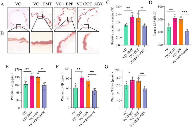
Methods: Differences in the intestinal microbiota were analyzed by 16S ribosomal RNA gene sequencing. Metabolites were analyzed using liquid chromatography-mass spectrometry. Faecal microbiota transplantation and antibiotic treatment experiments were performed to evaluate the functions of the intestinal microbiota in VC.
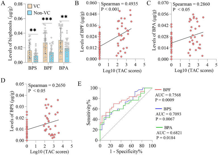
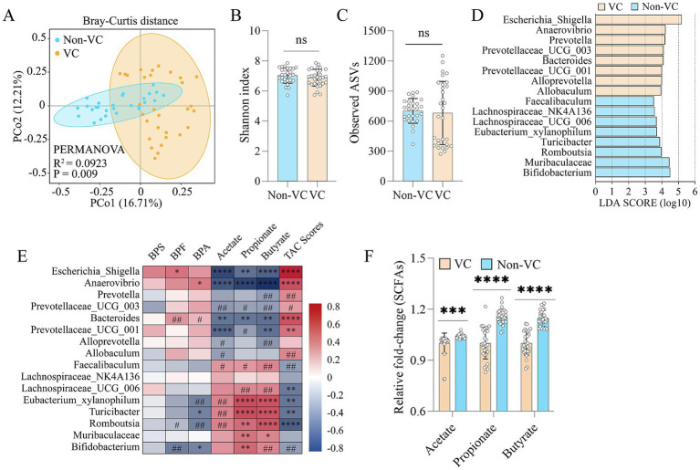
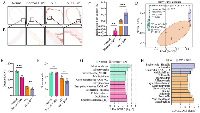
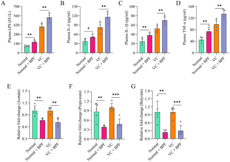
Results: We enrolled consecutively 57 patients. Patients were assigned to a calcification group (30 patients) and a non-calcification group (27 patients) based on the presence or absence of calcification in the thoracic aorta wall. The results showed that patients with vascular calcification (VC) had higher levels of bisphenol F (BPF), bisphenol S (BPS) and bisphenol A (BPA) in the fecal samples than patients without VC. The thoracic aortic calcification score was significantly positively correlated with the BPF (Spearman r = 0.4935, p < 0.001), BPA (Spearman r = 0.2860, p < 0.05) and BPS (Spearman r = 0.2650, p < 0.05). We then explored the effects of BPF exposure on normal and vitamin D3 + nicotine (VDN)-treated rats. BPF exposure induced mild VC in normal rats and aggravated VC in VDN-treated rats. BPF exposure disturbed the gut microbiota and promoted inflammatory responses.
Conclusion: The results here elucidate the mechanism underlying BPF-triggered or BPF-aggravated VC through the gut-vascular axis and provide a theoretical basis for cardiovascular disease risk assessment in humans.
Keywords: bisphenol F; faecal microbiota transplantation; gut microbiota; inflammation; short-chain fatty acids; vascular calcification.
Copyright © 2025 Yan, Pan, Liu, Yuan, Chen, Gao, Lin, Lin, Wang, He, Wang, Xu, Li, Zhang, Lan, Shao, Pang, Yin, Sun and Luo.
Conflict of interest statement
The authors declare that the research was conducted in the absence of any commercial or financial relationships that could be construed as a potential conflict of interest.
Figures
Similar articles
-
Use of high-resolution metabolomics to assess the biological perturbations associated with maternal exposure to Bisphenol A and Bisphenol F among pregnant African American women.Environ Int. 2022 Nov;169:107530. doi: 10.1016/j.envint.2022.107530. Epub 2022 Sep 17. Environ Int. 2022. PMID: 36148711 Free PMC article.
-
Targeted Metabolomics and Widely Targeted Quantitative Lipidomics Reveal Synergistic Metabolic Interference in Adipose Tissue After the Combined Chronic Exposure to Bisphenol A Substitutes, Bisphenol S and Bisphenol F, and Fructose in Male Rats.J Biochem Mol Toxicol. 2025 Aug;39(8):e70440. doi: 10.1002/jbt.70440. J Biochem Mol Toxicol. 2025. PMID: 40787705
-
Estimation of BPA, BPS, and BPF exposure levels in the general Korean population using the physiologically-based toxicokinetic model and human urinary biomonitoring for risk assessment.Int J Hyg Environ Health. 2025 Jul 28;269:114623. doi: 10.1016/j.ijheh.2025.114623. Online ahead of print. Int J Hyg Environ Health. 2025. PMID: 40729838
-
Biomonitoring of occupational exposure to bisphenol A, bisphenol S and bisphenol F: A systematic review.Sci Total Environ. 2021 Aug 20;783:146905. doi: 10.1016/j.scitotenv.2021.146905. Epub 2021 Apr 5. Sci Total Environ. 2021. PMID: 33865140
-
Bisphenol S and F: A Systematic Review and Comparison of the Hormonal Activity of Bisphenol A Substitutes.Environ Health Perspect. 2015 Jul;123(7):643-50. doi: 10.1289/ehp.1408989. Epub 2015 Mar 16. Environ Health Perspect. 2015. PMID: 25775505 Free PMC article.
References
-
- Ardissino D., Berzuini C., Merlini P. A., Mannuccio Mannucci P., Surti A., Burtt N., et al. (2011). Influence of 9p21.3 genetic variants on clinical and angiographic outcomes in early-onset myocardial infarction. J. Am. Coll. Cardiol. 58, 426–434. doi: 10.1016/j.jacc.2010.11.075, PMID: - DOI - PubMed