. 2024 Feb 15:4:1266279.
doi: 10.3389/fsysb.2024.1266279. eCollection 2024.

Computational inference of chemokine-mediated roles for the vagus nerve in modulating intra- and inter-tissue inflammation

Affiliations

Computational inference of chemokine-mediated roles for the vagus nerve in modulating intra- and inter-tissue inflammation

Ashti M Shah et al. Front Syst Biol. .

Abstract

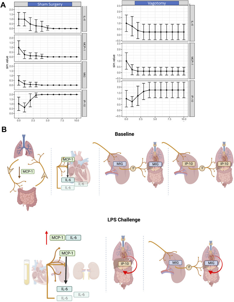
Introduction: The vagus nerve innervates multiple organs, but its role in regulating cross-tissue spread of inflammation is as yet unclear. We hypothesized that the vagus nerve may regulate cross-tissue inflammation via modulation of the putatively neurally regulated chemokine IP-10/CXCL10. Methods: Rate-of-change analysis, dynamic network analysis, and dynamic hypergraphs were used to model intra- and inter-tissue trends, respectively, in inflammatory mediators from mice that underwent either vagotomy or sham surgery. Results: This analysis suggested that vagotomy primarily disrupts the cross-tissue attenuation of inflammatory networks involving IP-10 as well as the chemokines MIG/CXCL9 and CCL2/MCP-1 along with the cytokines IFN-γ and IL-6. Computational analysis also suggested that the vagus-dependent rate of expression of IP-10 and MIG/CXCL9 in the spleen impacts the trajectory of chemokine expression in other tissues. Perturbation of this complex system with bacterial lipopolysaccharide (LPS) revealed a vagally regulated role for MIG in the heart. Further, LPS-stimulated expression of IP-10 was inferred to be vagus-independent across all tissues examined while reducing connectivity to IL-6 and MCP-1, a hypothesis supported by Boolean network modeling. Discussion: Together, these studies define novel spatiotemporal dimensions of vagus-regulated acute inflammation.

Keywords: chemokines; inflammation; systems biology; vagotomy; vagus nerve.


Conflict of interest statement

YV is a co-founder of, and stakeholder in, Immunetrics, Inc. and a member of the advisory board of Anuna AI. Neither of these relationships impacted the work contained in this manuscript. The remaining authors declare that the research was conducted in the absence of any commercial or financial relationships that could be construed as a potential conflict of interest. The author(s) declared that they were an editorial board member of Frontiers, at the time of submission. This had no impact on the peer review process and the final decision.

Figures

FIGURE 1
FIGURE 1
Inter-tissue inflammatory network analysis 7-day after sham surgery or vagotomy. (A) IL-6 is significantly elevated in the plasma, lung, and kidney following vagotomy compared to sham surgery. The edges of the box-and-whisker plot show data at the 25th and 75th percentile and the whiskers extend to the most extreme data points. (B–E) DyHyp nodes (tissue) are indicated by colored boxes. Edges surrounding one or more nodes are indicated as solid lines around nodes. Black lines indicate that the edge has a positive rate of change and red lines indicate that the edge has a negative rate-of-change. Intra-tissue networks (DyNA) are depicted within each DyHyp node to indicate that the inflammatory network is occurring within a single tissue. Thick black arrows indicate that inflammatory mediators are significantly positively correlated (r ≥ 0.95) and thin black arrows indicate that inflammatory mediators are positively, but not significantly, correlated (r > 0.90). (F) DyNA network complexity within each organ by experimental condition. (G) Graph of edge distribution, which indicates the number of edges surrounding each node in the DyHyp model.
FIGURE 2
FIGURE 2
Rate of change analysis of IFN-γ, IP-10, MIG, MCP-1, and IL-6 in distinct organs and the systemic circulation. (A–J) The rate of change for each inflammatory mediator was calculated in each organ. Organs are organized on the x-axis by increasing rate-of-change for the respective inflammatory mediator graphed. Negative rates-of-change indicate that the expression of the inflammatory mediator is decreasing with time whereas positive rates-of-change indicate that the expression of the inflammatory mediator is increasing with time.
FIGURE 3
FIGURE 3
DyNA and DyHyp models of inter- and intra-tissue inflammation in sham and vagotomized mice in response to LPS challenge. (A, B) DyNA networks highlight intra-tissue correlations between inflammatory mediators (|r| > 0.95) in mice that had received sham surgery and were subsequently challenged with LPS. (C–F) DyHyp inflammatory networks highlight inter-tissue trends in inflammatory mediators whose rate-of-change is outstanding across one or more tissues.
FIGURE 4
FIGURE 4
Rate-of-change of IFN-γ, IP-10, MIG, MCP-1 and IL-6 in tissues following LPS challenge in mice previously subjected to sham surgery or vagotomy. (A–J) The rate of change of inflammatory mediator expression in response to LPS challenge was calculated. Organs are organized on the x-axis by increasing rate-of-change for the respective inflammatory mediator graphed.
FIGURE 5
FIGURE 5
In silico and conceptual models of the long-term effects of vagotomy on IL-6, MCP-1, and IP-10 expression. (A) Inflammatory response to sham surgery and vagotomy as predicted by a Boolean network. The effect of vagotomy was modeled by excluding intra-nodal connections involving MIG. (B) A conceptual model of the inter-tissue impact of the vagus nerve inferred from experimental and computational analyses. At baseline, the vagus nerve downregulates MCP-1 across the lung-gut axis. In parallel, the rate of MIG and IP-10 expression across tissues is set by the vagally tuned rate of MIG and IP-10 expression in the spleen. Above a certain pro-inflammatory threshold induced by LPS, MCP-1 stimulates the expression of IL-6 and this is inhibited in the heart by the vagus nerve. Vagal innervation has minimal effect on the kinetics of IP-10 expression in response to LPS. Further, MIG expression across tissues is in part influenced by LPS as well as the vagally tuned rate of MIG expression by the spleen. In parallel, MCP-1 induces expression of IL-6 in the heart, spleen, and kidney and this cytokine also appears (or is expressed by cells in) the systemic circulation.

Similar articles

References

    1. Albert I., Thakar J., Li S., Zhang R., Albert R. (2008). Boolean network simulations for life scientists. Source Code biol.med. 3, 16. 10.1186/1751-0473-3-16 - DOI - PMC - PubMed
    1. Azhar N., Namas R. A., Almahmoud K., Zaaqoq A., Malak O. A., Barclay D., et al. (2021). A putative “chemokine switch” that regulates systemic acute inflammation in humans. Sci. Rep. 11 (1), 9703. 10.1038/s41598-021-88936-8 - DOI - PMC - PubMed
    1. Bonaz B., Sinniger V., Pellissier S. (2016). Anti-inflammatory properties of the vagus nerve: potential therapeutic implications of vagus nerve stimulation. J. Physiol. 594 (20), 5781–5790. 10.1113/jp271539 - DOI - PMC - PubMed
    1. Bone R. C. (1996). Toward a theory regarding the pathogenesis of the systemic inflammatory response syndrome: what we do and do not know about cytokine regulation. Crit. Care Med. 24 (1), 163–172. 10.1097/00003246-199601000-00026 - DOI - PubMed
    1. Borovikova L. V., Ivanova S., Zhang M., Yang H., Botchkina G. I., Watkins L. R., et al. (2000). Vagus nerve stimulation attenuates the systemic inflammatory response to endotoxin. Nature 405 (6785), 458–462. 10.1038/35013070 - DOI - PubMed

LinkOut - more resources