. 2025 Aug 4:34:102168.
doi: 10.1016/j.mtbio.2025.102168. eCollection 2025 Oct.

Neutrophils loaded NAD+ impede TLR4/NF-κB/NLRP3 pathway for sepsis treatment

Affiliations

Neutrophils loaded NAD+ impede TLR4/NF-κB/NLRP3 pathway for sepsis treatment

Yingchun Zhao et al. Mater Today Bio. .

Abstract

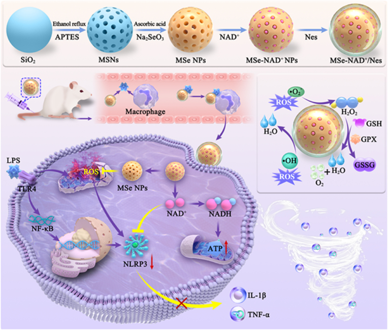
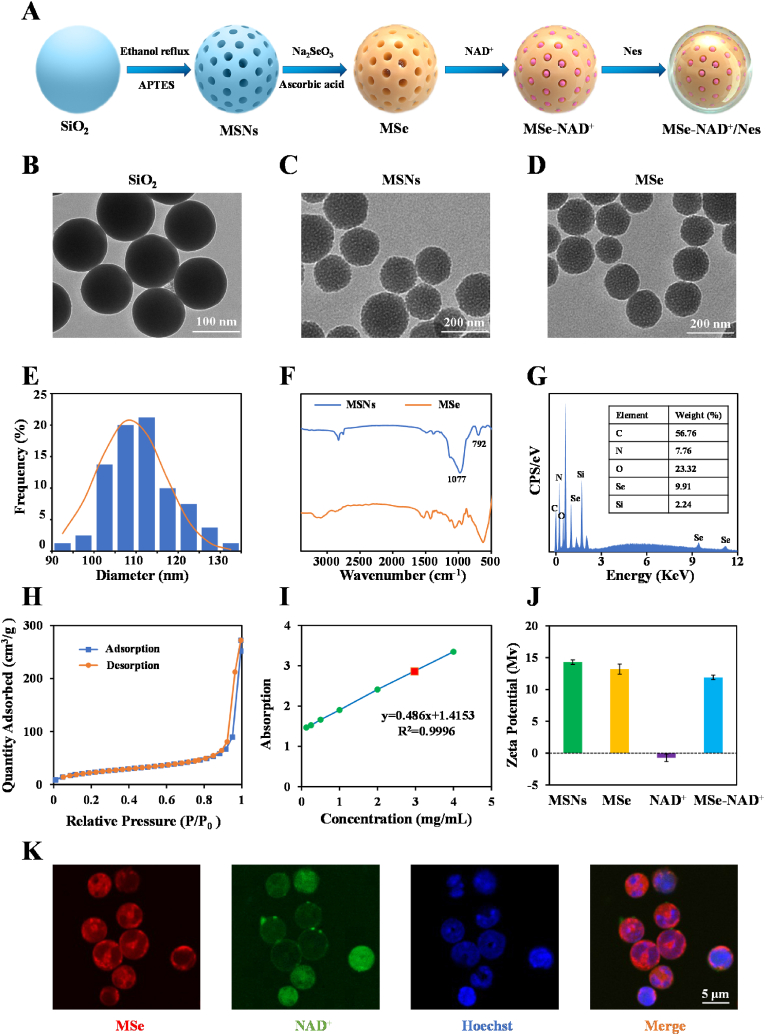
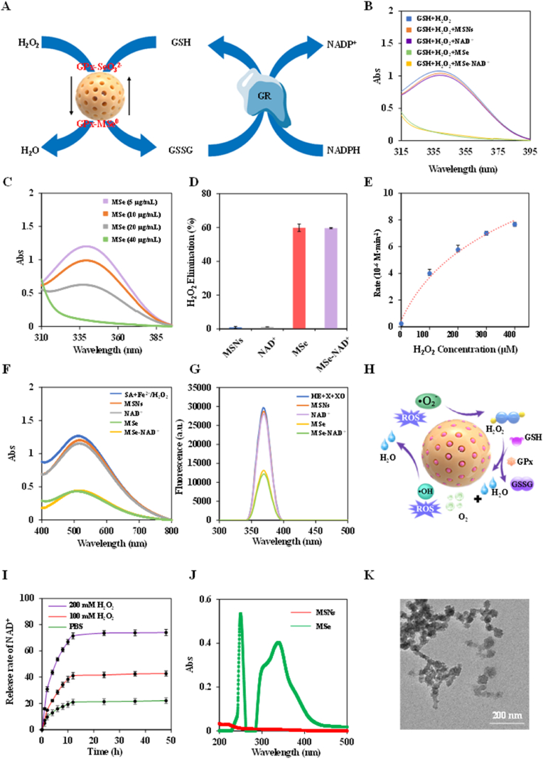
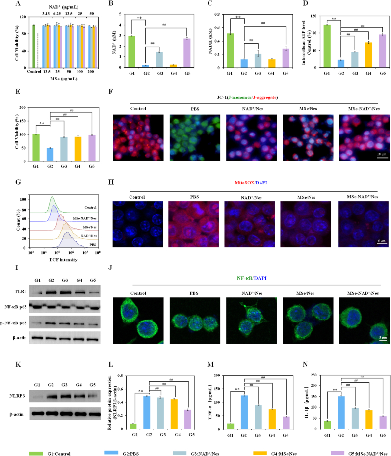
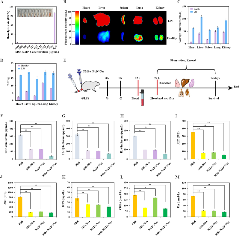
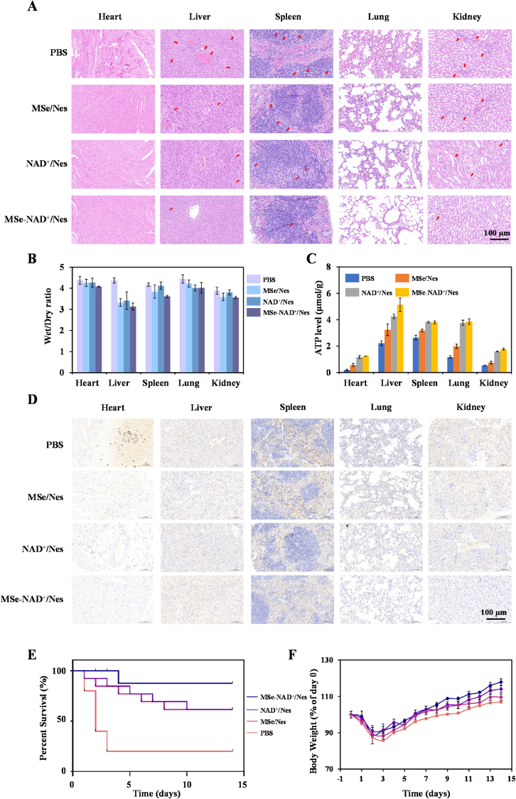
Systemic inflammation, excessive reactive oxygen species (ROS) and mitochondrial impairment are the main cause of multi-organ dysfunction syndrome in sepsis. Nevertheless, the pharmaceuticals currently in development focus solely on a single mechanism of disease, which is evidently inadequate. Herein, a precision nanodrug delivery system (MSe-NAD+/Nes) has been designed, incorporating mesoporous selenium nanozymes (MSe NPs) and leveraging a neutrophil-targeting strategy, to accomplish accurate delivery and mitigate inflammation. Upon reaching the inflammatory region, MSe NPs destroys selenium bonds and releases NAD+ under the action of ROS, which in turn supplements the NAD+ pool and promotes the recovery of mitochondrial function. Moreover, MSe NPs are capable of efficiently eliminating ROS by mimicking the activity of glutathione peroxidase (GPx), thus preventing the activation of the NLRP3 inflammasome. In vivo administration has indicated that MSe-NAD+/Nes efficiently alleviates organ oxidative stress, restores ATP levels, attenuates systemic hyperinflammation, and facilitates rapid organ repair. This study presents a potential modality of inflammation remission via ROS scavenging and mitochondrial repairment for the reliable and safe therapy of sepsis.

Keywords: Inflammation; NAD+; Neutrophil; ROS; Sepsis.


Conflict of interest statement

The authors declare that they have no known competing financial interests or personal relationships that could have appeared to influence the work reported in this paper.

Figures

Image 1
Graphical abstract
Fig. 1
Fig. 1
Schematic diagram of MSe-NAD+/Nes nanoplatforms as neutralize ROS and attenuate the inflammatory response for sepsis. Leveraging a neutrophil-targeting strategy to accomplish accurate delivery and mitigate inflammation. Upon reaching the inflammatory region, MSe NPs destroys selenium bonds and releases NAD+ under the action of ROS, which enhance mitochondrial homeostasis and cellular energy supply by effectively delivering NAD+ into cells. Meanwhile, MSe NPs are capable of efficiently eliminating ROS by mimicking the activity of glutathione peroxidase (GPx), thus preventing the activation of the NLRP3 inflammasome activation and attenuate the level of inflammatory factors, reduce the production of inflammatory factor storms, alleviate the condition of sepsis and effectively improve organ dysfunction.
Fig. 2
Fig. 2
Synthesis and characterization of MSe-NAD+/Nes. Scheme of the preparation of MSe-NAD+/Nes (A); The TEM image of SiO2 (B), MSNs (C), MSe NPs (D); The statistical graph of particle size distribution of TEM image of MSe NPs (E); FTIR spectrum of MSNs and MSe NPs (F); The elements quantitative analysis of MSe NPs by EDS elemental analysis (G); Nitrogen adsorption desorption isotherms for MSe NPs (H); UV–vis spectroscopy calibration curve for measuring NAD + loading efficiency (I); Zeta potential of MSNs, MSe NPs, NAD+, MSe-NAD+ NPs during the preparation process (J); CLSM images of MSe-NAD+/Nes after culturing at 37 °C. The nuclei of neutrophils were stained with Hoechst 33342 (Blue), MSe NPs were stained with rhodamine B (Red), and NAD+ was labeled with FITC (Green) (K).
Fig. 3
Fig. 3
The Wide-ranging ROS scavenging activity of MSe-NAD+/Nes. Schematic diagram of MSe NPs simulating GPx in the elimination of H2O2 (A); The GPx-like activity of MSNs, NAD+, MSe NPs and MSe-NAD+ NPs (B); The GPx-like activity of different concentration MSe-NAD+ NPs (C); The H2O2 elimination efficiency of MSNs, NAD+, MSe NPs and MSe-NAD+ NPs (D); Michaelis-Menten plot with varying concentration of H2O2 (0–400 μM) for MSe NPs (E); UV–vis spectra of SA after reacting with ·OH, in presence of MSNs, NAD+, MSe NPs and MSe-NAD+ NPs respectively (F); Fluorescent spectra of HE after reacting with X and XO, in presence of MSNs, NAD+, MSe NPs and MSe-NAD+ NPs, respectively (G); Schematic diagram of the mechanism of multiple reactive oxygen species scavenging by MSe NPs (H); NAD+ release behaviors of MSe-NAD+ NPs. The release of NAD+ was observed with time in the presence of 0 μM, 100 μM, 200 μM H2O2 in PBS buffer at pH 7.4 (I); UV–vis spectra in spectra of MSNs solution and MSe NPs solution at room temperature (J); TEM image of MSe-NAD+ NPs responsiveness in simulated media (200 μM H2O2 in PBS buffer at pH 7.4) (K).
Fig. 4
Fig. 4
ROS scavenging and mitochondrial repairment of MSe-NAD+/Nes in vitro. RAW264.7 cells were incubated with different concentrations of control, MSe NPs, NAD+, and MSe-NAD+ NPs for 24 h, and the cell viability (A); The intracellular NAD+ (B) and NADH (C) level of RAW264.7 cells in NAD+/Nes, MSe/Nes, and MSe-NAD+/Nes in the LPS-induced energy consumption model, Raw264.7 cells not treated with LPS as a negative control; Quantification of intracellular ATP level (D) and cell viability (E) in an LPS-mediated energy depletion model; Fluorescence images of RAW264.7 cells stained with JC-1 were used to analyze the depolarization of mitochondrial membrane (F); Flow cytometry analysis of ROS in control, PBS, MSe/Nes, NAD+/Nes and MSe-NAD+/Nes treated RAW264.7 cells (G); MitoSOX staining analysis of RAW264.7 cells in control, PBS, MSe/Nes, NAD+/Nes and MSe-NAD+/Nes treatment groups (H); The expression level of TLR4, p-NF-κB, NF-κB protein detected by Western blot (I); The nuclear localization of NF-κB (p65) observed by CLSM (J); The expression level of NLRP3 protein detected by Western blot (K,L); The expression level of pro-inflammatory cytokines TNF-α (M)and IL-1β (N) detected by ELISA in the culture supernatant of RAW264.7 cells. The group without LPS stimulation was taken as the control group. RAW264.7 cells were treated with LPS (100 ng/mL) and then treated with PBS, MSe/Nes, NAD+/Nes and MSe/NAD+/Nes respectively (equivalent to 25 μg/mL MSe NPs or 6.5 μg/mL NAD+). G1 respresents Control group; G2 respresents PBS group; G3 respresents NAD+/Nes group; G4 respresents MSe/Nes group; G5 respresents MSe-NAD+/Nes group. Values are expressed as mean ± S.D. ∗∗p < 0.01, ∗p < 0.05 vs. control group, ##p < 0.01, #p < 0.05 vs. PBS group.
Fig. 5
Fig. 5
In vivo therapeutic effects of MSe-NAD+/Nes on sepsis model. Relative hemolysis rates of MSe-NAD+ NPs at different concentrations (A); Biodistribution of Cy5-labeled MSe-NAD+/Nes in healthy and septic mice after MSe-NAD+/Nes administration for 24 h (B); Quantitative analysis of the mean fluorescence intensity of the organ or tissue shown in the ex vivo images (C); ICP-MS analysis of selenium content in the dissected tissues of mice treated with MSe-NAD+/Nes after 24 h of administration (the data are expressed as the percentage of ID/g of the injected dose) (D); Experimental flowchart of the treatment process (E); Pro-inflammatory cytokine TNF-α (F), IL-1β (G), IL-6 (H) levels in the serum of septic mice receiving PBS, MSe/Nes, NAD+/Nes and MSe-NAD+/Nes treatments were measured after LPS injection 12 h; Serum ALT (I), AST (J), BUN (K), CREA (L) and UA (M) levels in each group; Values are expressed as mean ± S.D. ∗∗p < 0.01, ∗p < 0.05 vs. PBS group.
Fig. 6
Fig. 6
MSe-NAD+/Nes Alleviates Multiple-Organ Dysfunction and Inflammation of Sepsis Mice. The H&E staining of heart, liver, spleen, lung and kidney of PBS, MSe/Nes, NAD+/Nes and MSe-NAD+/Nes groups after treatment of sepsis mice (A); The wet/dry ratio of heart, Liver, Spleen, Lung and Kidney in sepsis mice (B); ATP levels in different organs of the mice treated with PBS, MSe/Nes, NAD+/Nes and MSe-NAD+/Nes groups (C); The immunohistochemical changes in NLRP3 in the heart, liver, spleen, lung, spleen, and kidney (D); Survival rate of sepsis mice within 14 days, data are mean ± SD, n = 10 (E); Body weight charts for 14 days of sepsis mice (F).

Similar articles

References

    1. Shi C., Wang X., Wang L., Meng Q., Guo D., Chen L., Dai M., Wang G., Cooney R., Luo J. A nanotrap improves survival in severe sepsis by attenuating hyperinflammation. Nat. Commun. 2020;11:3384. - PMC - PubMed
    1. Huang B., Zhang N., Qiu X., Zeng R., Wang S., Hua M., Li Q., Nan K., Lin S. Mitochondria-targeted SkQ1 nanoparticles for dry eye disease : inhibiting NLRP3 inflammasome activation by preventing mitochondrial DNA oxidation. J. Contr. Release. 2024;365:1–15. - PubMed
    1. Nandi D., Debnath M., Iii J.F., Pandey A., Bharadwaj H., Patel R., Kulkarni A. Nanoparticle-mediated co-delivery of inflammasome inhibitors provides protection against sepsis. Nanoscale. 2024;16:4678–4690. - PubMed
    1. Liu F., Zhang K., Lu B., Wang X., Dong Q., Xue T., Tan Y., Wang X., Du J. Oxygen-vacancy-rich monolayer BiO 2- x nanosheets for bacterial sepsis management via dual physically antibacterial and chemically anti-inflammatory functions. Adv. Healthcare Mater. 2024;13 - PubMed
    1. Wendel M., Heller A.R. Mitochondrial function and dysfunction in sepsis, Wien. Med. Wochenschr. 2010;160:118–123. - PubMed

LinkOut - more resources