Review
. 2025 Jul 22;2(3):ugaf026.
doi: 10.1093/narmme/ugaf026. eCollection 2025 Jul.

Aere perennius: how chromatin fidelity is maintained and lost in disease

Affiliations
Review

Aere perennius: how chromatin fidelity is maintained and lost in disease

Dustin R Fetch et al. NAR Mol Med. .

Abstract

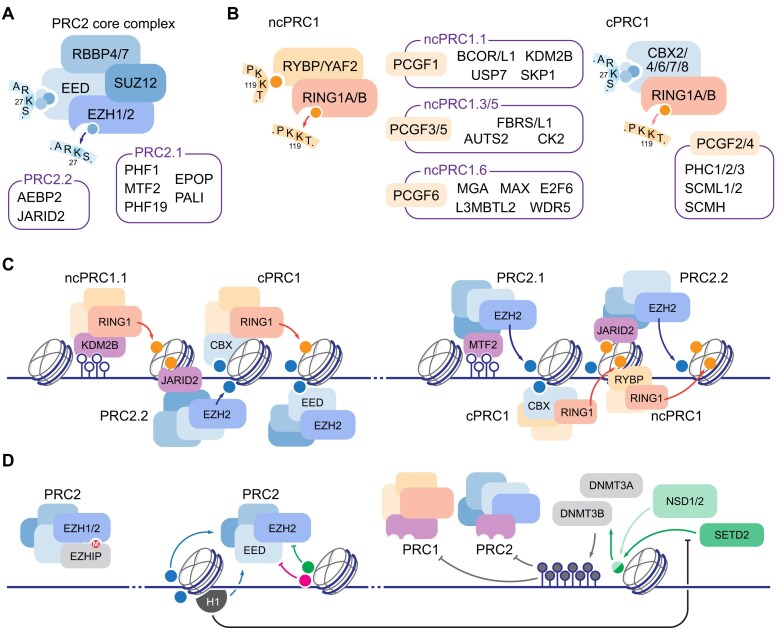
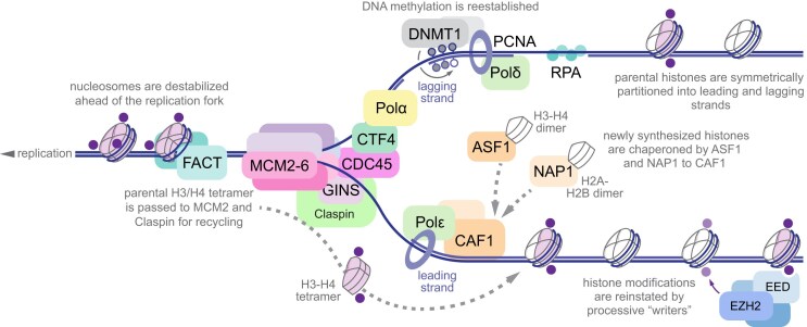
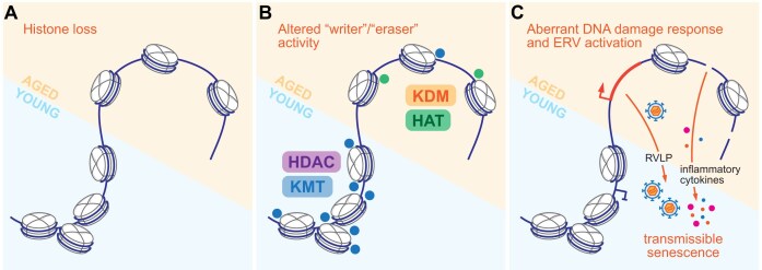
Multicellular organisms arise from a single genome template in the zygote, necessitating the cells of the developing embryo to up- and downregulate specific genes to establish and maintain their identity. This template is maintained, propagated, and interpreted as chromatin, a polymer of nucleic acids and associated structural and regulatory proteins. Recent genome-wide surveys documented a wealth of disease-associated mutations in chromatin factors, indicating their fundamental significance and potential for therapeutic targeting. However, chromatin factors exist in a complex balance, with a single deficiency often leading to pleiotropic downstream effects. Here, we review the mechanisms of chromatin regulation and partitioning, highlighting examples of how these processes are altered in human diseases. We argue that loss of chromatin fidelity, both locally at specific genes and regulatory elements, and globally at the megabase-scale, contributes to many pathological states and may thus represent an intriguing target for corrective interventions.


Conflict of interest statement

None declared.

Figures

Graphical Abstract
Graphical Abstract
Figure 1.
Figure 1.
Mechanisms of genome partitioning by Polycomb Group complexes. (A) Core PRC2 complex consists of four subunits, EED and EZH1/2 representing “reader” and “writer” modules for H3 K27 methylation. Sequence context of H3 tail is shown. Subunits associated with PRC2.1 and PRC2.2 variants are listed. (B) Two major PRC1 complexes are characterized by distinct subunit composition: non-canonical PRC1 (ncPRC1, left) contains H2A K119 “reader” RYBP/YAF2, and canonical PRC1 (cPRC1, right) contains one of several Chromobox proteins (CBX2/4/6/7/8) associated with H3 K27me3. Both complexes include RING1 E3 ubiquityl ligase “writer” module, with ncPRC1 characterized by higher enzymatic activity, and distinct complement of additional factors, anchored by PCGF subunits, listed in corresponding boxes. Sequence context of H3 and H2A tails are shown. (C) Recruitment and spreading of PcG domains in the vertebrate genome: left, unmethylated CpG islands are recognized by ncPRC1.1 subunit KDM2B, leading to initial H2A K119 ubiquitylation (orange circles) recognized by PRC2.2 via JARID2 subunit; methylated H3 K27 is in turn recognized by cPRC1 for compaction and/or further H2A ubiquitylation, and processive spreading of H3 K27 methylation is established by coordinate EED “reader” and EZH2 “writer” activity of PRC2; right, unmethylated CpGs associate with PRC2.1 subunit MTF2; initial H3 K27 methylation in turn recruits cPRC1 via CBX subunits, and PRC2 recruitment is propagated via H2A K119 recognition via JARID2, or H3 K27me3 recognition via EED. (D) Positive feedback regulation of PcG is balanced by incorporation of negative regulator subunits (EZHIP, left), incorporation of histone modifications that prevent H3 K27 methylation in cis (green and magenta circles correspond to H3 K36 and H3 K4 methylations; other modifications, including H3 phosphorylations, may act in similar fashion [53], middle), and activity of DNA methyltransferases, recruited in part by H3 K36 di- and tri-methylation by NSD1/2 and SETD2 enzymes, respectively (right). Direct stimulation of PRC2 by H1 incorporation is thus reinforced by reducing NSD1/2 activity in chromatin [6, 52].
Figure 2.
Figure 2.
Chromatin memory at the replication fork. Replication fork is shown progressing right to left, with lagging strand on the top and leading strand on the bottom. Parental histones are shown in light purple, with pre-existing post-translational modifications in dark purple. Nucleosomes are destabilized by FACT and parental H3/H4 tetramers are passed onto MCM2 and Claspin for symmetric recycling between the leading and lagging strands. Newly synthesized histones (white) are chaperoned by ASF1, NAP1 and CAF1 on both strands to maintain the nucleosome density. Histone modifications are reinstated by processive “reader”-“writer” activity of the cognate enzymes (light purple, only PRC2 is shown for clarity). DNA methylation is reestablished by maintenance methyltransferase DNMT1 via recognition of hemimethylated CpGs. Histone chaperones and DNMT1 operate on both strands but are omitted from lagging or leading strand for clarity.
Figure 3.
Figure 3.
Chromatin alterations in aging. (A) Histone loss and stochastic increase in chromatin accessibility are frequent in aging. (B) Young and aged cells are characterized by distinct activity of “writer” and “eraser” enzymes, with balance shift from lysine methyltransferases (KMT) and deacetylases (HDAC) in young cells and organisms to lysine demethylases (KDM) and acetyltransferases (HAT) in aged subjects. (C) Aberrant DDR and endogenous retroviruses are derepressed in aging cells, with shedded retrovirus-like particles (RVLPs) and secreted pro-inflammatory cytokines contributing to overall tissue senescence.
Figure 4.
Figure 4.
Histone H3 variant accumulation may alter redox capacity of the nucleosome. Replication-dependent H3.1 and replication independent H3.3 are shown schematically, with α-helices of histone fold represented as thicker bars. Five amino acids unique to each isoform are indicated, and H3.1-specific M90 and C96 are highlighted in magenta. Replacement of H3.1 with H3.3 in postnatal brain may reduce the redox buffering capacity in the aging post-mitotic cells.
Figure 5.
Figure 5.
Metabolic pathways are essential for chromatin fidelity. Key metabolic pathways directly implicated in chromatin regulation are shown above, with enzymes and additional crosstalk omitted for clarity. Left, polyamines are implicated in nucleosome organization and histone tail accessibility; middle, methionine and coupled folate cycle are the source of S-adenosyl-methionine (SAM) donor for histone and DNA methylation; right, TCA cycle is the source of α-KG cofactor for histone and DNA demethylases, with the exception of FAD-dependent LSD1 histone demethylase. Reciprocal relationships between modifications are illustrated below. dcSAM, decarboxylated S-adenosyl-methionine; SAH, S-adenosyl-homocysteine; THF, tetrahydrofolate; B2, B6, and B12 are critical cofactors of the folate cycle, with B12 coupling methionine and THF biosynthesis; 5,10-CH2-THF, 5,10-methylene-tetrahydrofolate; 5-CH3-THF, 5-methyl-tetrahydrofolate; Ac-CoA, acetyl-Coenzyme A; KMTs, histone lysine methyltransferases; DNMTs, DNA methyltransferases; KDMs, histone lysine demethylases; TETs, ten–eleven translocation (DNA demethylases); HDACs, histone lysine deacetylases; NAD+, nicotinamide adenine dinucleotide (oxidized form); NIC, nicotinamide.
Figure 6.
Figure 6.
Chromatin partitioning is altered in malignancy. (A) balanced activity of “writer” enzymes (A and B, left) establishes distinct chromatin domains (A and B, right). (B) inhibition of “writer” B by type 1 “oncohistones” in trans leads to expansion of domain A and may dilute the density of modifications established by A or associated “readers.” (C) reduced local activity of “writer” B due to effects of type 2 “oncohistones” in cis leads to stochastic disruption of domain B, with spurious accumulation of ectopic modifications. (D) altered compaction of chromatin fiber in H1 mutant malignancies drives redistribution of A and B-dependent modifications.

Similar articles

References

    1. Liberali P, Schier AF The evolution of developmental biology through conceptual and technological revolutions. Cell. 2024; 187:3461–95. 10.1016/j.cell.2024.05.053. - DOI - PubMed
    1. Lassar AB, Paterson BM, Weintraub H Transfection of a DNA locus that mediates the conversion of 10T1/2 fibroblasts to myoblasts. Cell. 1986; 47:649–56. 10.1016/0092-8674(86)90507-6. - DOI - PubMed
    1. Tapscott SJ, Davis RL, Thayer MJ et al. MyoD1: a nuclear phosphoprotein requiring a Myc homology region to convert fibroblasts to myoblasts. Science. 1988; 242:405–11. 10.1126/science.3175662. - DOI - PubMed
    1. Takahashi K, Yamanaka S Induction of pluripotent stem cells from mouse embryonic and adult fibroblast cultures by defined factors. Cell. 2006; 126:663–76. 10.1016/j.cell.2006.07.024. - DOI - PubMed
    1. Lambert SA, Jolma A, Campitelli LF et al. The Human transcription factors. Cell. 2018; 172:650–65. 10.1016/j.cell.2018.01.029. - DOI - PubMed

LinkOut - more resources