

Advancements in the non-invasive diagnosis of renal fibrosis
- PMID: 40809416
- PMCID: PMC12343605
- DOI: 10.3389/fmed.2025.1646412
Advancements in the non-invasive diagnosis of renal fibrosis
Abstract
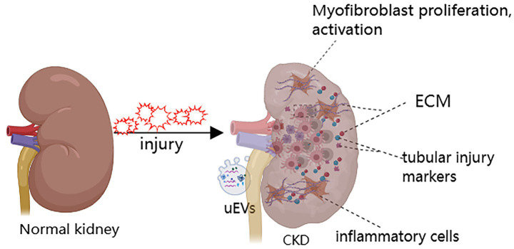
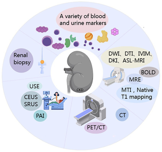
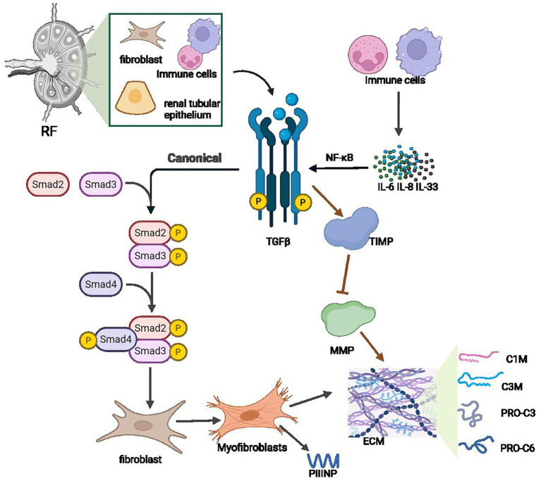
Renal fibrosis is the central pathological pathway by which various primary and secondary kidney diseases progress to end-stage renal disease. It is characterized by excessive extracellular matrix deposition and destruction of the native renal parenchyma, ultimately leading to irreversible loss of nephrons. Currently, percutaneous renal biopsy with histopathological evaluation remains the diagnostic gold standard for renal fibrosis, allowing semiquantitative scoring of renal interstitial fibrosis and glomerulosclerosis (e.g., Banff grading). However, this invasive procedure carries a risk of bleeding and is limited by sampling error and inter-observer variability, making it impractical for dynamic disease monitoring. In recent years, significant advances have been made in noninvasive diagnostic techniques. These include: (1) blood and urine biomarkers such as markers of ECM metabolism, inflammatory factors, tubular injury markers, and extracellular vesicles; (2) imaging modalities including novel ultrasound techniques, shear wave elastography, functional magnetic resonance imaging (MRI) methods such as diffusion-weighted imaging, blood oxygen level-dependent MRI, magnetic resonance elastography, and positron emission tomography/computed tomography using radiotracers targeting fibrosis-associated molecules such as 68Ga-FAPI. This review systematically summarizes the latest evidence on the above biomarkers and advanced imaging modalities, with an emphasis on their diagnostic performance (sensitivity/specificity), utility for dynamic monitoring, and bottlenecks in clinical translation. The aim is to develop a multimodal, noninvasive assessment system to enable earlier fibrosis detection, stratified disease management, and precise intervention targeting fibrogenic pathways, ultimately improving renal disease outcomes.
Keywords: biomarkers; chronic kidney disease; imaging techniques; non-invasive diagnosis; renal fibrosis.
Copyright © 2025 Yuan, Wang, Kang, Wu and Ou.
Conflict of interest statement
The authors declare that the research was conducted in the absence of any commercial or financial relationships that could be construed as a potential conflict of interest.
Figures
Similar articles
-
Management of urinary stones by experts in stone disease (ESD 2025).Arch Ital Urol Androl. 2025 Jun 30;97(2):14085. doi: 10.4081/aiua.2025.14085. Epub 2025 Jun 30. Arch Ital Urol Androl. 2025. PMID: 40583613 Review.
-
Prescription of Controlled Substances: Benefits and Risks.2025 Jul 6. In: StatPearls [Internet]. Treasure Island (FL): StatPearls Publishing; 2025 Jan–. 2025 Jul 6. In: StatPearls [Internet]. Treasure Island (FL): StatPearls Publishing; 2025 Jan–. PMID: 30726003 Free Books & Documents.
-
MarkVCID cerebral small vessel consortium: II. Neuroimaging protocols.Alzheimers Dement. 2021 Apr;17(4):716-725. doi: 10.1002/alz.12216. Epub 2021 Jan 21. Alzheimers Dement. 2021. PMID: 33480157 Free PMC article.
-
The Black Book of Psychotropic Dosing and Monitoring.Psychopharmacol Bull. 2024 Jul 8;54(3):8-59. Psychopharmacol Bull. 2024. PMID: 38993656 Free PMC article. Review.
-
Non-invasive diagnostic assessment tools for the detection of liver fibrosis in patients with suspected alcohol-related liver disease: a systematic review and economic evaluation.Health Technol Assess. 2012;16(4):1-174. doi: 10.3310/hta16040. Health Technol Assess. 2012. PMID: 22333291 Free PMC article.
References
Publication types
LinkOut - more resources
Full Text Sources