. 2025 Jul 30:12:1603161.
doi: 10.3389/fmed.2025.1603161. eCollection 2025.

Developing a procedure mimicking transvaginal mesh implantation in women in a modified POP rat model

Affiliations

Developing a procedure mimicking transvaginal mesh implantation in women in a modified POP rat model

Lulu Wang et al. Front Med (Lausanne). .

Abstract

Introduction: This study aims to establish a simple and reproducible transvaginal mesh surgery rat model based on the modified pelvic organ prolapse rat model.

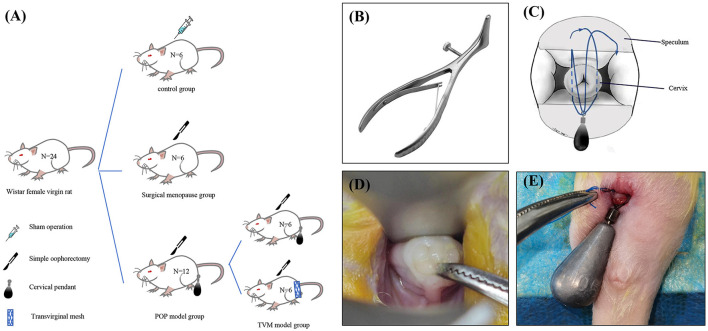
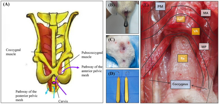
Methods: A total of 24 10-week-old female nulliparous Wistar rats were used in this study. The control group consisted of six rats with no interventions. The ovariectomy group included six rats that underwent bilateral ovariectomy. The pelvic organ prolapse group comprised 12 rats that underwent cervical pendant modeling 2 weeks after bilateral ovariectomy. Fourteen days post-modeling, six rats from the pelvic organ prolapse group underwent transvaginal mesh surgery. The rat pelvic organ prolapse quantification system was used to evaluate the prolapse condition of the rats before and after pelvic organ prolapse modeling, as well as after transvaginal mesh surgery. Vaginal wall tissue was collected to assess biomechanical changes before and after pelvic organ prolapse modeling. Additionally, vaginal wall and sacral ligament tissues were collected to evaluate structural changes and collagen alterations before and after pelvic organ prolapse modeling.

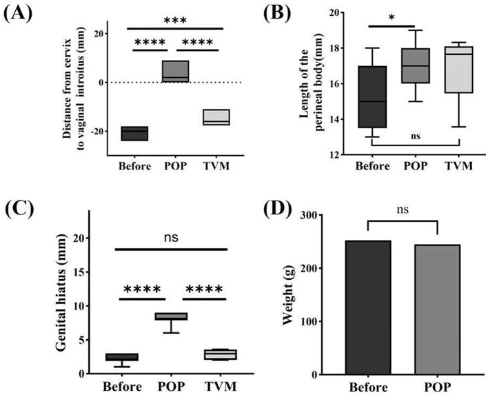
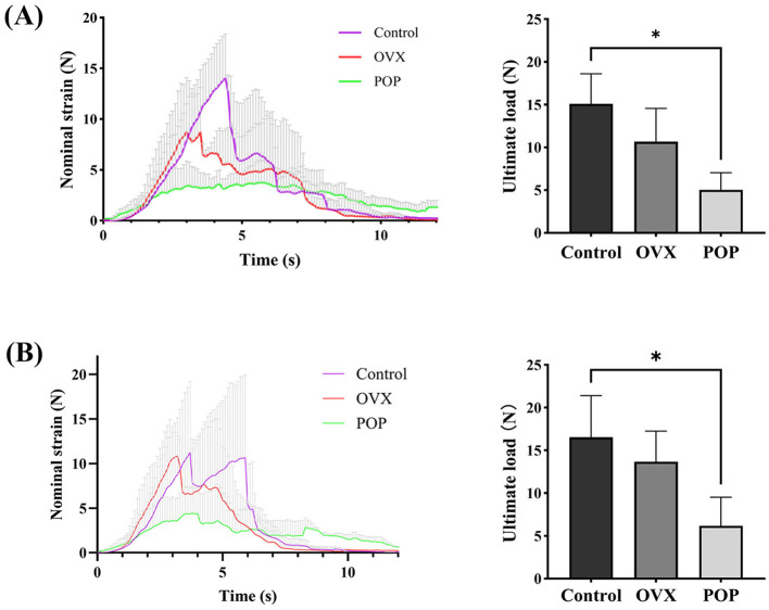
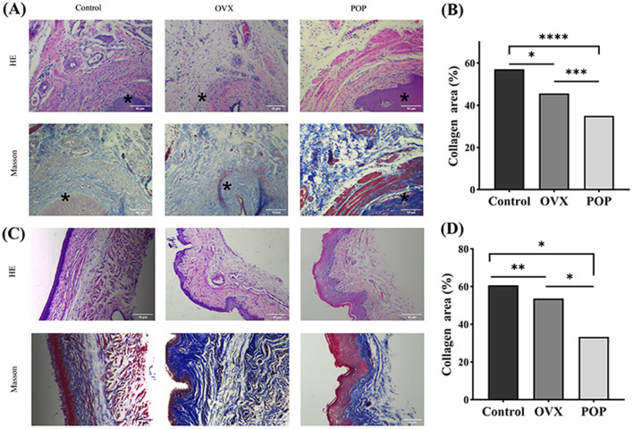
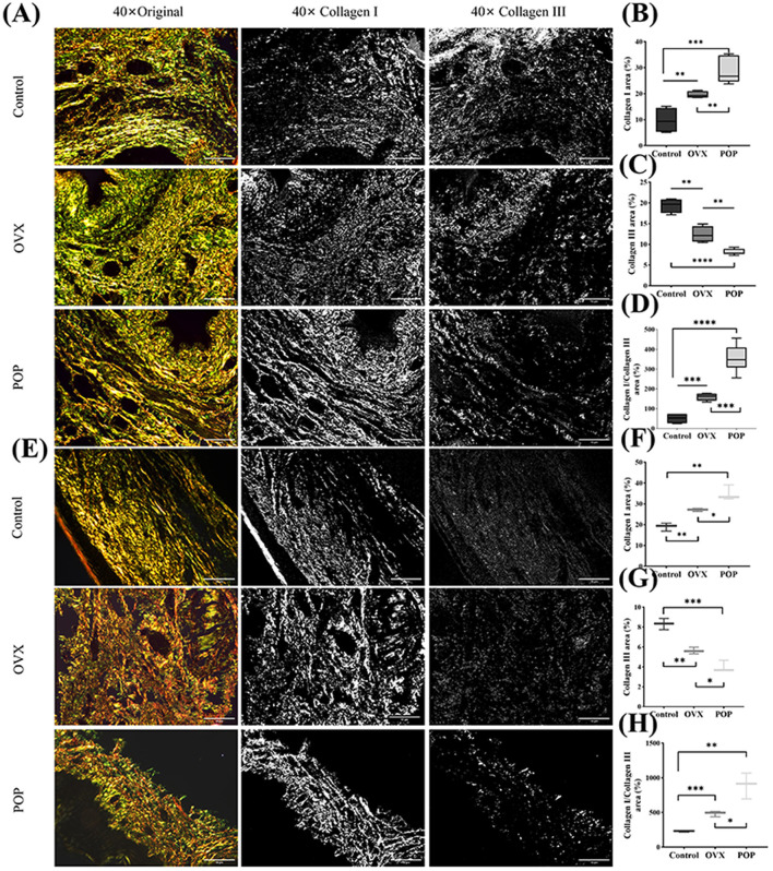
Results: The pelvic organ prolapse rat model exhibits anatomical prolapse, biomechanical changes, and pathological changes, including collagen fiber rupture and reduced collagen density. In contrast, the transvaginal mesh rat model demonstrates anatomical recovery in prolapsed rats.

Conclusion: This study successfully modified the pre-existing rat model of pelvic organ prolapse and effectively mimicked human transvaginal mesh surgery using this model.

Keywords: animal model; biomechanical; pelvic organ prolapse; rat; transvaginal mesh.


Conflict of interest statement

The authors declare that the research was conducted in the absence of any commercial or financial relationships that could be construed as a potential conflict of interest.

Figures

(A) Diagram of experimental groups with Wistar female virgin rats categorized into control, surgical menopause, POP model, and TVM model groups, each with specific procedures. (B) Illustration of a speculum tool. (C) Diagram showing the speculum's insertion with labeled cervix. (D) Close-up of a surgical procedure on a rat cervix. (E) Image of a transvaginal mesh procedure on a rat.
Figure 1
Schematic diagram illustrating the establishment of a rat model for POP and specific methodologies. (A) Experimental grouping; (B) modified vaginal speculum for rats; (C) schematic representation of the modeling technique; (D) rat cervix exposed under the speculum; (E) schematic depiction of the post-modeling cervix.
(A) Anatomical diagram of pelvic muscles showing coccygeal and pubococcygeal muscles with pathways of pelvic mesh. (B) Close-up of a tool inserted in an animal model. (C) Surgical site on an animal, showing stitches. (D) Two surgical tools with hook ends. (E) Surgical exposure of muscle layers with labeled areas PM, UF, VA, Re, and Coccygeus.
Figure 2
TVM protocol and comparison map. (A) Schematic diagram illustrating the anatomical structure of rat TVM and mesh alignment. (B) Preoperative perineal condition of rats undergoing TVM. (C) Postoperative perineum after TVM in rats. (D) Puncture guide needle. (E) Gross anatomical and mesh trajectory diagram following TVM surgery in rats. PM, Pubococcygeus muscle; MA, Anterior pelvic mesh trajectory path; MP, Posterior pelvic mesh trajectory path; Ur, Urethra; VA, Vagina; Re, Rectum.
Two panels labeled A and B depict graphs comparing nominal strain over time and bar charts of ultimate load. Both graphs show three lines: control (purple), OVX (red), and POP (green). The control group displays the highest nominal strain and ultimate load, followed by OVX and then POP. Bar charts indicate significant differences between control, OVX, and POP, with marked asterisks showing statistical significance.
Figure 3
Systematic evaluation of ROPQ in rats: (A) the distance between the cervix and vaginal orifice; (B) perineal body length; (C) reproductive hiatus size; (D) changes in weight before and after Pelvic Organ Prolapse Modeling. Significant differences are denoted by *P < 0.05; ***P < 0.001; ****P < 0.0001.
(A) Box plot showing the distance from cervix to vaginal introitus in millimeters before, during POP, and after TVM, with significant differences indicated by asterisks. (B) Box plot displaying the length of the perineal body, with significant differences noted between before and POP but not POP and TVM. (C) Box plot illustrating genital hiatus size, with significant changes before, POP, and TVM. (D) Bar graph comparing the weight in grams before and after POP, showing no significant difference.
Figure 4
Biomechanical testing of the vaginal wall: (A) strain and ultimate load of the anterior vaginal wall tissue; (B) strain and ultimate load of the posterior vaginal. Significant differences are denoted by *P < 0.05.
(A) Microscopic images showing tissue samples in Control, OVX, and POP groups stained with HE and Masson methods, indicating differences in structure. (B) Bar graph displaying collagen area percentages for Control, OVX, and POP groups, with significant differences marked by asterisks. (C) Additional tissue images from the same groups, also stained with HE and Masson. (D) Another bar graph showing collagen area percentages with significant differences indicated by asterisks.
Figure 5
The sacral ligament and vaginal wall tissue were subjected to 40 × microscopic HE staining and Masson staining. *, Cervix. (A) HE staining and Masson staining of the sacral ligament in the control group, OVX group, and POP group; (B) HE staining and HE staining of the sacral ligament in the control group, OVX group, and POP group; (C) statistical chart depicting the proportion of collagen in the sacral ligament; (D) Statistical plot illustrating the proportion of collagen in the anterior vaginal wall. Significant differences are denoted by *P < 0.05; **P < 0.01; ***P < 0.001; ****P < 0.0001.
Histological images and box plots depicting collagen levels. (A, E) Show original and stained tissue sections at forty times magnification for control, OVX, and POP groups. (B–D, F–H) Are box plots comparing collagen I and III areas among the three groups. Statistical significance is indicated by asterisks.
Figure 6
The collagen composition and arrangement in the sacral ligament and anterior vaginal wall were examined using polarized light microscopy after staining with Sirius red. (A) The Sirius red staining of the sacral ligament tissue; (B) collagen I in the POP group was significantly higher than that in the control and OVX groups; (C) collagen III in the POP group was significantly lower than that in the control and OVX groups; (D) the ratio of Col I/Col III in the POP group was significantly higher than that in the control and OVX groups; (E) the Sirius red staining of the anterior vaginal wall tissue; (F–H) the results of Col I, Col III, and Col I/Col in anterior vaginal wall were similar with the sacral ligament tissue. Significant differences are denoted by *P < 0.05; **P < 0.01; ***P < 0.001; ****P < 0.0001.

Similar articles

References

    1. Darzi S, Alappadan J, Paul K, Mazdumder P, Rosamilia A, Truong YB, et al. Immunobiology of foreign body response to composite PLACL/gelatin electrospun nanofiber meshes with mesenchymal stem/stromal cells in a mouse model: Implications in pelvic floor tissue engineering and regeneration. Biomater Adv. (2023) 155:213669. 10.1016/j.bioadv.2023.213669 - DOI - PubMed
    1. Drewes PG, Yanagisawa H, Starcher B, Hornstra I, Csiszar K, Marinis SI, et al. Pelvic organ prolapse in fibulin-5 knockout mice. Am J Pathol. (2007) 170:578–89. 10.2353/ajpath.2007.060662 - DOI - PMC - PubMed
    1. Paul K, Darzi S, O'Connell CD, Hennes DM, Rosamilia A, Gargett CE, et al. 3D printed mesh geometry modulates immune response and interface biology in mouse and sheep model: implications for pelvic floor surgery. Adv Sci. (2025) 12:2405004. 10.1002/advs.202405004 - DOI - PMC - PubMed
    1. Guo T, Du Z, Wang X-Q, Lang J-H, Sun Z-J. Ovariectomy with simulated vaginal delivery to establish a rat model for pelvic organ prolapse. Connect Tissue Res. (2023) 64:376–88. 10.1080/03008207.2023.2199091 - DOI - PubMed
    1. Mao M, Li Y, Zhang Y, Kang J, Zhu L. Tissue composition and biomechanical property changes in the vaginal wall of ovariectomized young rats. Biomed Res Int. (2019) 2019:8921284. 10.1155/2019/8921284 - DOI - PMC - PubMed

LinkOut - more resources