. 2025 Aug 13:8:0768.
doi: 10.34133/research.0768. eCollection 2025.

Mechanical Agitation-Assisted Transmembrane Drug Delivery by Magnetically Powered Spiky Nanorobots

Affiliations

Mechanical Agitation-Assisted Transmembrane Drug Delivery by Magnetically Powered Spiky Nanorobots

Xiaojia Liu et al. Research (Wash D C). .

Abstract

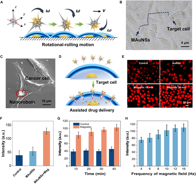
Breaking through cell membrane barriers is a crucial step for intracellular drug delivery in antitumor chemotherapy. Hereby, a magnetic nanorobot, capable of exerting mechanical agitation on cellular membrane to promote intracellular drug delivery, was developed. The main body of the nanorobots was composed of nano-scaled gold nanospikes that were deposited with Ni and Ti nanolayers for magnetic activation and biocompatibility, responsively. The nanorobots can be precisely navigated to target cancer cells under external magnetic field control. By virtue of the sharp nanospike structures, the magnetically powered rotation behavior of the nanorobots can impose mechanical agitation on the living cell membrane and thus improve the membrane permeability, leading to promoted transmembrane cargo delivery. Coarse-grained molecular dynamics simulation revealed that the mechanism of mechanical intervention regulated permeability of the bilayer lipid membrane, allowing for enhanced transmembrane diffusion of small cargo molecules. An in vitro study demonstrated that these nanorobots can markedly enhance the efficiency of drug entry into tumor cells, thus improving the effectiveness of tumor therapy under magnetic activation in vivo. This work paves a new way for overcoming cell membrane barriers for intracellular drug delivery by using a magnetic nanorobotic system, which is expected to promote further application of magnetically controlled nanorobot technology in the field of precision medicine.


Conflict of interest statement

Competing interests: The authors declare that they have no competing interests.

Figures

Fig. 1.
Fig. 1.
Scheme of the preparation of MAuNS robots, and their anti-cancer therapeutic applications by mechanically regulated cell membrane permeability for transmembrane drug delivery.
Fig. 2.
Fig. 2.
Characterization of MAuNS robots. (A) Scanning electron microscope (SEM) image of the MAuNSs. (B) Transmission electron microscope (TEM) image and (C) high-magnification image of the region marked by a red square of a single MAuNSs robot. (D) EDX mappings of a MAuNSs robot. (E) Plot showing the spine length and tip angle of the MAuNSs. (F) Statistics of the length of the MAuNSs. (G) UV–Vis spectrum of the MAuNSs suspended in deionized (DI) water.
Fig. 3.
Fig. 3.
Motion control of MAuNSs robots by external magnetic field. (A) Schematic of the translational motion of MAuNSs robots driven by gradient field. (B) Optical microscope screenshot of the trajectory of a MAuNSs robot under a gradient magnetic field guidance. (C) Velocity of MAuNSs under different gradient magnetic field strengths (error bars indicate standard deviation, n = 50). (D) Schematic of the rolling motion of a MAuNSs robot. (E) Optical microscope screenshot of the trajectory of a rolling nanorobot at different time intervals. (F) Velocity of MAuNSs’ rolling motion under different magnetic field frequencies (error bars indicate standard deviation, n = 50). (G) Schematic of the rotation of a MAuNS robot with an applied XOY magnetic field. (H) Optical microscope screenshot of rotation nanorobots at different time intervals. (I) Rotational angular velocity of magnetically controlled robot motion at different magnetic field frequencies (4 to 30 Hz) (error bars indicate standard deviation, n = 50). (J) Simulation results of the velocity and direction of the fluid field around the nanospike under translational motion and (K) rolling motion.
Fig. 4.
Fig. 4.
Magnetic field driven cell targeting and mechanical agitation enabled transmembrane drug delivery by MAuNSs robots. (A) Schematic diagram of a MAuNSs robot rolling forward driven by rotational magnetic field. (B) Optical microscopy screenshot capturing a MAuNSs robots targeting tumor cell (HepG-2) membranes in a cellular environment. (C) SEM image of cells (MAuNSs: 1 mg/ml; magnetic field exposure time: 30 min; red dotted circle indicates the presence of the MAuNSs). (D) Schematic diagram showing that mechanical agitation promoted transmembrane drug delivery. (E) Fluorescence images and (F) intensity analysis of drug molecules entering cells under different conditions. (G) Fluorescence intensity of drug molecules entering the cell at different times (10 to 40 s) and (H) different rotation frequencies (4 to 16 Hz) (error bars indicate standard deviation, n = 10).
Fig. 5.
Fig. 5.
Coarse-grained molecular dynamics simulations. (A) Simulation of diffusion of small molecules at different rotational frequencies and (B) the corresponding lipid number density on the cell membrane. (C) Pore area, (D) mean square displacement, and (E) penetrated NP number produced by small molecules crossing the cell membrane at different rotational frequencies. (F) Screenshots of the dynamic behavior of MAuNS robots crossing the cell membrane at different times (t0 = 0 s, t1 = 2 s, t2 = 4 s, and t3 = 8 s).
Fig. 6.
Fig. 6.
In vivo cancer therapy by synergetic antitumor effect of the MAuNSs robots. (A) Schematic illustration of the antitumor animal experiments. HepG-2 cells and MAuNSs were subcutaneously injected in turn into normal mice followed by magnetotherapy. (B) Survival of mice at different days. (C)Tumor volume (tumor volume divided by initial volume) in various mice groups. (D) Change in weight of mice in different groups over time. (E) Representative photographs of mice. (F) Morphologies of tumors at the original cell injection site. (G) H&E-stained images of tumors at the original cell injection site. (H) Necrosis in various mice groups. (I) H&E-stained images of liver tissue (error bars indicate standard deviation, n = 10).

References

    1. Wang L, Zhu X, Xu C, Jin D, Ma X. Artificial breakthrough of cell membrane barrier for transmembrane substance exchange: A review of recent progress. Adv Funct Mater. 2024;34(13):2311920.
    1. Xie J, Shen Q, Huang K, Zheng T, Cheng L, Zhang Z, Yu Y, Liao G, Wang X, Li C. Oriented assembly of cell-mimicking nanoparticles via a molecular affinity strategy for targeted drug delivery. ACS Nano. 2019;13(5):5268–5277. - PubMed
    1. Tang K, Tang Z, Niu M, Kuang Z, Xue W, Wang X, Liu X, Yu Y, Jeong S, Ma Y, et al. Allosteric targeted drug delivery for enhanced blood-brain barrier penetration via mimicking transmembrane domain interactions. Nat Commun. 2025;16(1):3410. - PMC - PubMed
    1. Shi Z, Graber ZT, Baumgart T, Stone HA, Cohen AE. Cell membranes resist flow. Cell. 2018;175(7):1769–1779. - PMC - PubMed
    1. Oroojalian F, Beygi M, Baradaran B, Mokhtarzadeh A, Shahbazi MA. Immune cell membrane-coated biomimetic nanoparticles for targeted cancer therapy. Small. 2021;17(12):2006484. - PubMed

LinkOut - more resources