. 2025 Aug 6:53:801-818.
doi: 10.1016/j.bioactmat.2025.07.045. eCollection 2025 Nov.

Spore-inspired inhalation drug delivery system for asthma therapy

Affiliations

Spore-inspired inhalation drug delivery system for asthma therapy

Mengqi Tong et al. Bioact Mater. .

Abstract

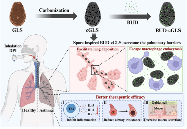
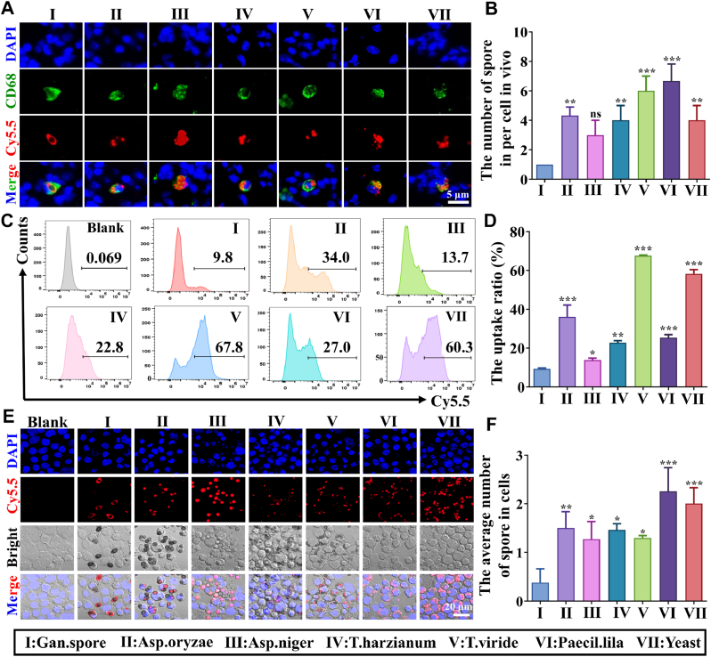
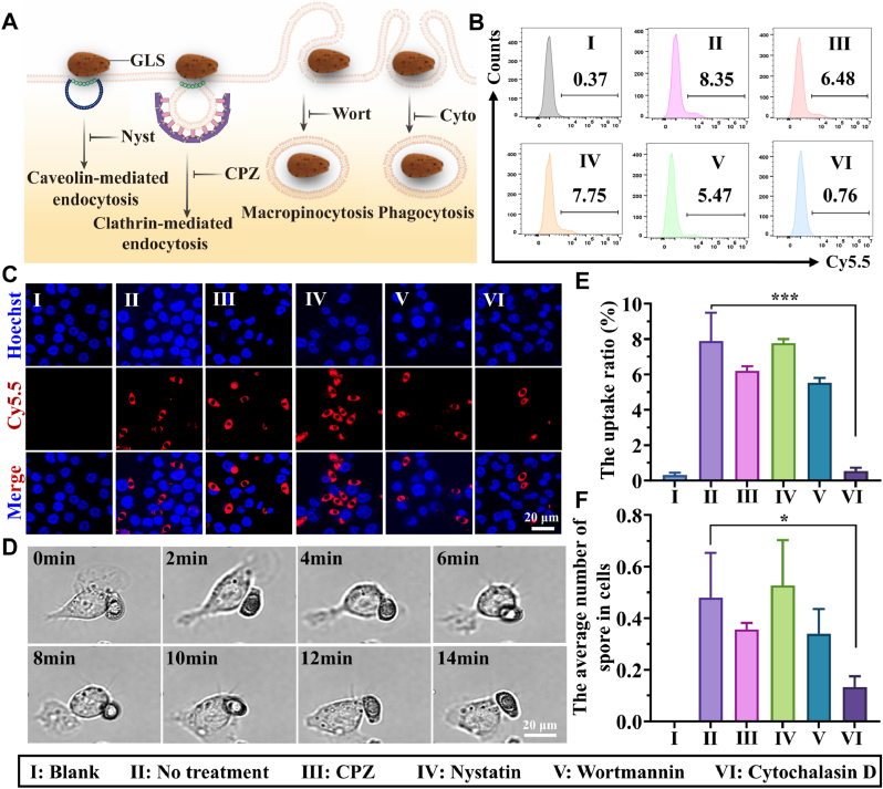
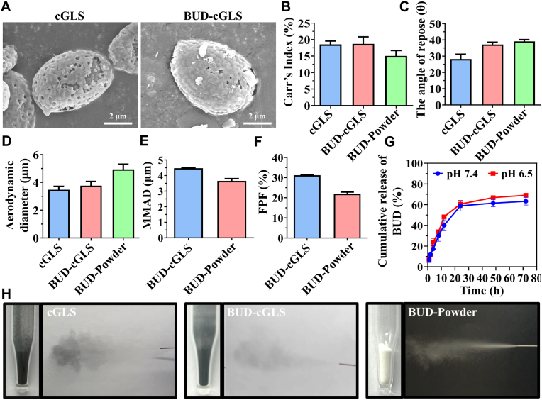
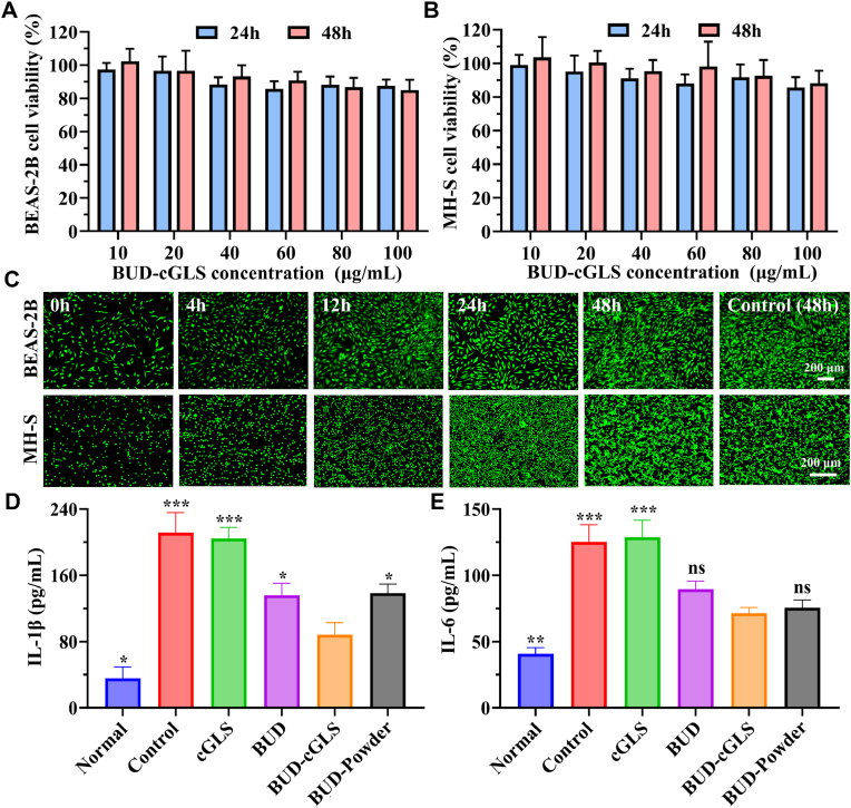
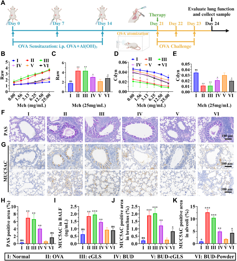
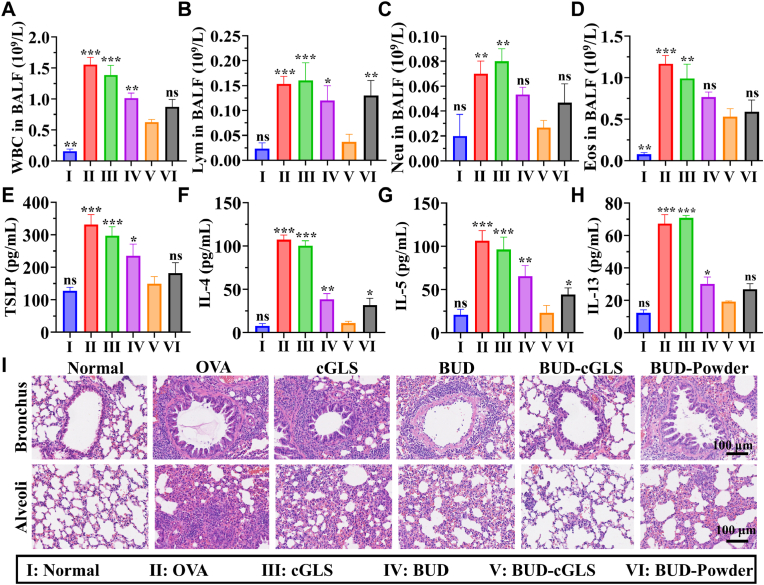
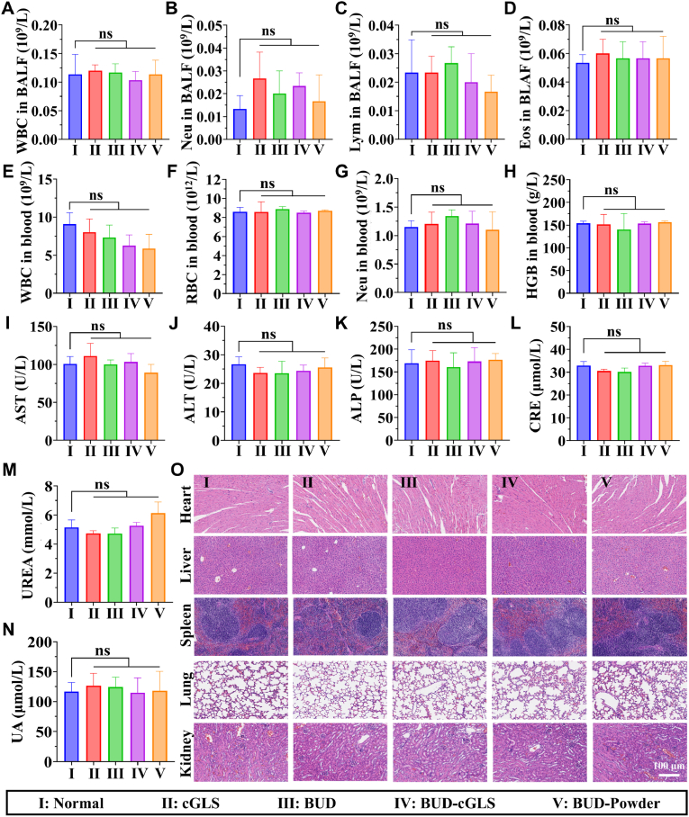
The delivery efficiency of drugs in the lung is crucial for inhaled therapies targeting pulmonary diseases. However, current inhalation carriers face challenges overcoming pulmonary barriers, leading to insufficient delivery efficiency. To tackle this limitation, we have developed a "spore-inspired" strategy. Ganoderma lucidum spores (GLS) provide dual delivery advantages: their natural morphology promotes bronchial-alveolar deposition while evading macrophage endocytosis, enhancing pulmonary retention. Using these features, a biomimetic carrier called carbonized GLS (cGLS) is created through precise carbonization, which preserves the spores' natural morphological benefits while reducing the immune response and increasing drug-loading capacity. Subsequently, we develop the spore-inspired inhalation drug delivery system BUD-cGLS by loading the asthma medication budesonide (BUD), which facilitates accurate regulation of the "deposition-escape-release" process. In the OVA-induced asthma model, BUD-cGLS significantly reduces airway resistance, suppresses mucin secretion, and decreases inflammatory cytokines. Overall, these findings highlight the potential of this spore-inspired carrier as a promising inhalation platform for delivering drugs to treat asthma and other pulmonary diseases.

Keywords: Asthma; Budesonide; Dry powder inhaler; Pulmonary delivery; Spore-inspired carrier.


Conflict of interest statement

The authors declare that no competing interest exists.

Figures

Image 1
Schematic illustration of the spore-inspired inhalation drug delivery system BUD-cGLS preparation procedures and their application in OVA-induced asthma.
Fig. 1
Fig. 1
Schematic illustration of the spore-inspired inhalation drug delivery system BUD-cGLS preparation procedures and their application in OVA-induced asthma. (Created in BioRender. Tong, M (2025) https://BioRender.com/wovsmft).
Fig. 2
Fig. 2
The characterization and evaluation of inhalability of different spores in vitro. (A) The SEM images of different spores; (B) The diameter of different spores; (C) The Carr's Index of different spores; (D) The angle of repose (θ) of different spores; (E) The aerodynamic diameter and morphology of different spores; (F) The quantitative aerodynamic diameter according to Fig. 2E; (G) The dry powder aerosol of different spores. The lactose is an inhalation carrier positive control. Gan.spore, Aspergillus oryzae (Asp.oryzae), Aspergillus niger (Asp.niger), Trichoderma harzianum (T.harzianum), Trichoderma viride (T.virde), Paecilomyces lilacinus (Paceil.lila), Yeast.
Fig. 3
Fig. 3
Biodistribution and lung accumulation after inhalation administration of different spores. (A) The representative IVIS images of lungs at different time points after inhalation administration of different Cy5.5-spores; (B) The corresponding quantitative fluorescence relative intensity of Fig. 3A; (C) Distribution of spores in bronchus and alveoli at 0.5 h after inhalation administration. Nuclei (DAPI, blue) and spores (Cy5.5, red), Scale bars:100 μm, 20 μm, and 20 μm (left to right); The corresponding quantitative data of spores in bronchus (D) and alveoli (E); (∗P < 0.05; ∗∗P < 0.01; ∗∗∗P < 0.001 vs. Gan. spore; ns represents no significant difference).
Fig. 4
Fig. 4
The macrophage endocytosis assay of spores in vivo and in vitro. (A) The representative fluorescent images of macrophage endocytosis of different Cy5.5-spores in lung tissue sections at 6 h after intratracheal administration, Macrophage (CD68, green), Nuclei (DAPI, blue), Spores (Cy5.5, red); (B) The corresponding quantitative of spores in macrophage; (C) The flow cytometry data of cellular uptake of MH-S cells to Cy5.5-spores in vitro; (D) The quantitative of MH-S cells uptake ratio of Cy5.5-spores according to Fig. 4C; (E) The CLSM images of MH-S cells uptake of Cy5.5-spores after incubation in vitro; (F) The corresponding quantitative of MH-S cells uptake number of Cy5.5-spores according to Fig. 4E; (∗P < 0.05; ∗∗P < 0.01; ∗∗∗P < 0.001 vs. Gan.spore; ns represents no significant difference).
Fig. 5
Fig. 5
The endocytosis pathway of Cy5.5-GLS in MH-S cells. (A) Schematic representation of the endocytic pathways used by cells to internalize molecules,(Nyst: Nystatin, CPZ: Chlorpromazine, Wort: Wortmannin, Cyto: Cytochalasin D). (B) The flow cytometry data of cellular uptake of Cy5.5-GLS when the cells were with various endocytosis inhibitors, including CPZ, Nystatin, Wortmannin, and Cytochalasin D; (C) The representative CLSM images of endocytosis inhibitors pre-treated MH-S cells after 3 h incubation with Cy5.5-GLS, Cy5.5 (red), nuclei (blue); (D) The representative images of the uptake process of MH-S cells to the GLS according to the video; (E) The corresponding quantitative values of Fig. 5B; (F) The corresponding quantitative of MH-S cells uptake number of Cy5.5-GLS according to Fig. 5C; (∗P < 0.05; ∗∗P < 0.01; ∗∗∗P < 0.001 vs. No treatment; ns represents no significant difference).
Fig. 6
Fig. 6
Characterization of BUD-cGLS. (A) The SEM images of cGLS and BUD-cGLS; (B) The Carr's Index of cGLS, BUD-cGLS, and BUD-Powder; (C) The angle of repose (θ) of cGLS, BUD-cGLS, and BUD-Powder; (D) The aerodynamic diameter of cGLS, BUD-GLS, and BUD-Powder; (E) MMAD of BUD-cGLS and BUD-Powder; (F) FPF of BUD-GLS and BUD-Powder; (G) In vitro release profiles of BUD-cGLS under neutral (pH 7.4) and mildly acidic (pH 6.5) conditions; (H) The morphology and aerosol of cGLS, BUD-cGLS and BUD-Powder.
Fig. 7
Fig. 7
The safety of BUD-cGLS on BEAS-2B cells and MH-S cells and theanti-inflammatory effect. Cell viability of BEAS-2B cells (A) and MH-S cells (B) with treatment of increasing concentrations of BUD-cGLS; (C) Live/Dead staining images of BEAS-2B cells and MH-S cells with treatment by BUD-cGLS; Expression of IL-1β (D) and IL-6 (E) in BEAS-2B cells stimulated by IL-13 with different treatments; (∗P < 0.05; ∗∗P < 0.01; ∗∗∗P < 0.001 vs. BUD-cGLS; ns represents no significant difference).
Fig. 8
Fig. 8
The effect of BUD-cGLS on lung function and mucus inhibition in the OVA-induced asthma model. (A) Schematic diagram of the asthma model establishment and treatment (Created in BioRender. Tong, M. (2025) https://BioRender.com/h7zvyr5); (B) Airway resistance (Raw) with increasing concentrations of methacholine (Mch). (C) Airway resistance with 25 mg/mL methacholine; (D) Dynamic compliance (Cdyn) with increasing concentrations of methacholine; (E) Dynamic compliance with 25 mg/mL methacholine; (F) The PAS staining of the lung sections with different treatments; (G) Immunohistochemical staining of MUC5AC in the lung tissue; (H) Quantitative analysis of PAS-positive area; (I) The levels of MUC5AC in the BALF were detected by ELISA; Quantitative analysis of MUC5AC expression in bronchus (J) and alveoli (K). (∗P < 0.05; ∗∗P < 0.01; ∗∗∗P < 0.001 vs. BUD-cGLS; ns represents no significant difference).
Fig. 9
Fig. 9
The inflammation-inhibiting effect of BUD-cGLS on the OVA-induced asthma model. The number of white blood cells (A), lymphocytes (B), neutrophils (C), and eosinophils (D) in the BALF; TSLP (E), IL-4 (F), IL-5 (G), and IL-13 (H) levels in the BALF determined by ELISA; (I) H&E staining images of bronchus and alveoli after different treatments. (∗P < 0.05; ∗∗P < 0.01; ∗∗∗P < 0.001 vs. BUD-cGLS; ns represents no significant difference).
Fig. 10
Fig. 10
In vivo biocompatibility assessment of cGLS, BUD, BUD-cGLS, and BUD-Powder. The blood routine tests of WBC (A), Neu (B), Lym (C), and Eos (D) in BALF; The blood routine tests of WBC (E), RBC (F), Neu (G), and HGB (H) in blood; Biochemical analysis results of AST(I), ALT (J), ALP (K), CRE (L), UREA (M), and UA (N); (O) Representative histology images of the major organs of mice after intratracheal administration by H&E staining. (vs. Normal; ns represents no significant difference).

Similar articles

References

    1. Natalini J.G., Singh S., Segal L.N. The dynamic lung microbiome in health and disease. Nat. Rev. Microbiol. 2023;21:222–235. - PMC - PubMed
    1. Li X., Cao X., Guo M., Xie M., Liu X. Trends and risk factors of mortality and disability adjusted life years for chronic respiratory diseases from 1990 to 2017: systematic analysis for the global burden of disease study 2017. BMJ. 2020;368 - PMC - PubMed
    1. Han M.K., Hanania N.A., Martinez F.J. Confronting the challenge of copd: what is new in the approaches to diagnosis, treatment, and patient outcomes. Chest. 2018;154:984–985. - PubMed
    1. Leung J.M., Niikura M., Yang C.W.T., Sin D.D. Covid-19 and copd. Eur. Respir. J. 2020;56 - PMC - PubMed
    1. Safiri S., Carson-Chahhoud K., Karamzad N., Sullman M.J.M., Nejadghaderi S.A., Taghizadieh A., Bell A.W., Kolahi A.A., Ansarin K., Mansournia M.A., Collins G.S., Kaufman J.S. Prevalence, deaths, and disability-adjusted life-years due to asthma and its attributable risk factors in 204 countries and territories, 1990-2019. Chest. 2022;161:318–329. - PubMed

LinkOut - more resources