

A simultaneous presentation of uncommon, pigmented lesions: Oral melanoacanthoma and moderate oral epithelial dysplasia
- PMID: 40809527
- PMCID: PMC12341500
A simultaneous presentation of uncommon, pigmented lesions: Oral melanoacanthoma and moderate oral epithelial dysplasia
Abstract
Introduction: Melanin-containing mucosal lesions can present a diagnostic challenge due to the presence of a wide range of lesions with a similar clinical appearance.
Case description: This is a report of a unique case of the simultaneous presentation of 2 pigmented lesions in a 68-year-old white female patient.
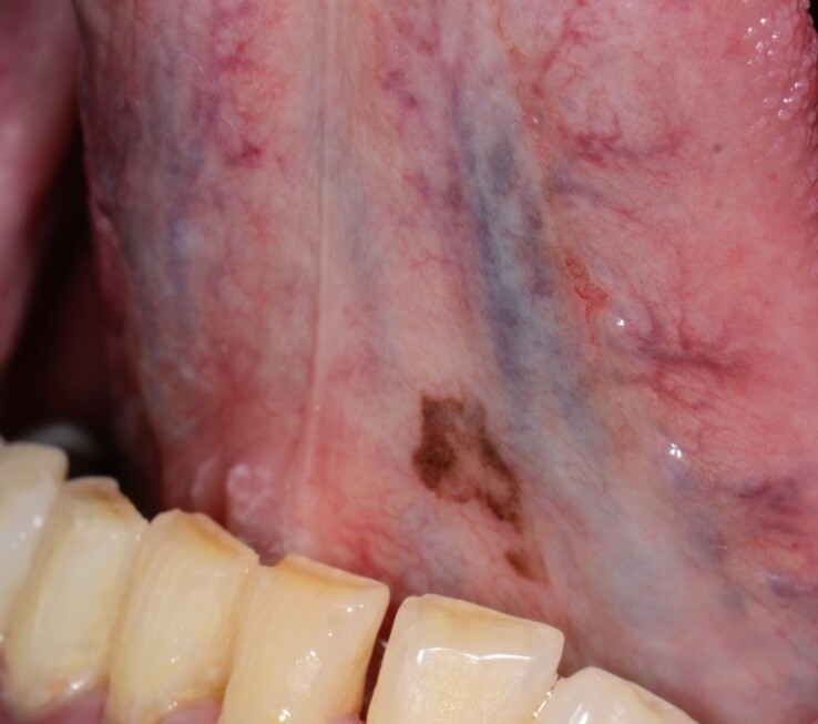
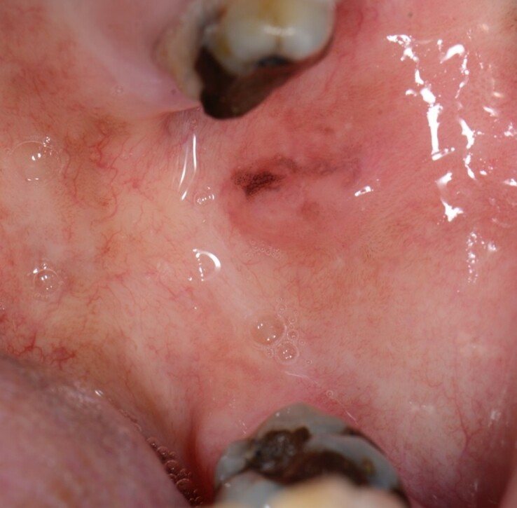
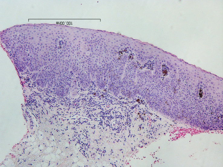
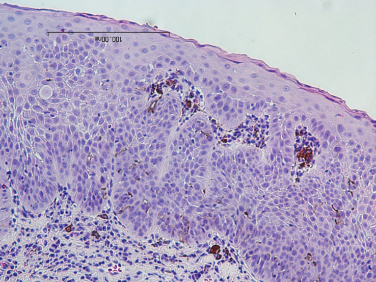
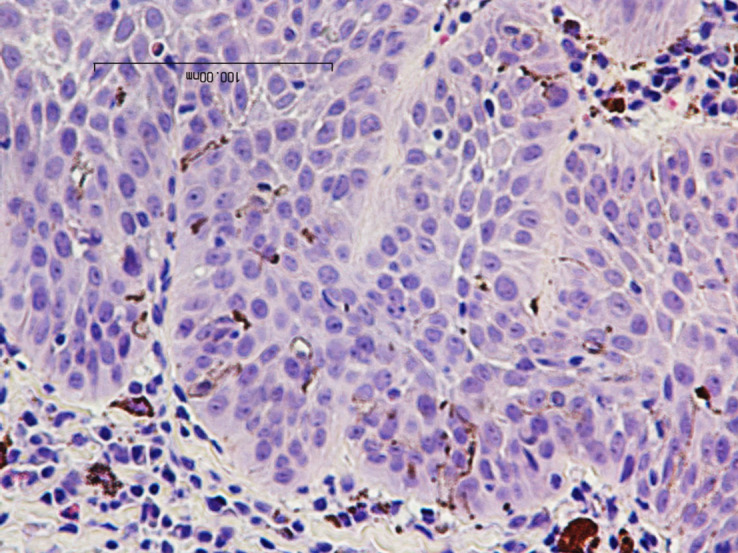
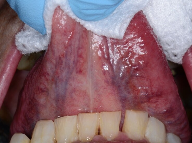
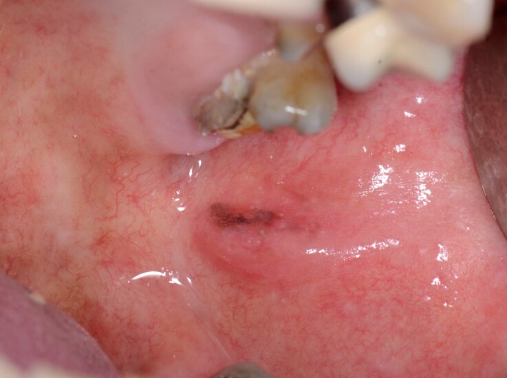
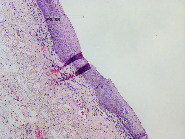
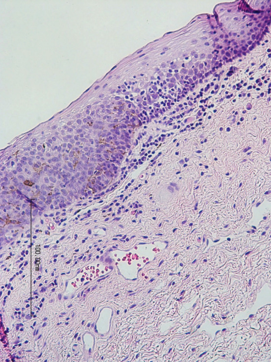
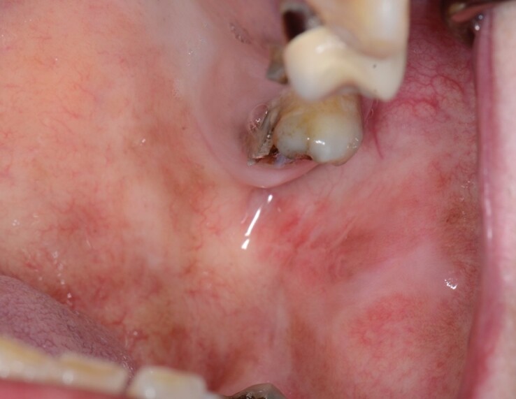
Discussion: The first pigmented lesion on the ventral tongue was diagnosed as melanoacanthoma, which does not have any potential for malignant transformation. The ventral tongue is a very unusual location for a melanoacanthoma. Interestingly, another lesion on the buccal mucosa with a similar appearance presented as moderate epithelial dysplasia with increased melanin pigment that has the potential for malignant transformation. Pigmented moderate oral epithelial dysplasia is a very rare condition. The simultaneous presence of a melanoacanthoma would have led to a mistaken assumption of multifocal melanoacanthoma which, although rare, has been described in the literature.
Conclusion: These findings reinforce that it is prudent to perform multiple biopsies of all suspicious oral lesions.
Introduction : Les lésions muqueuses contenant de la mélanine peuvent présenter un défi diagnostique en raison de la présence d’un large éventail de lésions ayant un aspect clinique similaire.
Description du cas : Le présent rapport fait état d’un cas unique de la présentation simultanée de 2 lésions pigmentées chez une patiente blanche âgée de 68 ans.
Discussion : Le diagnostic de la première lésion pigmentée sur la langue ventrale est celui d’un mélanoacanthome, qui n’a pas de potentiel de transformation maligne. Il est très rare qu’un mélanoacanthome se trouve sur la partie ventrale de la langue. Fait intéressant, une autre lésion de la muqueuse buccale d’apparence similaire s’est révélée être une dysplasie épithéliale modérée avec une augmentation du pigment mélanique qui présente un potentiel de transformation maligne. La dysplasie épithéliale buccale modérée pigmentée est une affection très rare. La présence simultanée d’un mélanoacanthome aurait mené à l’hypothèse erronée d’un mélanoacanthome multifocal qui, bien que rare, a été décrit dans la documentation.
Conclusion : Ces résultats confirment qu’il est prudent d’effectuer des biopsies multiples de toutes les lésions buccales suspectes.
Keywords: mouth; oral; oral melanoacanthoma; pigmented lesions; pigmented moderate oral epithelial dysplasia.
Copyright © 2025 CDHA | ACHD.
Conflict of interest statement
The authors declare that they have no conflicts of interest.
Figures
Similar articles
-
Prescription of Controlled Substances: Benefits and Risks.2025 Jul 6. In: StatPearls [Internet]. Treasure Island (FL): StatPearls Publishing; 2025 Jan–. 2025 Jul 6. In: StatPearls [Internet]. Treasure Island (FL): StatPearls Publishing; 2025 Jan–. PMID: 30726003 Free Books & Documents.
-
The Black Book of Psychotropic Dosing and Monitoring.Psychopharmacol Bull. 2024 Jul 8;54(3):8-59. Psychopharmacol Bull. 2024. PMID: 38993656 Free PMC article. Review.
-
[Volume and health outcomes: evidence from systematic reviews and from evaluation of Italian hospital data].Epidemiol Prev. 2013 Mar-Jun;37(2-3 Suppl 2):1-100. Epidemiol Prev. 2013. PMID: 23851286 Italian.
-
Systemic pharmacological treatments for chronic plaque psoriasis: a network meta-analysis.Cochrane Database Syst Rev. 2017 Dec 22;12(12):CD011535. doi: 10.1002/14651858.CD011535.pub2. Cochrane Database Syst Rev. 2017. Update in: Cochrane Database Syst Rev. 2020 Jan 9;1:CD011535. doi: 10.1002/14651858.CD011535.pub3. PMID: 29271481 Free PMC article. Updated.
-
Systemic pharmacological treatments for chronic plaque psoriasis: a network meta-analysis.Cochrane Database Syst Rev. 2020 Jan 9;1(1):CD011535. doi: 10.1002/14651858.CD011535.pub3. Cochrane Database Syst Rev. 2020. Update in: Cochrane Database Syst Rev. 2021 Apr 19;4:CD011535. doi: 10.1002/14651858.CD011535.pub4. PMID: 31917873 Free PMC article. Updated.
References
-
- Carlos-Bregni R , Contreras E , Netto AC , et al. Oral melanoacanthoma and oral melanotic macule: a report of 8 cases, review of the literature, and immunohistochemical analysis Med Oral Patol Oral Cir Bucal 2007 ; 12 ( 5 ): E374 – E379 - PubMed
LinkOut - more resources
Full Text Sources