

Anatomical Insights Into Profound Pulmonary Venous Malformation in an Elderly Individual: A Report of a Rare Cadaveric Case
- PMID: 40809595
- PMCID: PMC12348210
- DOI: 10.7759/cureus.87905
Anatomical Insights Into Profound Pulmonary Venous Malformation in an Elderly Individual: A Report of a Rare Cadaveric Case
Abstract
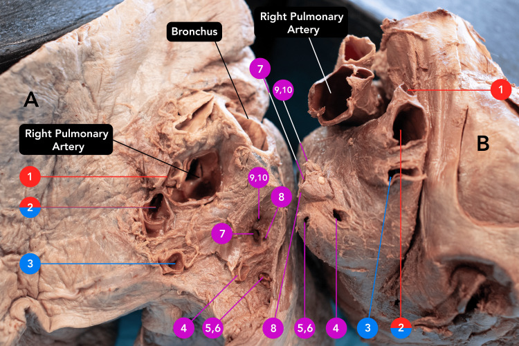
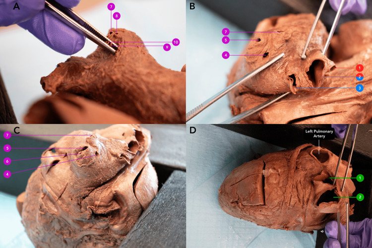
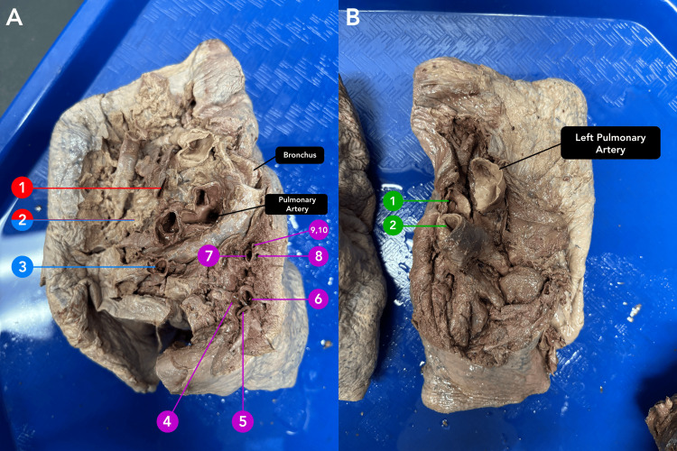
A male cadaver, 87 years of age, was received through the Saint Louis University Gift of Body Program. Significant pulmonary vein (PV) variations were observed following routine dissection. The donor displayed two normal left PVs and 10 significantly undersized right PVs. By caliper measurement, the left PVs had long axis diameters of 19 mm and 20 mm, while the right PVs ranged from 1 mm to 11 mm. The total cross-sectional area of the 10 right PVs was 173 mm2 which was significantly less than the 429 mm2 total cross-sectional area of the two left PVs. This was unexpected since the total diameter and cross-sectional area of right PVs are usually reported to be larger. While it is not uncommon for patients to have slight variations in the pulmonary venous structure, there are no known reports of individuals with 12 asymmetric PVs. Anatomical variation in the quantity and dimensions of PVs results in abnormal blood drainage and increased fluid resistance in the vessels, which could potentially contribute to cardiopulmonary sequelae. Appreciation of variations and their possible developmental underpinnings could allow for a better understanding of pathology and appropriate treatment for patients with anomalous vessels and cardiovascular diseases. For example, recent correlations between PV defects, atrial fibrillation, and ectopic heartbeats have been elucidated. Individuals with abnormal PVs have been found to express higher rates of ectopic foci resulting in atrial fibrillation. Successful resolution of ectopic foci via radiofrequency catheter ablation requires accessory PVs to be effectively identified and accounted for when evaluating treatment options. As such, a comprehensive understanding of both typical and atypical pulmonary vasculature prompts refinement of interventional procedures for maximal safety and efficacy for these patients.
Keywords: accessory pulmonary veins; embryology; pulmonary abnormalities; pulmonary vein variations; vessel anomalies.
Copyright © 2025, Schwab et al.
Conflict of interest statement
Human subjects: All authors have confirmed that this study did not involve human participants or tissue. Conflicts of interest: In compliance with the ICMJE uniform disclosure form, all authors declare the following: Payment/services info: All authors have declared that no financial support was received from any organization for the submitted work. Financial relationships: All authors have declared that they have no financial relationships at present or within the previous three years with any organizations that might have an interest in the submitted work. Other relationships: All authors have declared that there are no other relationships or activities that could appear to have influenced the submitted work.
Figures
Similar articles
-
Prescription of Controlled Substances: Benefits and Risks.2025 Jul 6. In: StatPearls [Internet]. Treasure Island (FL): StatPearls Publishing; 2025 Jan–. 2025 Jul 6. In: StatPearls [Internet]. Treasure Island (FL): StatPearls Publishing; 2025 Jan–. PMID: 30726003 Free Books & Documents.
-
The Black Book of Psychotropic Dosing and Monitoring.Psychopharmacol Bull. 2024 Jul 8;54(3):8-59. Psychopharmacol Bull. 2024. PMID: 38993656 Free PMC article. Review.
-
[Volume and health outcomes: evidence from systematic reviews and from evaluation of Italian hospital data].Epidemiol Prev. 2013 Mar-Jun;37(2-3 Suppl 2):1-100. Epidemiol Prev. 2013. PMID: 23851286 Italian.
-
Intravenous magnesium sulphate and sotalol for prevention of atrial fibrillation after coronary artery bypass surgery: a systematic review and economic evaluation.Health Technol Assess. 2008 Jun;12(28):iii-iv, ix-95. doi: 10.3310/hta12280. Health Technol Assess. 2008. PMID: 18547499
-
Sexual Harassment and Prevention Training.2024 Mar 29. In: StatPearls [Internet]. Treasure Island (FL): StatPearls Publishing; 2025 Jan–. 2024 Mar 29. In: StatPearls [Internet]. Treasure Island (FL): StatPearls Publishing; 2025 Jan–. PMID: 36508513 Free Books & Documents.
References
-
- Lung segments from anatomy to surgery. Wąsik J, Tubbs RS, Zielinska N, Karauda P, Olewnik Ł. Folia Morphol (Warsz) 2024;83:20–34. - PubMed
-
- Anomalous pulmonary venous connections. Katre R, Burns SK, Murillo H, Lane MJ, Restrepo CS. Semin Ultrasound CT MR. 2012;33:485–499. - PubMed
-
- Development and structures of the venous pole of the heart. Anderson RH, Brown NA, Moorman AF. Dev Dyn. 2006;235:2–9. - PubMed
-
- Hegazy A. Lap Lambert Academic Publishing; 2014. Clinical Embryology for Medical Students and Postgraduate Doctors.
Publication types
LinkOut - more resources
Full Text Sources
Research Materials