. 2025 Jul 30:13:1585106.
doi: 10.3389/fcell.2025.1585106. eCollection 2025.

Exosomal miR-2137 from cadmium-treated hepatocytes drives renal ferroptosis via GPX4 suppression and is alleviated by selenium

Affiliations

Exosomal miR-2137 from cadmium-treated hepatocytes drives renal ferroptosis via GPX4 suppression and is alleviated by selenium

Qian Wen et al. Front Cell Dev Biol. .

Abstract

Cadmium (Cd) is a toxic heavy metal that primarily affects the liver and kidneys. Despite greater Cd accumulation in the liver, Cd-induced oxidative damage is more pronounced in the kidney, suggesting the involvement of hepatorenal communication. However, the underlying mechanism remains unclear. To investigate Cd-induced hepatorenal toxicity, we established a Cd-exposed mouse model and assessed ferroptosis-related liver and kidney injury. Exosomes derived from Cd-exposed hepatocytes were isolated, and miRNAs targeting GPX4 were screened and identified. The role of GPX4-targeting miRNAs in mediating renal toxicity induced by hepatocyte-derived exosomes was evaluated in vivo using antagomirs. The protective effect of selenium (Se) supplementation against Cd-induced hepatic and renal damage was also examined. Cd exposure induced significant liver and kidney injury through GPX4-downregulated ferroptosis. Mechanistically, exosomes from Cd-treated hepatocytes were enriched in miR-2137, which targets renal GPX4 and promotes ferroptosis in the kidney. Injection of hepatocyte-derived exosomes alone reduced renal GPX4 levels in vivo, an effect that was reversed by miR-2137 antagomir treatment. Furthermore, Se supplementation restored GPX4 expression and protected both liver and kidney tissues from Cd-induced damage. These findings reveal a novel exosome-mediated hepatorenal communication pathway under Cd exposure, wherein hepatocyte-derived exosomal miRNAs contribute to distant renal injury. Targeting specific exosomal miRNAs or enhancing GPX4 expression via selenium may offer therapeutic strategies against Cd toxicity.

Keywords: GPx4; cadmium; ferroptosis; hepatorenal communication; selenium.


Conflict of interest statement

The authors declare that the research was conducted in the absence of any commercial or financial relationships that could be construed as a potential conflict of interest.

Figures

FIGURE 1
FIGURE 1
Cd induces liver and kidney damage and is rescued by Se. (A) Experimental strategy: 8w mice were given SeMet-supplemented diet for 3months, and CdCl2 was administered intraperitoneally at the same time for the last 7 days (B) HE staining of pathological sections of mouse liver and kidney. The black arrows indicate damaged hepatocytes and renal tubular cells. Liver scale: 100 μm, Kidney scale: 30 μm. (C–E) Mouse Liver Function Test: The kits detect mouse serum AST, ALT and AKP levels. (F–H) Mouse Kidney Function Test: The kits detect serum BUN, CRE and UA levels in mice. (I) Myeloperoxidase (MPO) levels were assayed in the liver and kidney of differently treated mice. (J) Mass spectrometric detection of cadmium accumulation in liver and kidney of mice under different treatments. Symbol colors represent treatment groups: black = control, blue = Cd, green = Cd + Se, and red = Se. Data are expressed as mean ± SEM. Pairwise comparisons between groups were conducted using two-tailed non-paired Student’s t tests. ns, not significant (p ≥ 0.05); *p < 0.05; **p < 0.01; ***p < 0.001.
FIGURE 2
FIGURE 2
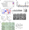
Ferroptosis contributes to Cd-induced liver damage. (A) Liver KEGG enrichment analysis and (B) Heat map of anti-ferroptosis genes in mice in the cadmium-treated and selenium-cadmium co-treated groups. (C) Expression of anti-ferroptosis genes in mouse liver by reverse transcription-polymerase chain reaction (RT-PCR). Values were normalized to the mRNA level of the housekeeping gene Actin in the respective sample. (D) Immunoblotting of GPX4 and Hsp90 in mouse liver. The right panel shows the quantitative analysis of GPX4 protein expression. (E) Malondialdehyde (MDA) levels and (F) GSH/GSSG levels were detected in the livers of mice. (G) Transmission electron microscopy of mitochondrial ferroptosis in AML12 cells. Representative mitochondria are shown in white boxes. Scale: 1 μm. N. nucleus; M. mitochondria. The white arrows indicate mitochondria exhibiting typical morphological features of ferroptosis, including reduced mitochondrial volume, increased membrane density, and decreased or vanished cristae structures. (H) Representative images of mouse liver organoids treated with 1.25 μM CdCl2 and 10 μM SeMet alone or in combination for 12 days. Statistical graph of class organ diameters (right panel). Scale: 100 μm. Data are expressed as mean ± SEM. Pairwise comparisons between groups were conducted using two-tailed non-paired Student’s t tests. ns, not significant (p ≥ 0.05); *p < 0.05; **p < 0.01; ***p < 0.001.
FIGURE 3
FIGURE 3
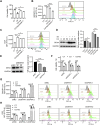
Severe ferroptosis is observed in Cd-exposed kidney tissue. (A) Expression of anti-ferroptosis genes in mouse kidney by reverse transcription-polymerase chain reaction (RT-PCR). Values were normalized to the mRNA level of the housekeeping gene Actin in the respective sample. (B) Immunoblotting of GPX4 and GAPDH in mouse kidney. The figure below shows the quantitative analysis of GPX4 protein expression. (C) CCK8 assay for renal HK2 cell survival under SeMet, CdCl2 and ferroptosis inhibitor liproxstain-1 treatment. (D) Immunoblotting for detection of GPX4 in SeMet and CdCl2-treated HK2 cells. The right panel shows the quantitative analysis of GPX4 protein expression. (E–F) Detection of BODIPY C11 (E) and (F) ROS level of HK2 cell under different treatments. Data are expressed as mean ± SEM. Pairwise comparisons between groups were conducted using two-tailed non-paired Student’s t tests. ns, not significant (p ≥ 0.05); *p < 0.05; **p < 0.01; ***p < 0.001.
FIGURE 4
FIGURE 4
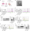
Cd-induced hepatorenal communication exacerbates renal ferroptosis via exosomes. (A) Pattern diagram: liver primary cells were isolated from mice and treated with PBS/CdCl2 for 24 h. Cell supernatants were collected and exosomes were extracted. HK2 cells were treated with CT/CdCl2 exosomes in combination with SeMet. (B) Representative images of the morphology of cellular supernatant exosomes observed by transmission electron microscopy. Scale: 100 μm. (C) Detection of BODIPY C11 and (D) ROS level of HK2 cells. HK2 cells were treated with CT/CdCl2 exosomes and SeMet for 24 h. (E) Immunoblotting for detection of GPX4 and GAPDH in CT/CdCl2 exosomes and SeMet HK2 cells. The right figure shows the quantitative analysis of GPX4 protein expression. (F) Detection of BODIPY C11 and (G) ROS level of HK2 cells. HK2 cells were transfected with either empty vector (EV) or a GPX4-overexpressing plasmid, followed by treatment with exosomes isolated from the supernatant of CT/CdCl2 treated Huh7 cells. (H) Western blot analysis of GPX4 and Actin expression in HK2 cells. The processing conditions are in accordance with those of (F) and (G). The right figure shows the quantitative analysis of GPX4 protein expression. Data are expressed as mean ± SEM. Pairwise comparisons between groups were conducted using two-tailed non-paired Student’s t tests. ns, not significant (p ≥ 0.05); *p < 0.05; **p < 0.01; ***p < 0.001.
FIGURE 5
FIGURE 5
Antagonistic effect of Se on Cd-induced ferroptosis via upregulation of GPX4. (A) CCK8 assay for AML12 cell survival under SeMet, CdCl2 and ferroptosis inhibitor liproxstain-1 treatment. (B) Detection of BODIPY C11 and (C) ROS level of AML12 cell under different treatments. (D) Immunoblotting for detection of GPX4 in SeMet and CdCl2-treated AML12 cells. The right panel shows the quantitative analysis of GPX4 protein expression. (E) Immunoblotting for detection of GPX4 knockdown efficiency in AML12 cells. The right panel shows the quantitative analysis of GPX4 protein expression. (F) CCK8 assay cell survival of cell lines with AML12 knockdown of GPX4 after cadmium and/or selenium treatment. (G) Detection of BODIPY C11 and (H) ROS level of cell lines with AML12 knockdown of GPX4 after cadmium and/or selenium treatment. Data are expressed as mean ± SEM. Pairwise comparisons between groups were conducted using two-tailed non-paired Student’s t tests. ns, not significant (p ≥ 0.05); *p < 0.05; **p < 0.01; ***p < 0.001.
FIGURE 6
FIGURE 6
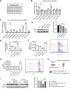
Cd-induced liver exosomal miR-2137 mediates renal cell ferroptosis and is blunted with Se. (A) Prediction of miRNAs targeting GPX4 using TargetScan. (B) Detection of serum levels of miRNAs targeting predicted miRNAs in control and CdCl2-exposed mice. (C) Detection of levels of miRNAs targeting predicted miRNAs in supernatant exosomes of PBS/CdCl2-treated Huh7 cells. (D) GPX4 and Actin levels were detected by immunoblotting after transfection of 293T for 48 h using miRNAs. The right panel shows the quantitative analysis of GPX4 protein expression. (E) Regulation of GPX4 3‘UTR by miRNAs was detected using dual luciferase in 293T cells. (F) Detection of BODIPY C11 and (G) ROS level in HK2 cells transfected with different miRNAs. (H) Pattern diagram: liver primary cells were treated with PBS/CdCl2 for 24 h, supernatants were collected and exosomes were isolated. Exosomes were injected into mice by tail vein and NC/miR-2137 antagomir treatment was given twice a week for a total of 2 weeks. (I) Immunoblotting to detect the expression levels of GPX4 and GAPDH in mouse kidney. The right panel shows the quantitative analysis of GPX4 protein expression. Data are expressed as mean ± SEM. Pairwise comparisons between groups were conducted using two-tailed non-paired Student’s t tests. ns, not significant (p ≥ 0.05); *p < 0.05; **p < 0.01; ***p < 0.001.
FIGURE 7
FIGURE 7
Model. Cd-induced renal ferroptosis by liver exosomes is rescued by Se supplementation. During Cd exposure, miR-2137 from hepatic exosomes targeted renal GPX4 and aggravated renal ferroptosis. Se supplementation alleviated renal ferroptosis exacerbated by hepatorenal communication by upregulating GPX4.

Similar articles

References

    1. An X., Yu W., Liu J., Tang D., Yang L., Chen X. (2024). Oxidative cell death in cancer: mechanisms and therapeutic opportunities. Cell Death Dis. 15 (8), 556. 10.1038/s41419-024-06939-5 - DOI - PMC - PubMed
    1. Bautista C. J., Arango N., Plata C., Mitre-Aguilar I. B., Trujillo J., Ramírez V. (2024). Mechanism of cadmium-induced nephrotoxicity. Toxicology 502, 153726. 10.1016/j.tox.2024.153726 - DOI - PubMed
    1. Bebelman M. P., Smit M. J., Pegtel D. M., Baglio S. R. (2018). Biogenesis and function of extracellular vesicles in cancer. Pharmacol. Ther. 188, 1–11. 10.1016/j.pharmthera.2018.02.013 - DOI - PubMed
    1. Biagioli M., Pifferi S., Ragghianti M., Bucci S., Rizzuto R., Pinton P. (2008). Endoplasmic reticulum stress and alteration in calcium homeostasis are involved in cadmium-induced apoptosis. Cell Calcium 43 (2), 184–195. 10.1016/j.ceca.2007.05.003 - DOI - PubMed
    1. Dixon S. J., Lemberg K. M., Lamprecht M. R., Skouta R., Zaitsev E. M., Gleason C. E., et al. (2012). Ferroptosis: an iron-dependent form of nonapoptotic cell death. Cell 149 (5), 1060–1072. 10.1016/j.cell.2012.03.042 - DOI - PMC - PubMed

LinkOut - more resources