Review
. 2025 Aug 5;10(1):bpaf058.
doi: 10.1093/biomethods/bpaf058. eCollection 2025.

Application of dicentric chromosome assay for evaluation of radioprotective effect

Affiliations
Review

Application of dicentric chromosome assay for evaluation of radioprotective effect

Marcela Milanová et al. Biol Methods Protoc. .

Abstract

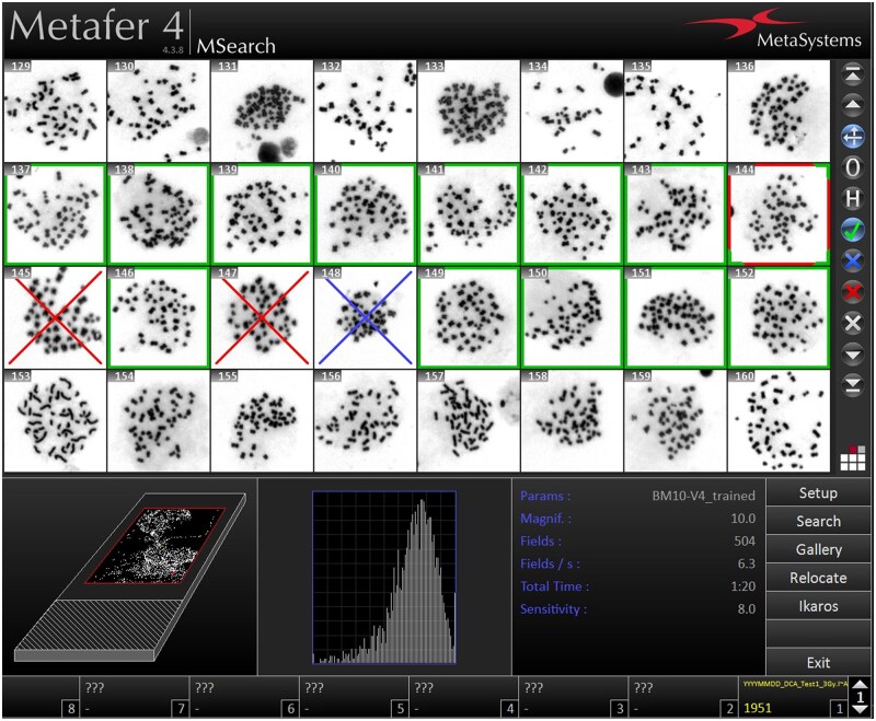
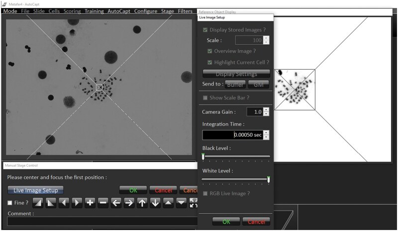
The dicentric chromosome assay is a well-established biodosimetric method used to assess absorbed ionizing radiation doses by detecting dicentric chromosomal aberrations. Here, we present a detailed, reproducible protocol for applying the dicentric chromosome assay for in vitro evaluation of radioprotective agents, including novel piperazine derivatives compared with amifostine and its active metabolite WR-1065. The protocol covers all key steps-blood sample preparation, in vitro irradiation, lymphocyte culture, metaphase preparation, and scoring of dicentric chromosomes. It highlights critical stages that affect data quality and reproducibility. Integrating manual scoring with automated analysis using the Metafer system ensures accurate and efficient assessment. Thus, this protocol bridges the fields of biological dosimetry and preclinical screening of radioprotective agents, providing a reliable framework for emergency radiation dose estimation and the development of new radiation medical countermeasures.

Keywords: amifostine; chromosome aberrations; cytogenetics; ionizing radiation; radioprotective agent.


Figures

Figure 1.
Figure 1.
Optimal dripping position on the microscope slide for automated scanning (left) with the optimal spread and number of metaphases (right).
Figure 2.
Figure 2.
Menu for the slide setup. Recommended parameters for the first metaphase capture using MSearch/TL mode.
Figure 3.
Figure 3.
The focus position setup and the reference object selection (magnification 10x, Metafer 4 - MSearch).
Figure 4.
Figure 4.
Sample metaphases selection meeting all parameters in analysis window (green frame—marked/accepted cell; red frame—active position; blue cross—rejected cell; and red cross—deleted cell).
Figure 5.
Figure 5.
Focusing and centering of the first reference metaphase (magnification 63x, Metafer 4 - Autocapt).
Figure 6.
Figure 6.
Metaphases inappropriate for evaluation: (A) metaphase with excessively spread chromosomes, (B) more than one captured metaphase, (C) a metaphase with an incomplete set of chromosomes.
Figure 7.
Figure 7.
Example of metaphase in a non-irradiated control sample without DCs (A), metaphase after 3 Gy irradiation with DCs (B); and graphical representation of the number of dicentric chromosomes (DC) per cell after exposure to IR (3 Gy) alone or after 1-h pretreatment with the compounds at optimal non-toxic concentration [C] and maximum tolerated concentration [D]. The incubation interval was 48 h, and then the cells were subjected to DCA. Error bars represent the standard deviation. The asterisks above the individual bars represent statistical significance (*P < .05, **P < .01, ***P < .001, ****P < .0001) related to 3 Gy. Reproduced from Chmil et al. 2024 [7] with permission from the Royal Society of Chemistry under CC BY 3.0.

Similar articles

References

    1. Thiruvengadam NR, Miranda J, Kim C et al. Burden of ionizing radiation in the diagnosis and management of necrotizing pancreatitis. Clin Transl Gastroenterol 2021;12:e00347. 10.14309/ctg.0000000000000347 - DOI - PMC - PubMed
    1. Chang ML, Hou JK. Cancer risk related to gastrointestinal diagnostic radiation exposure. Curr Gastroenterol Rep 2011;13:449–57. 10.1007/s11894-011-0214-8 - DOI - PubMed
    1. Herfarth H, Palmer L. Risk of radiation and choice of imaging. Dig Dis 2009;27:278–84. 10.1159/000228561 - DOI - PubMed
    1. De Ruysscher D, Niedermann G, Burnet NG et al. Radiotherapy toxicity. Nat Rev Dis Primers 2019;5:13. 10.1038/s41572-019-0064-5 - DOI - PubMed
    1. Holdstock D, Waterston L. Nuclear weapons, a continuing threat to health. Lancet 2000;355:1544–7. 10.1016/S0140-6736(00)02176-0 - DOI - PubMed

LinkOut - more resources