. 2025 Jul 11;10(1):bpaf056.
doi: 10.1093/biomethods/bpaf056. eCollection 2025.

Innovative approach for the qualitative-quantitative assessment of neurodevelopment biomarkers research in placenta tissue using immunohistochemistry digital image analysis

Affiliations

Innovative approach for the qualitative-quantitative assessment of neurodevelopment biomarkers research in placenta tissue using immunohistochemistry digital image analysis

Caroline Camilo et al. Biol Methods Protoc. .

Abstract

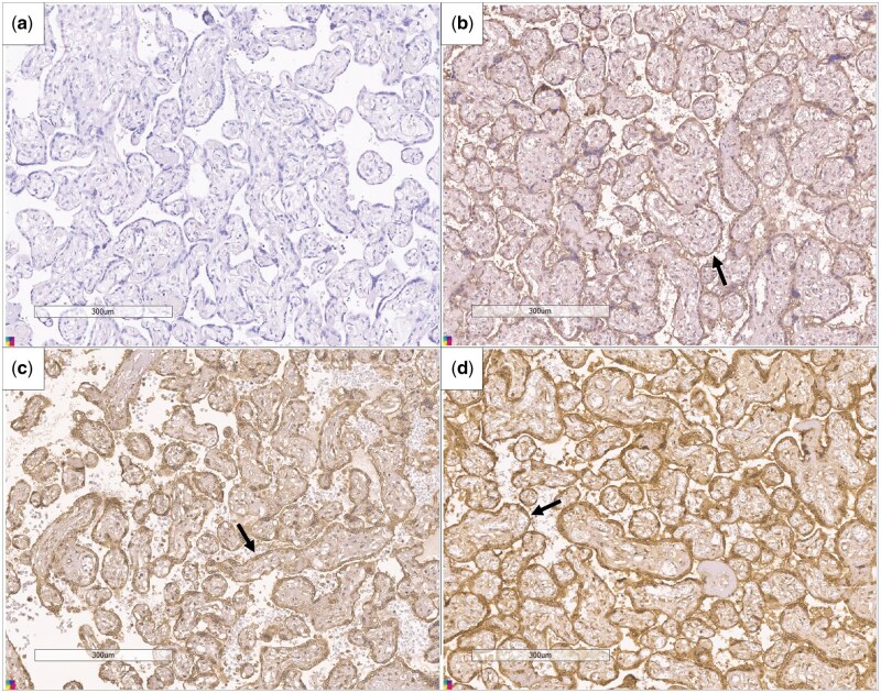
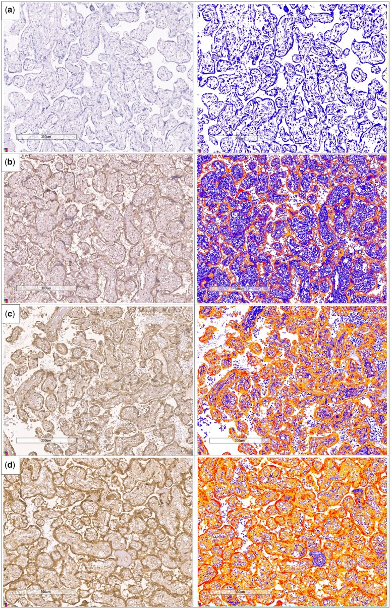
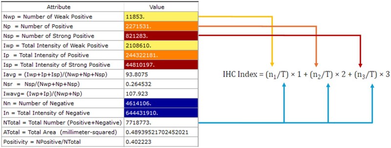
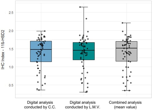
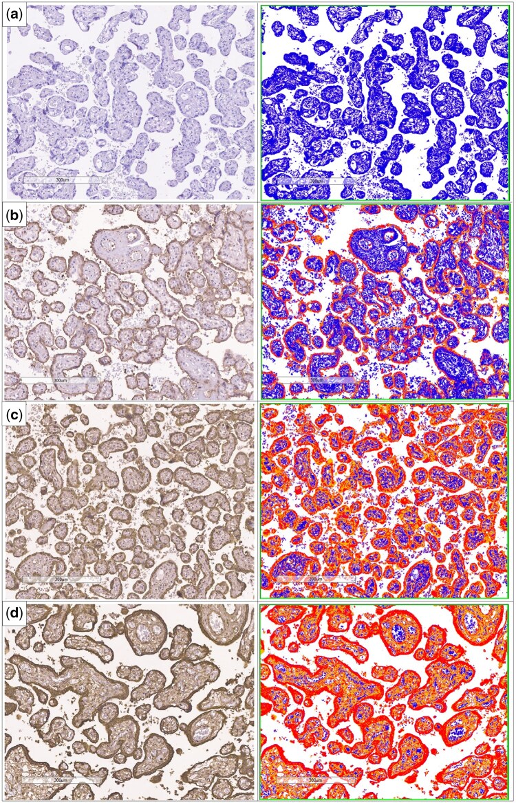
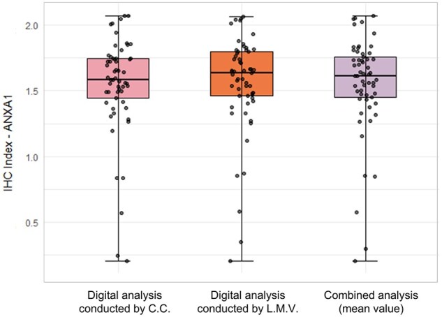
We aimed to develop and validate a standardized, qualitative-quantitative protocol for digital IHC analysis to assess neurodevelopmental biomarkers in placental tissue. Placental tissues from 60 births were obtained from the Western Region Birth Cohort (ROC), and IHC staining was performed using NovolinkTM Polymer System. The primary antibody against 11βHSD2 protein was used for protocol development, and ANXA1 was employed for validation. Slides were digitized using the Aperio ScanScope XT, and image analysis was conducted using the Positive Pixel Count V9 algorithm. Protein expression levels were calculated using the IHC Index formula. Protocol steps included combined optical and digital evaluation, representative fields per slide, intra- and interobserver validation, and assessment of reproducibility. Digital analysis of three random fields (scale bar: 300 µm) showed strong concordance with optical microscopy assessments for 11βHSD2 placental expression. Intraobserver validation showed a strong correlation (τ: 0.70, P < .001) and a substantial concordance (kw: 0.67; P-value < .001), while interobserver comparisons also yielded substantial agreement (kw: 0.61, P < .001), confirming the protocol's reliability. Validation using ANXA1 expression revealed moderate intra- and interobserver concordance (kw: 0.50 and kw: 0.48, respectively; both P < .001), reinforcing the protocol's applicability across different proteins. In conclusion, we established a reproducible digital IHC analysis protocol that enhances reliability in exploratory research. This approach optimizes image quantification, minimizes observer bias, and contributes to advances in developmental biology research and digital pathology focused on placental neurodevelopment biomarkers.

Keywords: digital analysis; immunohistochemistry; neurodevelopment biomarkers; placenta; protein expression; protocol.


Figures

Figure 1
Figure 1
Detection of 11βHSD2 protein expression in human placentas using the primary antibody anti-HSD11B2 (ab203132; Abcam) (Magnification ×20; scale bar: 200 µm). Positive staining in syncytiotrophoblasts is indicated by a brown color. Counterstaining: Mayer’s hematoxylin. (a) Negative control—Performed by excluding the primary antibody; (b) weak staining intensity (++); (c) moderate staining intensity (+++); (d) strong staining intensity (++++). Black arrows indicate more intense staining along the syncytiotrophoblast margins
Figure 2
Figure 2
Results of 11βHSD2 protein expression digital analysis in three placental samples using Positive Pixel Count V9 algorithm in ImageScope™ software (×8 magnification; scale bar: 300 µm) with the primary antibody anti-HSD11B2 (ab203132; Abcam). (a) Negative control—performed by excluding the primary antibody; (b) weak staining intensity; (c) moderate staining intensity; (d) strong staining intensity. Left: positive staining in syncytiotrophoblasts is indicated by a brown color—counterstaining: Mayer’s hematoxylin; Right: digital image analysis—staining intensity colors: strongly positive (red), positive (orange), weakly positive (yellow), and negative (blue)
Figure 3
Figure 3
Correspondence between positive pixel count V9 algorithm results (Left) and IHC Index equation values (Right)
Figure 4
Figure 4
Boxplots of IHC Index values for 11βHSD2 protein expression obtained from digital image analyses conducted by C.C. and L.M.V., and the mean value of both analyses
Figure 5
Figure 5
Results of ANXA1 protein expression digital analysis in three placental samples using positive pixel count V9 algorithm in ImageScope™ software (×8 magnification; scale bar: 300 µm) with the primary antibody anti-ANXA1 (#71-3400, Invitrogen). (a) Negative control—performed by excluding the primary antibody; (b) weak staining intensity; (c) moderate staining intensity; (d) strong staining intensity. Left: positive staining in syncytiotrophoblasts is indicated by a brown color—counterstaining: Mayer’s hematoxylin; Right: digital image analysis—staining intensity colors: strongly positive (red), positive (orange), weakly positive (yellow), and negative (blue)
Figure 6
Figure 6
Boxplots of IHC Index values for ANXA1 protein expression obtained from digital image analyses conducted by C.C. and L.M.V., and the mean value of both analyses

Similar articles

References

    1. Escobar Díaz Guerrero R, Carvalho L, Bocklitz T et al. Software tools and platforms in digital pathology: a review for clinicians and computer scientists. J Pathol Inform 2022;13:100103. 10.1016/j.jpi.2022.100103 - DOI - PMC - PubMed
    1. Kadir AB, Singh NR. The role of digital pathology in enhancing diagnostic accuracy in oncology. J Bras Patol Med Lab 2024;60:1–7. 10.1900/JBPML.2024.60.01.001 - DOI
    1. Aeffner F, Adissu HA, Boyle MC et al. Digital microscopy, image analysis, and virtual slide repository. ILAR J 2018;59:66–79. 10.1093/ilar/ily007 - DOI - PMC - PubMed
    1. Maity S, Nauhria S, Nayak N et al. Virtual versus light microscopy usage among students: a systematic review and meta-analytic evidence in medical education. Diagnostics (Basel) 2023;13:558. 10.3390/diagnostics13030558 - DOI - PMC - PubMed
    1. Aeffner F, Zarella MD, Buchbinder N et al. Introduction to digital image analysis in whole-slide imaging: a white paper from the digital pathology association. J Pathol Inform 2019;10:9. 10.4103/jpi.jpi_82_18 - DOI - PMC - PubMed