. 2025 Aug 12:9:25424823251358414.
doi: 10.1177/25424823251358414. eCollection 2025 Jan-Dec.

Potential link of high fat diet and mRNA expression of Alzheimer's disease-related genes in the enteric mucosa of a rat model of Alzheimer's disease

Affiliations

Potential link of high fat diet and mRNA expression of Alzheimer's disease-related genes in the enteric mucosa of a rat model of Alzheimer's disease

Kuppan Gokulan et al. J Alzheimers Dis Rep. .

Abstract

Background: High-fat diet (HFD) consumption is linked to Alzheimer's disease (AD). Identifying changes in the mRNA expression due to the ingestion of HFD in the intestine-often called the second brain due to its dense enteric neurons-could offer insights into AD development and progression.

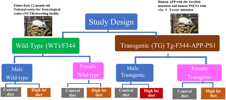
Objective: This study assesses whether the introduction of HFD at adult-age influence expression of AD-related genes in the intestines of Wild-type (WT) or the amyloid precursor protein/presenilin1 (APP/PS1)-overexpressing Transgenic (TG) rats.

Methods: Twelve-month-old WT and TG rats (male and female) were fed a control diet (CD; 8% energy from fat), or HFD (45% energy from fat) for six months. Ileal tissues were assessed for the mRNA expression of genes responsible for development/progression of AD.

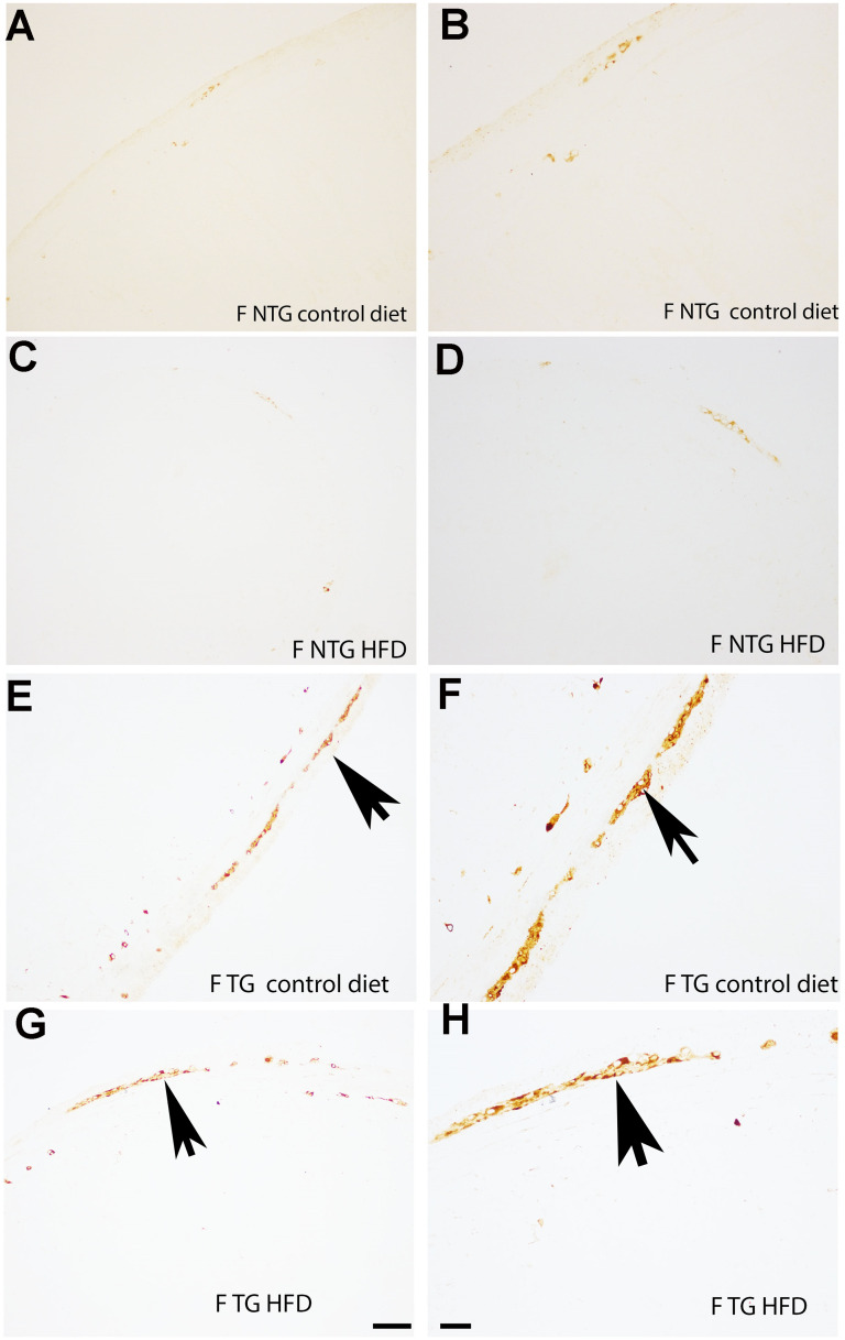
Results: The WT HFD-fed rats (compared to CD-fed rats) showed increased mRNA expression of genes involved in the development of AD. In contrast, the TG HFD-fed female group, showed a higher number of upregulated genes compared to their respective CD-fed TG group. In TG HFD-fed rats there was higher mRNA expression of genes crucial for synaptic transmission such as Brain-derived neurotrophic factor in females and Choline acetyltransferase in males. Expression of Plasminogen was higher in HFD-fed TG female rats and HFD-fed WT male rats. Overall, the HFD-fed WT male showed mRNA expression of genes involved in the development of AD. However, HFD-fed TG females were more vulnerable for the progression of AD. It is likely that the enteric Plasminogen plays a major role in gut-brain axis for the development of AD in WT male, and progression of AD in TG female during the consumption of HFD.

Conclusions: The consumption of HFD perturbed the expression of enteric genes known to be involved in amyloid-β generation, clearance, and degradation, in a sex-dependent manner.

Keywords: APP/PS1-overexpressing transgenic; Alzheimer's disease; gastrointestinal tract; gut-brain axis; high fat diet; mRNA expression; rat transgenic model.


Conflict of interest statement

The authors declared no potential conflicts of interest with respect to the research, authorship, and/or publication of this article.

Figures

Figure 1.
Figure 1.
Schematic diagram of the study design. Twelve-month-old male and female Wild-type [Fischer 344 (F344); WT] or the amyloid precursor protein/presenilin1 (APP/PS1)-overexpressing Transgenic (TG) rats were used in this study (n = 4 per experimental group). This schematic figure shows the experimental plan for the 12 months old WT and TG rats that were fed Control diet (CD) or high-fat diet (HFD) for following six months. At the end of six months rats were sacrificed, to assess the changes in the mRNA expression of Alzheimer's disease related genes in the ileal mucosa.
Figure 2.
Figure 2.
Demonstration of amyloid-β protein precursor immunohistochemistry in ileal mucosa of intestine tissue of male rats. Sparse amyloid-β protein precursor (AβPP) immunoreactivity [APP (E5X2B) Rabbit mAb that recognizes endogenous levels of human AβPP] was present in the intestine of male non-transgenic animals [M-NTG (or WT); control diet or high fat diet (HFD)] (A-D). However, in male transgenic animals [M-TG; control diet or high fat diet (HFD)], (E-H) widespread immuno-positive staining for AβPP is demonstrated in Auerbach's nerve plexus, located between longitudinal and circular smooth muscle layers of the intestinal wall, as well as multifocal staining in Meissner's nerve plexus, located in the submucosa. Bar magnification (A, C, E, G = 100 µm; B, D, F, H = 50 µm). Arrow indicates AβPP immunostaining in the nerve plexus. Each figure is representative of images from n = 4 animals.
Figure 3.
Figure 3.
Demonstration of amyloid-β protein precursor immunohistochemistry in ileal mucosa of intestine tissue of female rats. Very scant amyloid-β protein precursor (AβPP) immunoreactivity [APP (E5X2B) Rabbit mAb that recognizes endogenous levels of human AβPP] was present in the intestine of female non-transgenic animals [F-NTG (or WT); control diet or high fat diet (HFD)] (A-D). However, in female transgenic animals [F-TG; control diet or high fat diet (HFD)], (E-H), widespread immuno-positive staining for AβPP is demonstrated in Auerbach's nerve plexus, located between longitudinal and circular smooth muscle layers of the intestinal wall, as well as multifocal staining in Meissner's nerve plexus, located in the submucosa. Bar magnification (A, C, E, G = 100 µm; B, D, F, H = 50 µm). Arrow indicates AβPP immunostaining in the nerve plexus. Each figure is representative of image n = 4 in each experimental group.
Figure 4.
Figure 4.
Effect of high fat diet on the differential expression of Alzheimer's disease-related genes in the ileal tissue. The bar in each experimental group female-Wild Type-high fat diet (F-WT-HFD), male-Wild Type-high fat diet (M-WT-HFD), female-Transgenic-high fat diet (F-TG-HFD), and male-Transgenic-high fat diet (M-TG-HFD) are compared with their respective control diet (CD)-fed rats female-Wild Type-control diet (F-WT-CD), male-Wild Type- control diet (M-WT-CD), female-Transgenic- control diet (F-TG-CD), and male-Transgenic- control diet (M-TG-CD). Each experimental group consisted of n = 4. Rat Alzheimer's Disease RT² Profiler PCR plates were used to assess the expression of 84 genes that are thought to be important in the development and progression of Alzheimer's disease.
Figure 5.
Figure 5.
Venn diagram showing common and sex-specific genes in high fat diet-fed male and female rats (as compared to control diet-fed rats) in ileal tissue. (A) This Venn diagram shows that expression of same 14 genes was high in high fat diet (HFD)-fed male and female Wild Type WT) rats. (B) In HFD-fed Transgenic (TG) rats, only 2 genes were common between male and female. In contrast, sex specific genes were in higher in HFD-fed female (13 genes). All genes presented here had a 2-fold change, as compared to their respective CD fed group, along with p value less than 0.05. Each experimental group consisted of n = 4.
Figure 6.
Figure 6.
This Venn diagram shows genes expressed common and specific to high fat diet-fed wild type and transgenic rats in ileal tissue. (A) High fat diet (HFD) caused differential expression of 30 Alzheimer's disease (AD)-related genes in Wild Type (WT)-rats, but in Transgenic (TG) male rats the effect is minimal (3 AD-related genes). (B) The HFD-fed female WT rats showed higher number of AD-related genes in comparison to HFD-fed TG-female rats. However, 8 AD-related genes are common between WT and TG females. All genes presented here had a 2-fold change, as compared to their respective CD fed group, along with p value less than 0.05. Each experimental group consisted of n = 4.
Figure 7.
Figure 7.
Effects of high fat diet on the mRNA expression of the neuroinflammatory pathway genes in ileal tissue. The mRNA expression of different genes in the high fat diet (HFD)-fed rats compared to rats received control diet (CD) in same genotype (Wild Type (WT) or Transgenic (TG)) for various pathways are shown in the figure as fold regulation (as compared to respective CD). These pathways include amyloid-β generation, oligomerization, clearance & degradation pathway genes [A (female) and B (male)]; G-Protein Coupled Receptor (signaling) related genes [C (female) and D (male)]; cell cycle related genes [E (female) and F (male)]; and apoptotic genes (G). The horizontal line in each graph represents the gene expression in CD (value = 1). p-value in graphs is depicted by either ‘*’ (≤ 0.05) or ‘†’ (≤ 0.005). Each experimental group consisted of n = 4.
Figure 8.
Figure 8.
Effects of high fat diet on the mRNA expression of the oxidative stress pathway related genes in ileal tissue. The mRNA expression of different genes (as fold regulation) in high fat diet (HFD)-fed rats compared to rats received control diet (CD) are shown in A (Female) or B (male) for oxidoreductases and oxidative Stress related pathways and in C (female) and 8D (male) for protease inhibitors, protein kinases, synaptic formation transcriptional regulation related genes. The horizontal line in each graph represents the gene expression in CD (value = 1). p-value in graphs is depicted by either ‘*’ (≤ 0.05) or ‘†’ (≤ 0.005). Each experimental group consisted of n = 4.
Figure 9.
Figure 9.
Possible molecular mechanisms of high fat diet for signaling of the gut-brain axis in control and disease model of male AD. Differentially expressed Alzheimer's disease (AD)-related genes in high fat diet (HFD)-fed wild type (WT) males and transgenic (TG) males are shown in left and right panel, respectively. It is possible that long-term consumption of HFD may affect the expression of several AD-related genes including genes involved in amyloid-β (Αβ) generation (Apbb1, Aplb2, Ache, and Apba3) and metal dys-homeostasis (Mt3), which are known to be cause neuroinflammation in male WT, while a co-ordination between Chat and Snca may show a protective role in TG male for the progression of AD.
Figure 10.
Figure 10.
Possible molecular mechanisms of HFD for signaling of the gut-brain axis in control and disease model of female AD. Differentially expressed Alzheimer's disease (AD)-related genes in high fat diet (HFD)-fed wild type (WT) females and transgenic (TG) females are shown in left and right panel, respectively. Increased expression of several genes involved in the tau and amyloid-β (Aβ) pathology may lead to neuroinflammation in the HFD-fed WT female rats. On the other hand, the pattern and profile of gene expressed due to HFD in TG female may compromise the integrity of the intestinal barrier that may damage the blood-brain barrier.

References

    1. Matthews KA, Xu W, Gaglioti AH, et al. Racial and ethnic estimates of Alzheimer's disease and related dementias in the United States (2015–2060) in adults aged ≥65 years. Alzheimers Dement 2019; 15: 17–24. - PMC - PubMed
    1. Clapp M, Aurora N, Herrera L, et al. Gut microbiota's effect on mental health: the gut-brain axis. Clin Pract 2017; 7: 987. - PMC - PubMed
    1. Ngah WZW, Ahmad HF, Ankasha SJ, et al. Dietary strategies to mitigate Alzheimer's disease: insights into antioxidant vitamin intake and supplementation with microbiota-gut-brain axis cross-talk. Antioxidants (Basel) 2024; 13: 1504. - PMC - PubMed
    1. Waheed A, Ghaffar M, Mustafa S, et al. Nutrigenomics and neurological disorders: exploring diet-brain interactions for cognitive health. Neurogenetics 2024; 26: 10. - PubMed
    1. Malesza IJ, Malesza M, Walkowiak J, et al. High-fat, western-style diet, systemic inflammation, and gut microbiota: a narrative review. Cells 2021; 10: 3164. - PMC - PubMed

LinkOut - more resources