. 2025 Aug 7;31(29):107066.
doi: 10.3748/wjg.v31.i29.107066.

Tetrahydroxylated bile acids prevents malignant progression of Barret esophagus in vitro by inhibiting the interleukin-1β-nuclear factor kappa-B pathway

Affiliations

Tetrahydroxylated bile acids prevents malignant progression of Barret esophagus in vitro by inhibiting the interleukin-1β-nuclear factor kappa-B pathway

Anatolii Mamchur et al. World J Gastroenterol. .

Abstract

Background: Barrett esophagus (BE), a metaplastic adaptive process to gastrointestinal reflux, is associated with a higher risk of developing esophageal adenocarcinoma. However, the factors and mechanism that drive the malignant progression of BE is not well understood.

Aim: To investigate the role of bile acids, a component of the reflux fluid, in the malignant progression of BE.

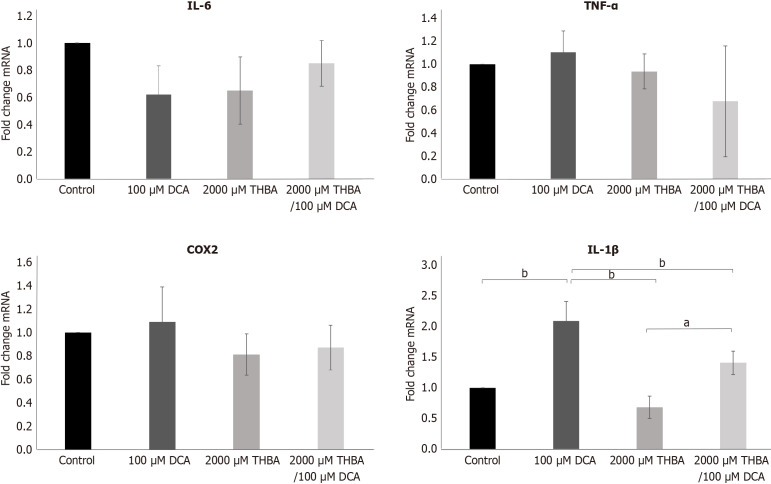
Methods: Using engineered green fluorescent protein- labeled adult tissue-resident stem cells isolated from BE clinical biopsies (BE-ASCs) as the target, we studied the effect of hydrophobic deoxycholic acid (DCA) and hydrophilic tetrahydroxylated bile acids (THBA) on cell viability by fluorescence intensity analysis, mucin production by dark density measurement, tissue structure by pathology analysis, expression of different pro-inflammatory factors gene by quantitative polymerase chain reaction and proteins by Western blot.

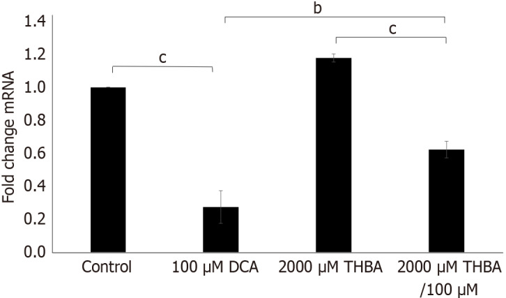
Results: We found that hydrophobic DCA has cytotoxic and proinflammatory effects through activation of interleukin-1β (IL-1β)-nuclear factor kappa-B (NF-κB) inflammatory pathway on BE-ASCs. This action results in impaired cell viability, tissue intactness, reduced mucin production, and increased transition to disorganized atypical cells without intestinal features. In contrast, co-culture with hydrophilic THBA inhibited the IL-1β-NF-κB inflammatory pathway with maintenance of mature intestinal type cellular and histomorphology.

Conclusion: Our data indicates that the hydrophilic bile acid THBA can counteract the cytotoxic and proinflammatory effect of hydrophobic DCA and prevent the malignant progression of BE by inhibiting the IL-1β-NF-κB pathway.

Keywords: Barrett esophagus; Cell viability; Deoxycholic bile acid; Histology; Inflammation; Metaplasia; Mucin; Pathway; Tetrahydroxylated bile acids.


Conflict of interest statement

Conflict-of-interest statement: The authors declare that they have no conflict of interest.

Figures

Figure 1
Figure 1
Scheme of cultivation of adult tissue-resident stem cells isolated from Barrett esophagus clinical biopsies and cytotoxicity investigation using different concentrations of deoxycholic acid, tetrahydroxylated bile acids and their combinations. BE-ASCs: Barrett esophagus clinical biopsies; DCA: Deoxycholic acid; THBA: Tetrahydroxylated bile acids.
Figure 2
Figure 2
Effect of deoxycholic acid, tetrahydroxylated bile acids and their combination on adult tissue-resident stem cells isolated from Barrett esophagus clinical biopsies cytotoxicity. Representative green fluorescent protein-labeled (GFP) fluorescent images of Barrett esophagus clinical biopsies (BE-ASCs) in transwell inserts after 15 days in culture with various deoxycholic acid (DCA) and hydrophilic tetrahydroxylated bile acids (THBA) concentrations. 1 × 10⁶ BE-ASCs were plated in 12 Matrigel coated transwell inserts on the feeder layer of irradiated 3T3 mouse embryonic fibroblasts (1 × 10⁵ fibroblasts per transwell). A: BE-ASCs cultured with 100 μM DCA showed slight cytotoxicity and significant decreasing of viability (22-fold) after treatment with 150 μM DCA or higher; B: BE-ASCs treated with 3000 μM THBA or higher demonstrated significant decreases in viability; C: BE-ASCs co-treated with 2000 μM THBA/150 μM DCA showed 17-fold higher viability in comparison to cells treated with DCA alone; D-F: Viability levels of BE-ASCs treated with DCA (D), THBA (E), their combination (F), were correlated with GFP fluorescence intensity measured using ImageJ software. Averages from triplicates are presented as mean ± SD (n = 3). cP < 0.001. dP < 0.0001. DCA: Deoxycholic acid; THBA: Tetrahydroxylated bile acids.
Figure 3
Figure 3
Effects of deoxycholic acid and tetrahydroxylated bile acids co-exposure on adult tissue-resident stem cells isolated from Barrett esophagus clinical biopsies mucin production. Representative bright field images of Barrett esophagus clinical biopsies (BE-ASCs) in transwell inserts. A: BE-ASCs treated with 2000 μM tetrahydroxylated bile acids restored mucin production (the level of secretion was 17-fold higher in comparison to the cells treated with 150 μM deoxycholic acid alone); B: Mucin levels were correlated with dark density measured by ImageJ software. Data are presented as mean ± SD (n = 3). dP < 0.0001. DCA: Deoxycholic acid; THBA: Tetrahydroxylated bile acids.
Figure 4
Figure 4
Effects of deoxycholic acid and tetrahydroxylated bile acids co-exposure on adult tissue-resident stem cells isolated from Barrett esophagus clinical biopsies intestinal differentiation. Representative pictures of hematoxylin and eosin stained air liquid interface Barrett esophagus clinical biopsies tissue sections (× 20 magnification). Samples treated with 2000 μM tetrahydroxylated bile acids (THBA), co-treated with THBA and deoxycholic acid (DCA) (2000 μM THBA/150 μM DCA, 2000 μM THBA/100 μM DCA) and control demonstrated high level intestinal differentiation with distinct interspersed goblet-like cells between well polarized columnar intestine-like cells resembling mature intestinal mucosa. Samples treated with DCA alone showed attenuation of the cell layer, reduced number of goblet dells and nuclear atypia. (1goblet-like cells, 2columnar polarized intestine-like cells). DCA: Deoxycholic acid; THBA: Tetrahydroxylated bile acids; Scale bar = 250 μm.
Figure 5
Figure 5
Effects of deoxycholic acid and tetrahydroxylated bile acids co-exposure on MUC2 gene expression in adult tissue-resident stem cells isolated from Barrett esophagus clinical biopsies. Barrett esophagus clinical biopsies cells treated with deoxycholic acid (DCA) showed decreased level of MUC2 expression by 2.3-fold in comparison to control cells, while tetrahydroxylated bile acids co-treatment significantly restored MUC2 expression in DCA treated cells to over 50 percent of control. Data are presented as mean ± SD (n = 3). bP < 0.01. cP < 0.001. DCA: Deoxycholic acid; THBA: Tetrahydroxylated bile acids.
Figure 6
Figure 6
Effects of deoxycholic acid and tetrahydroxylated bile acids co-exposure on proinflammatory (interleukin-6, interleukin-1β, tumor necrosis factor-α, cyclooxygenase-2) genes expression in adult tissue-resident stem cells isolated from Barrett esophagus clinical biopsies. Barrett esophagus clinical biopsies treated with deoxycholic acid, tetrahydroxylated bile acids, their combination did not demonstrate significant difference in the gene expression interleukin-6, cyclooxygenase-2, and tumor necrosis factor-α. At the same time, these cells demonstrated significant 2-fold increase in interleukin-1β gene expression after cultivation with 100 μM deoxycholic acid. Data are presented as mean ± SD (n = 3). aP < 0.05. bP < 0.01. IL: Interleukin; TNF-α: Tumor necrosis factor-α; COX2: Cyclooxygenase-2; DCA: Deoxycholic acid; THBA: Tetrahydroxylated bile acids.
Figure 7
Figure 7
Effect of deoxycholic acid and tetrahydroxylated bile acids co-exposure on interleukin-1β-nuclear factor kappa-B pathway activity in adult tissue-resident stem cells isolated from Barrett esophagus clinical biopsies. Representative immunoblots demonstrating effects of deoxycholic acid (DCA) and tetrahydroxylated bile acids (THBA) co-exposure on interleukin-1β (IL-1β)-nuclear factor kappa-B (NF-κB) pathway activity in Barrett esophagus clinical biopsies (BE-ASCs). A: Cells treated with 100 μM DCA showed significant 2-fold increase of IL-1β protein expression in comparison to untreated cells. THBA and DCA co-exposure resulted in significant decrease of IL-1β expression vs DCA alone; B: Expression levels of IL-1β in BE-ASCs treated with 100 μM DCA, 2000 μM THBA and their combination-2000 μM THBA/100 μM DCA, were correlated with density levels measured by using ImageJ software; C: BE-ASCs treated with DCA demonstrated significant higher level of NF-κB phosphorylation in comparison to untreated cells and treated with combination THBA/DCA, THBA; D: Expression levels of NF-κB in BE-ASCs treated with 100 μM DCA, 2000 μM THBA and their combination-2000 μM THBA/100 μM DCA, were correlated with density levels measured by using ImageJ software. Data are presented as mean ± SD (n = 3). aP < 0.05. bP < 0.01. IL: Interleukin; DCA: Deoxycholic acid; THBA: Tetrahydroxylated bile acids; GAPDH: Glyceraldehyde-3-phosphate dehydrogenase; NF-κB: Nuclear factor kappa-B.

Similar articles

References

    1. BARRETT NR. Chronic peptic ulcer of the oesophagus and 'oesophagitis'. Br J Surg. 1950;38:175–182. - PubMed
    1. Stawinski PM, Dziadkowiec KN, Kuo LA, Echavarria J, Saligram S. Barrett's Esophagus: An Updated Review. Diagnostics (Basel) 2023;13:321. - PMC - PubMed
    1. Morales TG, Sampliner RE. Barrett's esophagus: update on screening, surveillance, and treatment. Arch Intern Med. 1999;159:1411–1416. - PubMed
    1. Chang JT, Katzka DA. Gastroesophageal reflux disease, Barrett esophagus, and esophageal adenocarcinoma. Arch Intern Med. 2004;164:1482–1488. - PubMed
    1. Guillem PG. How to make a Barrett esophagus: pathophysiology of columnar metaplasia of the esophagus. Dig Dis Sci. 2005;50:415–424. - PubMed

MeSH terms

Supplementary concepts