

Exploring minocycline's effect on retinal degeneration following N-methyl- N-nitrosourea exposure in rats
- PMID: 40809949
- PMCID: PMC12342126
- DOI: 10.17221/122/2024-VETMED
Exploring minocycline's effect on retinal degeneration following N-methyl- N-nitrosourea exposure in rats
Abstract
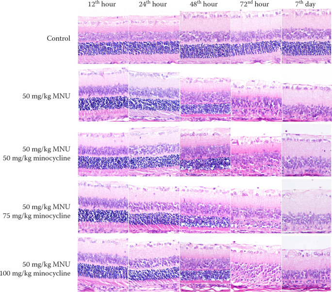
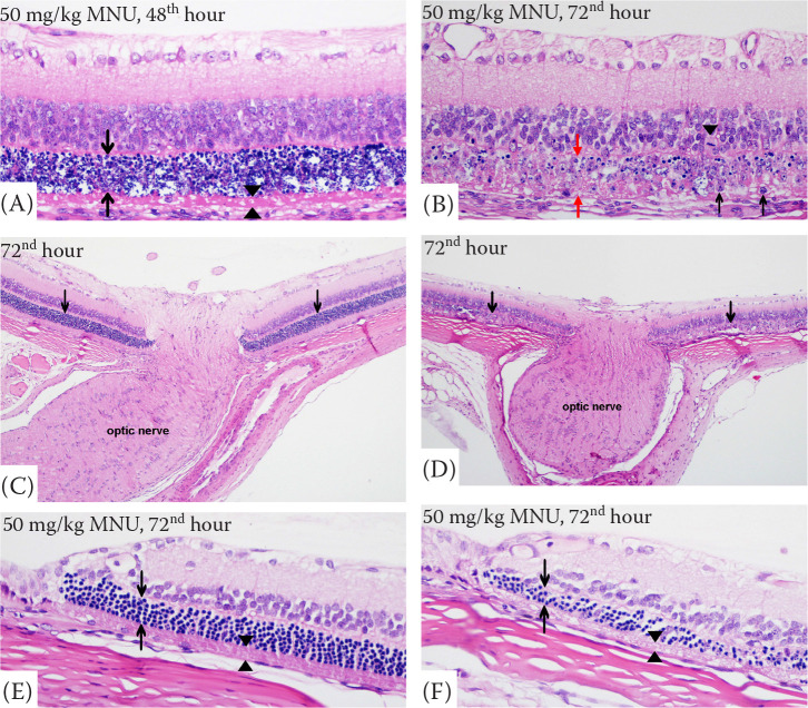
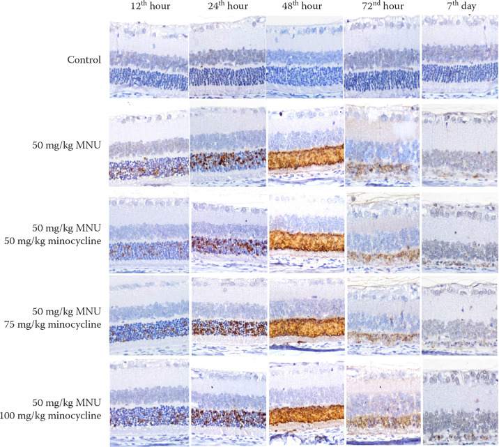
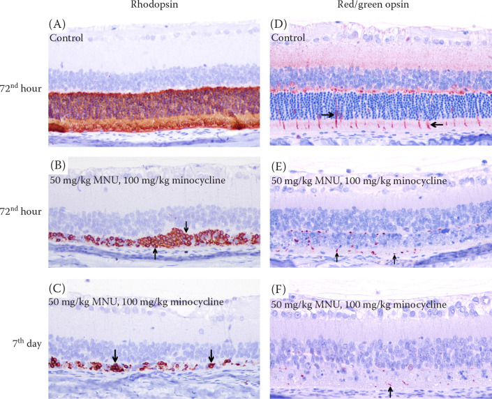
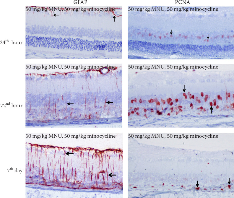
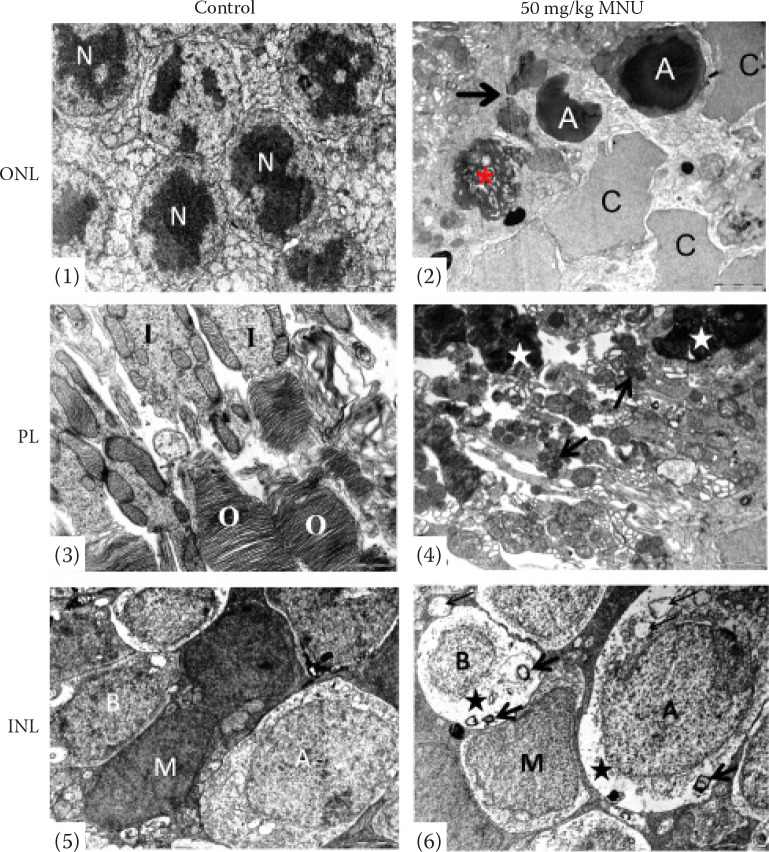
Retinal degeneration (RD) is often associated with deficiencies or the inaccurate production of photoreceptor-specific proteins, which are encoded by various genes and characterised by the apoptotic and ongoing death of photoreceptor cells. This study involved administering a single intraperitoneal (i.p.) dose of 50 mg/kg of N-methyl-N-nitrosourea (MNU) to rats to induce RD. Some of these rats also received intraperitoneal minocycline at varying doses to prevent RD. Euthanasia was conducted at five intervals: at 12, 24, 48, and 72 h, and on the 7th day; and eye samples were taken. These samples were analysed using histopathology, immunohistochemistry, and electron microscopy. Significant RD was observed in the MNU-treated groups, with photoreceptor cell apoptosis demonstrated by the TUNEL method. Compared with those in the control group, there was a progressive thinning of the photoreceptor layer and outer nuclear layer, along with increased levels of glial fibrillary acidic protein (GFAP) and proliferating cell nuclear antigen (PCNA), and reduced levels of rhodopsin and red/green opsin starting from the 12th hour in the experimental groups. Electron microscopy revealed that amacrine and bipolar cells, in addition to photoreceptors, were also affected. The minocycline treatment did not show significant differences in retinal layer thickness or the staining levels of PCNA, GFAP, and opsins in the MNU-induced RD model.
Keywords: minocycline; retinal degeneration; retinitis pigmentosa.
Copyright: © 2025 Karabulut et al.
Conflict of interest statement
The authors declare no conflict of interest.
Figures
Similar articles
-
Prescription of Controlled Substances: Benefits and Risks.2025 Jul 6. In: StatPearls [Internet]. Treasure Island (FL): StatPearls Publishing; 2025 Jan–. 2025 Jul 6. In: StatPearls [Internet]. Treasure Island (FL): StatPearls Publishing; 2025 Jan–. PMID: 30726003 Free Books & Documents.
-
The Black Book of Psychotropic Dosing and Monitoring.Psychopharmacol Bull. 2024 Jul 8;54(3):8-59. Psychopharmacol Bull. 2024. PMID: 38993656 Free PMC article. Review.
-
NTP Developmental and Reproductive Toxicity Technical Report on the Prenatal Development Studies of 2-((1-(4-Phenoxyphenoxy)propan-2-yl)oxy)pyridine (CASRN 95737-68-1) in Sprague Dawley (Hsd:Sprague Dawley® SD®) Rats and New Zealand White (Hra:NZW SPF) Rabbits: DART Report 07 [Internet].Research Triangle Park (NC): National Toxicology Program; 2022 Jan. Research Triangle Park (NC): National Toxicology Program; 2022 Jan. PMID: 35593777 Free Books & Documents. Review.
-
Sertindole for schizophrenia.Cochrane Database Syst Rev. 2005 Jul 20;2005(3):CD001715. doi: 10.1002/14651858.CD001715.pub2. Cochrane Database Syst Rev. 2005. PMID: 16034864 Free PMC article.
-
Neuraminidase inhibitors for preventing and treating influenza in adults and children.Cochrane Database Syst Rev. 2014 Apr 10;2014(4):CD008965. doi: 10.1002/14651858.CD008965.pub4. Cochrane Database Syst Rev. 2014. PMID: 24718923 Free PMC article.
References
-
- Aplin FP, Luu CD, Vessey KA, Guymer RH, Shepherd RK, Fletcher EL. ATP-induced photoreceptor death in a feline model of retinal degeneration. Invest Ophthalmol Vis Sci. 2014 Nov 25;55(12):8319-29. - PubMed
-
- Bonelli RM, Heuberger C, Reisecker F. Minocycline for Huntington’s disease: An open label study. Neurology. 2003 Mar 11;60(5):883-4. - PubMed
-
- Carter JH, Douglass LE, Deddens JA, Colligan BM, Bhatt TR, Pemberton JO, Konicek S, Hom J, Marshall M, Graff JR. Pak-1 expression increases with progression of colorectal carcinomas to metastasis. Clin Cancer Res. 2004 May 15;10(10):3448-56. - PubMed
-
- Chang CJ, Cherng CH, Liou WS, Liao CL. Minocycline partially inhibits caspase-3 activation and photoreceptor degeneration after photic injury. Ophthalmic Res. 2005 Jul-Aug;37(4):202-13. - PubMed
-
- Danulescu R, Costin D. Use of blood markers in early diagnosis of oxidative stress in age related macular degeneration. Rev Med Chir Soc Med Nat Iasi. 2012 Oct-Dec;116(4):1136-42. - PubMed
LinkOut - more resources
Full Text Sources
Miscellaneous