Review
. 2025 Jul 23;16(8):3349-3377.
doi: 10.1364/BOE.566801. eCollection 2025 Aug 1.

Application of quantum imaging in biology

Affiliations
Review

Application of quantum imaging in biology

Hoda Lotfipour et al. Biomed Opt Express. .

Abstract

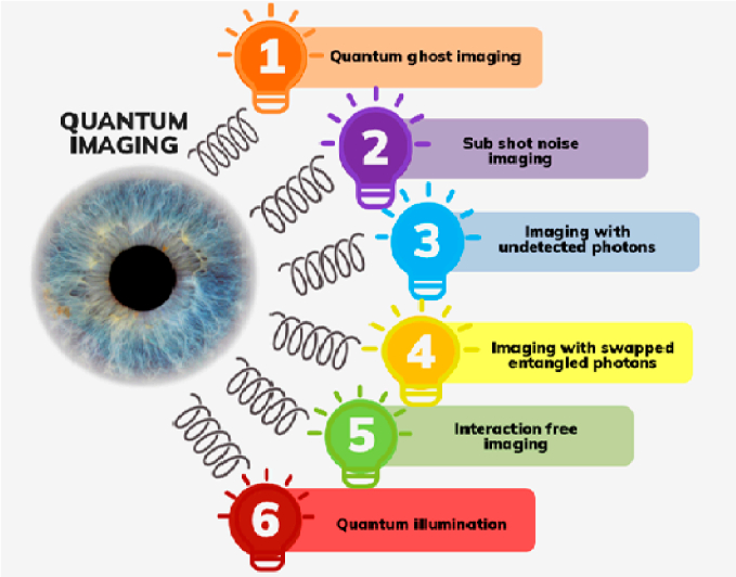
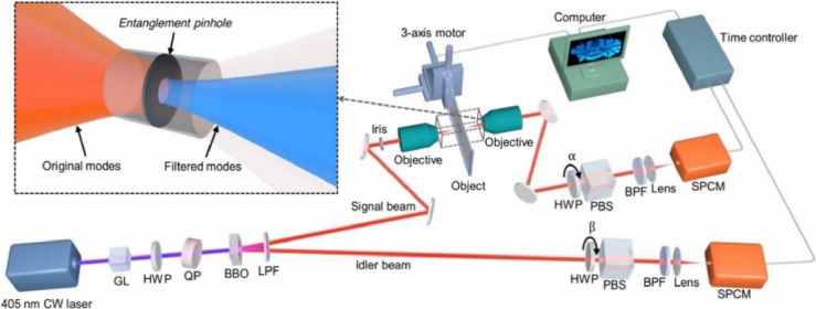
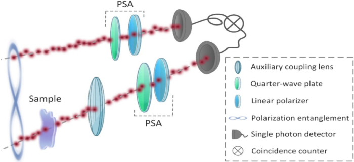
Application of quantum imaging in biology In biology, one of the main challenges is achieving a balance between high precision and minimal invasiveness in measurements. With cutting-edge techniques, the primary limitations to experimental accuracy often stem not from device-related noise but from fundamental physical constraints. Since nature is the underlying source of these constraints, it makes sense to go to quantum mechanics, the most basic theory of matter, for a solution. Improved measurement performance may be possible through the application of quantum effects, particularly those pertaining to coherence. It offers useful tools that, at the very least, offer interesting technical solutions even when they don't fully display cohesive behaviors. One of the primary applications of quantum technologies is quantum metrology, which uses the non-classical state of light to measure physical properties with great resolution and sensitivity. For biological applications, the quantum state of light may be utilized for precision enhancement and quantum noise reduction. This explains how quantum metrology, and particularly quantum imaging, can be used to enhance picture quality, measure shifts in quantum scales in biological systems, and boost imaging precision and resolution using quantum light sources, devices, and protocols. In this study, we give a summary of the possible uses of quantum technology in biology and medicine. This review presents a comprehensive overview of how quantum technologies can be applied in biology and medicine. It also explores the latest developments in quantum biological imaging, quantum microscopy, and quantum materials, while discussing the challenges and opportunities these emerging technologies bring.


Conflict of interest statement

The authors declare no conflicts of interest.

Figures

Fig. 1.
Fig. 1.
Various quantum imaging schemes [45].
Fig. 2.
Fig. 2.
The experimental scheme of quantum differential ghost imaging (QDGI) [13].
Fig. 3.
Fig. 3.
Schematic representation of the experimental set-up and the images of biological samples (a-d): Spirulina filaments and (e-h): fruit fly wing at different scan areas and step size [61].
Fig. 4.
Fig. 4.
three physical phenomena (induced coherence (IC), the interaction-free measurement (IFM), single-pixel imaging (SPI)) in the schematic setup (top)- The experimental setup of interaction-free, single-pixel QIUP (Down) [66].
Fig. 5.
Fig. 5.
The schematic experimental setup of imaging with entangled photons [5].
Fig. 6.
Fig. 6.
Conceptual sketch of the optical setup used in the experiments conducted by Zhang and colleagues. Reproduced from ref. [92].
Fig. 7.
Fig. 7.
Nitrogen and boron dual-doped GQDs for NIR-II bioimaging. (a) TEM image showing the monodisperse particles (b) photoluminescence spectrum of N–B-GQDs exhibiting NIR-II emission when excited with an 808 nm laser source. The insets display an optical image a photoluminescence image of N–B-GQDs in aqueous solution. (c) In vitro cytotoxicity study of N–B-GQDs performed by assessing the viability of SF763, 4T1, and B16F10 cells 72 h after incubation with N–B-GQDs. (d) In vivo NIR-II imaging of live mice. Adapted with permission from Ref. [103]. Copyright 2019 Elsevier.
Fig. 8.
Fig. 8.
Fluorescence images obtained at 20X, representing the fluorescence enrichment of compound f-WS2-QDs in the gut tissues of Drosophila third instar larvae. Upper panels (B, C) exhibit no fluorescence considered as control. Lower panel (E, F) demonstrates fluorescence in gut tissues of Drosophila third instar larvae. (A and D) shows BF (Bright Field) images of their respective panel. Reproduced with permission from Ref. [104]. Copyright © 2023 American Chemical Society.
Fig. 9.
Fig. 9.
NDSiV-polymer for living cell thermometry and intracellular tracking. (a) Custom-built confocal image of a living A549 cell with uptaken NDSiV-polymer nanoparticles. (b) Position of ZPL peaks of NDSiV-polymer at 25 and 37 °C. (c) Trajectory of NDSiV-polymer tracked in intracellular space [105].

Similar articles

References

    1. Casacio C. A., Madsen L. S., Terrasson A., et al. , “Quantum-enhanced nonlinear microscopy,” Nature 594(7862), 201–206 (2021). 10.1038/s41586-021-03528-w - DOI - PubMed
    1. Park J., Brady D. J., Zheng G., et al. , “Review of bio-optical imaging systems with a high space-bandwidth product,” Adv Photonics 3(04), 044001 (2021). 10.1117/1.AP.3.4.044001 - DOI - PMC - PubMed
    1. Berg M., Holroyd N., Walsh C., “Challenges and opportunities of integrating imaging and mathematical modeling to interrogate biological processes,” The International Journal of Biochemistry & Cell Biology 146, 106195 (2022). 10.1016/j.biocel.2022.106195 - DOI - PMC - PubMed
    1. Vernekar S., Xavier J., “Quantum correlation enhanced optical imaging,”
    1. Zhang Y., He Z., Tong X., et al. , “Quantum imaging of biological organisms through spatial and polarization entanglement,” Sci. Adv. 10(10), eadk1495 (2024). 10.1126/sciadv.adk1495 - DOI - PMC - PubMed

LinkOut - more resources