Lang-chuang-ding restores bone homeostasis in systemic lupus erythematosus associated osteoporosis by targeting NF-κB signaling: a network pharmacology and experimental study
- PMID: 40810073
- PMCID: PMC12343237
- DOI: 10.3389/fendo.2025.1639261
Lang-chuang-ding restores bone homeostasis in systemic lupus erythematosus associated osteoporosis by targeting NF-κB signaling: a network pharmacology and experimental study
Abstract
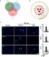
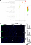
Systemic lupus erythematosus (SLE) is frequently associated with secondary osteoporosis (OP), substantially compromising patients' quality of life. Although Lang-chuang-ding (LCD), a traditional Chinese medicine formulation, has demonstrated efficacy in suppressing SLE progression, its therapeutic potential for SLE-associated OP remains uninvestigated. This study investigated the therapeutic effects and underlying pharmacological mechanisms of LCD on SLE-associated OP through in vivo experimental validation using MRL/lpr mouse model in conjunction with network pharmacology analysis. Our findings demonstrated that LCD significantly attenuated bone loss in the distal femur by improving bone morphometric parameters, including bone mineral density (BMD), trabecular number (Tb.N), and trabecular bone separation (Tb.Sp), while simultaneously suppressing osteoclast activity and promoting osteogenesis. Network pharmacological analysis identified 63 overlapping targets among LCD components, SLE-related genes, and OP-associated targets, with inflammatory mediators TNF-α, IL-6, and IL-1β emerging as pivotal hub targets. KEGG enrichment analysis revealed significant NF-κB pathway enrichment among the core therapeutic targets. Experimental validation demonstrated that LCD effectively suppressed inflammatory responses by markedly reducing pro-inflammatory cytokines IL-1β, TNF-α, and IL-6 expression while simultaneously inhibiting NF-κB pathway activation through downregulation of p-IκB, P65, and p-P65 in the distal femur. Collectively, these findings demonstrate that LCD effectively ameliorates SLE-associated OP through modulation of inflammatory cytokine networks and the NF-κB signaling pathway, establishing its therapeutic potential as a mechanism-based intervention for SLE-associated OP.
Keywords: Lang-chuang-ding; SLE-associated osteoporosis; bone homeostasis; inflammatory cytokines; mechanism.
Copyright © 2025 Luo, Zhou, Shen, Zeng and Ruan.
Conflict of interest statement
The authors declare that the research was conducted in the absence of any commercial or financial relationships that could be construed as a potential conflict of interest.
Figures


Similar articles
-
The potential mechanism of Huangqin for treatment of systemic lupus erythematosus based on network pharmacology, molecular docking and molecular dynamics simulation.PeerJ. 2025 Jun 26;13:e19536. doi: 10.7717/peerj.19536. eCollection 2025. PeerJ. 2025. PMID: 40585331 Free PMC article.
-
The mechanism of action in Mussaenda pubescens (Yuye Jinhua) against influenza A virus: Evidence from in vitro and in vivo studies.Phytomedicine. 2025 Sep;145:157070. doi: 10.1016/j.phymed.2025.157070. Epub 2025 Jul 13. Phytomedicine. 2025. PMID: 40684492
-
Investigation of the effect and mechanism of Fei Re Pu Qing powder in treating acute lung injury (ALI) by modulating macrophage polarization via serum pharmacology and network pharmacology.J Ethnopharmacol. 2025 Jul 24;351:120089. doi: 10.1016/j.jep.2025.120089. Epub 2025 Jun 9. J Ethnopharmacol. 2025. PMID: 40499803
-
The path less traveled: the non-canonical NF-κB pathway in systemic lupus erythematosus.Front Immunol. 2025 Jul 2;16:1588486. doi: 10.3389/fimmu.2025.1588486. eCollection 2025. Front Immunol. 2025. PMID: 40672954 Free PMC article. Review.
-
[Retrospective study on intervention of traditional Chinese medicine in osteoporosis and related pain diseases].Zhongguo Zhong Yao Za Zhi. 2025 Jun;50(11):3180-3188. doi: 10.19540/j.cnki.cjcmm.20250213.702. Zhongguo Zhong Yao Za Zhi. 2025. PMID: 40686186 Review. Chinese.
References
MeSH terms
Substances
LinkOut - more resources
Full Text Sources
Medical
Miscellaneous

