. 2025 Aug 19;122(33):e2514837122.
doi: 10.1073/pnas.2514837122. Epub 2025 Aug 14.

Stress granule-mediated ZBP1 activation drives necroptotic cell death in non-obstructive azoospermia and testicular aging

Affiliations

Stress granule-mediated ZBP1 activation drives necroptotic cell death in non-obstructive azoospermia and testicular aging

Hongen Lei et al. Proc Natl Acad Sci U S A. .

Abstract

Male infertility remains a major unmet medical challenge, with poorly defined molecular mechanisms and no effective therapies. Here, we identify a stress granule-mediated necroptotic pathway as a key driver of non-obstructive azoospermia, a severe form of male infertility marked by the loss of spermatogenesis. Environmental or physiological stress activates eIF2α kinases, inducing stress granule formation and the recruitment of ZBP1 and RIPK3 into a cytoplasmic complex. This assembly triggers RIPK3 activation, MLKL phosphorylation, and necroptotic death of spermatogonia and Sertoli cells. Genetic ablation of Zbp1 or Ripk3 protects mice from heat-induced testicular degeneration, establishing their essential role in stress-induced testicular damage. Importantly, activation of this pathway is also observed in aged human testes, linking stress-responsive necroptosis to both pathological infertility and the broader process of reproductive aging. These findings reveal an unrecognized mechanism that couples cellular stress responses to regulated cell death in the male reproductive system.

Keywords: ZBP1; necroptosis; non-obstructive azoospermia; stress granule; testis aging.


Conflict of interest statement

Competing interests statement:Xiaodong Wang is the co-founder of Sironax Inc., a biotech start-up developing therapeutic agents for degenerative diseases, Xiaodong Wang own 5.5% of Sironax stock.

Figures

Fig. 1.
Fig. 1.
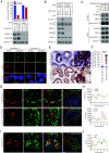
Phospho-MLKL(p-MLKL) were detected in the seminiferous tubules of NOA patients’ testes. (A–C) Hematoxylin and Eosin (H&E) staining of testis sections from human testicular torsion (n = 17) and azoospermia patients (n = 50) in (A). Johnsen Score evaluation of testicular torsion and azoospermia patients based on (A) and shown in (B). The number of seminiferous tubules with sperm was counted based on (A) and quantification in (C), seminiferous tubules with sperm were counted in five fields per testis. (Scale bar, 100 μm.) (D and E) Immunohistochemistry (IHC) analysis of human testicular torsion and NOA testis sections with p-MLKL antibody in (D). The number of seminiferous tubules with positive p-MLKL signal was counted based on IHC staining and quantification in (E). (Scale bar, 100 μm.) (F) Western blotting analysis of RIPK3, p-MLKL, and MLKL in the testis from two NOA patients, GAPDH was used as loading control. HeLa and HT29 cells treated with the combination of T/S/Z as negative and positive control for western blotting analysis, respectively. The asterisk (*) indicates nonspecific bands. (G) Immunofluorescence analysis of NOA testis sections with antibodies against p-MLKL (red) PIWIL4 (spermatogonium specific protein, green) and SOX9 (Sertoli cell–specific protein, green). (Scale bar, 50 μm.) Quantified data in (B, C, and E) represent the mean ± s.e.m. ****P < 0.0001. P values were determined by two-sided unpaired Student’s t tests.
Fig. 2.
Fig. 2.
Heat shock–induced necroptosis depends on ZBP1 and RIPK3. (AC) Schematic illustrating experiment design in (A). Cultured GC-2spd, 15P-1, and MA-10 cells were treated with DMSO or IFN-β for 18 h, then the cells were transferred to 1.5 mL Eppendorf Tubes (EP) and put into 37 °C or 43 °C water for 2 h. 6 h after heat shock (HS) cell viability as measured by Cell Titer-Glo in (B). The levels of p-MLKL, MLKL, and RIPK3 were analyzed by immunoblotting in (C), GAPDH was used as loading control. (D) Cultured L929 cells with wild type (WT), Ripk1, Ripk3, or Mlkl gene knocked out were treated with IFN-β and HS for 18 h and 1.5 h as described in SI Appendix, Fig. S6A, 2 h after heat shock cell viability as measured by Cell Titer-Glo. (E and F) Cultured L929 cells with wildtype or Zbp1 gene knocked out were treated with IFN-β and HS for 18 h and 1.5 h as described in SI Appendix, Fig. S6A, 2 h after heat shock cell viability as measured by Cell Titer-Glo in (E). The levels of p-MLKL, MLKL, and ZBP1 were analyzed by immunoblotting in (F), GAPDH was used as loading control. (G) Cultured L929(Ripk3−/−)-HA-3 × Flag-mRIPK3 cells were treated with IFN-β/HS as indicated. The cell extracts were prepared and subjected to immunoprecipitation with an anti-Flag antibody. The extracts (Input) and the immuno-precipitates (IP: Flag) were then subjected to western blotting analysis using antibodies as indicated. (HL) Schematic illustrating the experiment design in SI Appendix, Fig. S8A. The lower body of 12-wk-old Ripk3+/+, Ripk3−/− littermate, and Zbp1−/− male mice (n = 7 per each genotype) was put in 43 °C waters for 20 min every other day, for a total of three exposures. Mice were euthanized 7 d after the final treatment. Testes were collected for analysis of weight in (H), histology via H&E staining in (I), seminiferous tubules thickness quantification in (J), p-MLKL IHC staining in (K), and quantification of p-MLKL-positive cells in (L). (Scale bar, 100 μm.) Data in (B, D, and E) are mean ± SD of triplicate wells. Quantified data in (H, J, and L) represent the mean ± s.e.m. ****P<0.0001. P values were determined by two-sided unpaired Student’s t tests.
Fig. 3.
Fig. 3.
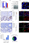
Stress granule is required for heat shock–induced necroptosis. (A and B) Cultured L929 cells with wild type or G3bp1 and G3bp2 double gene knocked out were treated with IFN-β and HS for 18 h and 1.5 h as described in SI Appendix, Fig. S6A, 2 h after heat shock cell viability as measured by Cell Titer-Glo in (A). The levels of p-MLKL, MLKL, ZBP1, G3BP1, and G3BP2 were analyzed by immunoblotting in (B), GAPDH was used as loading control. Data in (A) are mean ± SD of triplicate wells. (C) Cultured L929(Ripk3−/−)-HA-3 × Flag-mRIPK3 cells were treated with IFN-β/HS as indicated. The cell extracts were prepared and subjected to immunoprecipitation with an anti-Flag antibody. The extracts (Input) and the immuno-precipitates (IP: Flag) were then subjected to western blotting analysis using antibodies as indicated. The asterisk (*) indicates nonspecific bands. (D) Cultured L929(Zbp1+/+) and L929(Zbp1−/−) cells were treated with the indicated stimuli for 18 h (IFN-β) and 0.5 h (HS). The RIPK3 and G3BP1 were detected by immunofluorescence. (Scale bar, 10 μm.) (E and F) IHC analysis of human testicular torsion and NOA testis sections with G3BP1 antibody in (E). The number of seminiferous tubules with positive G3BP1 dot signal was counted based on IHC staining and quantification in (F). (Scale bar, 100 μm.) Data represent the mean ± s.e.m. ****P < 0.0001. P values were determined by two-sided unpaired Student’s t tests. (G and H) Immunofluorescence analysis of NOA testis sections (unknown cause of NOA, n = 5; cryptorchidism, n = 5) with antibodies against G3BP1(green) and RIPK3(red) in (G). (Scale bar, 50 μm.) Profiling of representative white dotted line traces the intensities of RIPK3 and G3BP1 signal based on (G) and analyzed in (H). (I and J) Immunofluorescence analysis of NOA testis sections (unknown cause of NOA, n = 5; cryptorchidism, n = 5) with antibodies against G3BP1(green) and ZBP1(red) in (I). (Scale bar, 50 μm.) Profiling of representative white dotted line traces the intensities of ZBP1 and G3BP1 signal based on (I) and analyzed in (J).
Fig. 4.
Fig. 4.
Stress kinases induce ZBP1 and RIPK3-dependent necroptosis in NOA. (A and B) Cultured L929 cells with wildtype or Pkr, Hri, Perk, and Gcn2 four gene knocked out (quadruple-KO) were treated with IFN-β and HS for 18 h and 1.5 h as described in SI Appendix, Fig. S6A, 2 h after heat shock cell viability as measured by Cell Titer-Glo (A). The levels of p-MLKL and MLKL were analyzed by immunoblotting in (B), GAPDH was used as loading control. Data in (A) are mean ± SD of triplicate wells. (C) Cultured L929(wildtype) and L929(quadruple-KO) cells were treated with the indicated stimuli for 18 h (IFN-β) and 0.5 h (HS). The RIPK3 and G3BP1 were detected by immunofluorescence. (Scale bar, 10 μm.) (D and E) IHC analysis of human testicular torsion and NOA testis sections with p-GCN2 antibody in (D). The number of seminiferous tubules with positive p-GCN2 signal was counted based on IHC staining and quantification in (E). (Scale bar, 100 μm.) Data represent the mean ± s.e.m. ****P < 0.0001. P values were determined by two-sided unpaired Student’s t tests. (F) Immunofluorescence analysis of NOA testis sections (n = 5) with antibodies against p-MLKL (red) and p-GCN2 (green). (Scale bar, 50 μm.) (G and H) IHC analysis of human testicular torsion and NOA testis sections with p-PKR antibody in (G). The number of seminiferous tubules with positive p-PKR signals was counted based on IHC staining and quantification in (H). (Scale bar, 100 μm.) Data represent the mean ± s.e.m. **P < 0.01. P values were determined by two-sided unpaired Student’s t tests. (I) Immunofluorescence analysis of NOA testis sections (n = 5) with antibodies against p-MLKL (red) and p-PKR (green). (Scale bar, 50 μm.) (J and K) Analysis of p-GCN2 and p-PKR-positive testis sections from the testicular torsion (n = 17) and NOA (n = 50) testes in (J). Analysis of NOA testis sections with single-positive p-GCN2, single-positive p-PKR, and double-positive p-GCN2 and p-PKR in (K).
Fig. 5.
Fig. 5.
p-MLKL signals were associated with stress biomarkers in aging testes. (A and B) H&E staining of testis sections from human testicular torsion (n = 17) and prostate cancer patients (aging testis, n = 30) in (A). Johnsen Score evaluation of testicular torsion and prostate cancer patients based on (A) and shown in (B). (Scale bar, 100 μm.) (C and D) Immunofluorescence analysis of human testicular torsion and aging testis sections with p-MLKL antibody in (C). The number of seminiferous tubules with positive p-MLKL signals was counted based on immunofluorescence staining and quantification in (D). (Scale bar, 100 μm.) (E and F) IHC analysis of human testicular torsion and aging testis sections with G3BP1 antibody in (E). The number of seminiferous tubules with positive G3BP1 dot signal was counted based on IHC staining and quantification in (F). (Scale bar, 100 μm.) Data represent the mean ± s.e.m. ****P < 0.0001. P values were determined by two-sided unpaired Student’s t tests. (G and H) IHC analysis of human testicular torsion and aging testis sections with p-GCN2 antibody in (G). The number of seminiferous tubules with positive p-GCN2 signal was counted based on IHC staining and quantification in (H). (Scale bar, 100 μm.) Data represent the mean ± s.e.m. ****P < 0.0001. P values were determined by two-sided unpaired Student’s t tests. (I) Immunofluorescence analysis of aging testis sections (n = 5) with antibodies against p-MLKL (red) and p-GCN2 (green). (Scale bar, 50 μm.) (J and K) Analysis of p-GCN2 and p-PKR-positive testis sections from the testicular torsion (n = 17) and aging testes (n = 30) in (J). Analysis of aging testis sections with single-positive p-GCN2, single-positive p-PKR, and double-positive p-GCN2 and p-PKR in (K).

References

    1. Agarwal A., Mulgund A., Hamada A., Chyatte M. R., A unique view on male infertility around the globe. Reprod. Biol. Endocrinol. 13, 37 (2015). 10.1186/s12958-015-0032-1. - DOI - PMC - PubMed
    1. Willott G. M., Frequency of azoospermia. Forensic Sci. Int. 20, 9–10 (1982). 10.1016/0379-0738(82)90099-8. - DOI - PubMed
    1. McLachlan R. I., Rajpert-De Meyts E., Hoei-Hansen C. E., de Kretser D. M., Skakkebaek N. E., Histological evaluation of the human testis–approaches to optimizing the clinical value of the assessment: Mini review. Hum. Reprod. 22, 10 (2007). - PubMed
    1. Agarwal A., et al. , Male infertility. Lancet 397, 319–333 (2021). 10.1016/S0140-6736(20)32667-2. - DOI - PubMed
    1. Jimbo M., et al. , Fertility in the aging male: A systematic review. Fertil. Steril. 118, 1022–1034 (2022). 10.1016/j.fertnstert.2022.10.035. - DOI - PMC - PubMed

MeSH terms

Substances