. 2025 Aug 14:blood.2025029430.
doi: 10.1182/blood.2025029430. Online ahead of print.

Cryo-EM structure of the tissue factor/factor VIIa complex with a factor X mimetic reveals a novel allosteric mechanism

Affiliations

Cryo-EM structure of the tissue factor/factor VIIa complex with a factor X mimetic reveals a novel allosteric mechanism

Josepha C Sedzro et al. Blood. .

Abstract

Blood clotting is triggered in hemostasis and thrombosis when the membrane-bound tissue factor (TF)/factor VIIa (FVIIa) complex activates factor X (FX). There are no structures of TF/FVIIa on membranes, with or without FX. Using cryo-EM to address this gap, we assembled TF/FVIIa complexes on nanoscale membrane bilayers (nanodiscs), bound to XK1 and an antibody fragment. XK1 is a FX mimetic whose protease domain is replaced by the first Kunitz-type (K1) domain of tissue factor pathway inhibitor, while 10H10 is a non-inhibitory, anti-TF antibody. We determined a cryo-EM structure of a TF/FVIIa/XK1/10H10/nanodisc complex with a resolution of 3.7 Å, allowing us to model all the protein backbones. TF/FVIIa extends perpendicularly from the membrane, interacting with a "handle shaped" XK1 at two locations: the K1 domain docks into FVIIa's active site, while the γ-carboxyglutamate-rich (GLA) domain binds to TF's substrate-binding exosite. The FX and FVIIa GLA domains also contact each other and the membrane surface. Except for a minor contact between the first epidermal growth factor (EGF)-like domain of XK1 and TF, the rest of the FX light chain does not interact with TF/FVIIa. The structure reveals a previously unrecognized, membrane-dependent allosteric activation mechanism between FVIIa and TF where a serine-rich loop in TF that partially obscures the TF exosite must undergo a shift to allow access of the FX GLA domain to its final binding location on the membrane-bound TF/FVIIa complex. This mechanism also provides a novel explanation for the otherwise puzzling phenomenon of TF encryption/decryption on cell surfaces.


Conflict of interest statement

Conflict-of-interest disclosure: All the authors declare no competing financial interests.

Figures

Figure 1.
Figure 1.. Biochemical characterization of TF/FVIIa/XK1/10H10 complexes.
(A) Schematic of the TF/FVIIa/XK1/10H10/nanodisc complex. FVIIa, light orange; TF, purple; MBP, gray; XK1, light sea green; and 10H10 Fab, blue; GLA, gamma (γ)-carboxyglutamate-rich domain; EGF, epidermal growth factor-like domain; K1, first Kunitz-type domain of TFPI. (B) TF exosite mutations K165A and K166A reduce the rates of FX activation by TF/FVIIa, with higher PS content moderating this effect. TF was incorporated into small unilamellar liposomes after which FVIIa and FX were added, and initial rates of FX activation were quantified. Bar graphs show FX activation rates (normalized to wild-type TF) observed when TF/liposomes contained 5% PS (left) or 20% PS (right). Fold-reductions are listed in supplemental Table S1. (C) Elevated XK1 concentrations are required to inhibit TF/FVIIa when TF exosite residues are mutated, with higher PS content moderating this effect. TF/FVIIa was assembled on TF/liposomes with 5% PS (left) or 20% PS (right), then pre-incubated with varying XK1 concentrations. The residual TF/FVIIa enzymatic activity was quantified (normalized to the FX activation rate without XK1) and plotted versus XK1 concentration, from which IC50 values were derived (listed in supplemental Table S1). Individual data points are graphed in panels B and C (n ≥ 3). ***, p<0.001.
Figure 2.
Figure 2.. Cryo-EM structure of the TF/FVIIa/XK1/10H10 complex.
(A) Four views of the cryo-EM density map (at contour level 0.1) together with the model of the TF/FVIIa/XK1/10H10 complex. The less structured portions of the XK1 density map (contour level 0.04) are shown in mesh, and the density for the nanodisc and MBP was subtracted for clarity. Views are rotated 90° around the y-axis. FVIIa is orange, TF is purple, XK1 is light sea green and 10H10 Fab is blue. (B) Views of the model of TF/FVIIa/XK1 complex, rotated 90° around the y-axis, with the same color scheme as in panel A. For clarity, the 10H10 Fab is not included in these views.
Figure 3.
Figure 3.. Protein–protein interactions between the FVIIa protease domain and the K1 domain of XK1.
(A) Cryo-EM density map (shown as a mesh) and model of the TF/FVIIa/XK1 complex, shown with density for the 10H10 Fab, nanodisc and MBP subtracted for clarity. Color scheme: FVIIa, orange; TF, purple; XK1, light sea green. Residues 31–41 of the XK1 K1 domain, which form a hairpin-like conformation, are in dark red. The cryo-EM density map is shown at contour level 0.1, while the density for EGF2 is visible at higher counter levels. (B) View of the interaction between the protease domain of FVIIa and the K1 domain of XK1 with the density map at contour level 0.1. Boxes indicate areas of the structure magnified in panels C and D. (C,D) Magnified views of specific regions of the FVIIa–XK1 interface boxed in panel B. Critical residues are shown as sticks and labeled, with carbon atoms colored according to the respective protein, oxygens in red, and nitrogens in blue. Selected atomic distances are indicated with gray dotted lines. The backbones of G365 and G375 are shown in gray. The cryo-EM density in panels C and D is shown at contour level 0.13.
Figure 4.
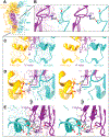
Figure 4.. Key protein–protein interactions between the TF/FVIIa complex and the FX light chain portion of XK1.
(A) The TF/FVIIa/XK1 cryo-EM density map, together with the atomic model, is repeated here from Figure 3A for orientation purposes. Color scheme: FVIIa, orange; TF, purple; XK1, light sea green. (B) Close-up view of the interaction between TF residue R200 and FX residue E51 (in the EGF1 domain of XK1), shown with (left panel) and without (right panel) the cryo-EM density map. (C) Close-up view of the GLA domains of FVIIa and XK1 in proximity to the TF 4×Ser loop (indicated by purple arrow), shown with (left panel) and without (right panel) the cryo-EM density map. (D) Another close-up view of GLA domain–GLA domain and GLA domain–TF interactions, with and without the density map. Interactions between TF residue K165, FVIIa residues γ35 and R36, and XK1 residue K45 are highlighted. (E) A close-up view (from a different angle relative to panels C and D), revealing interactions between the XK1 GLA domain and the TF exosite, shown with (left panel) and without (right panel) the cryo-EM density map. Interactions between XK1 residues γ14 and γ19 with TF residues K159, K166, and Y185 are highlighted.
Figure 5.
Figure 5.. Novel allosteric regulation of the TF exosite revealed by comparing the cryo-EM structure of TF/FVIIa/XK1/10H10 with the sTF/FVIIa crystal structure (3TH2).
(A) Superposition of 3TH2 crystal structure of sTF/FVIIa with the cryo-EM map and model of TF/FVIIa/XK1, aligned via their TF components. Insets highlight conformational differences in the FVIIa GLA domain (left box) and the TF 4×Ser loop (right box). The color scheme for the cryo-EM map (mesh) and model is: FVIIa in orange, TF in purple, and XK1 in light sea green. For the 3TH2 crystal structure, it is: FVIIa in light gray and sTF in black/dark gray. The map is contoured at 0.1. For clarity, the 10H10 density was subtracted from the cryo-EM map and omitted from the model. In the following close-up views (panels B-E), the map is contoured at 0.145 and key interacting residues are shown as sticks and labeled, with carbon atoms colored by molecule, nitrogen in blue, and oxygen in red. (B) Close-up view focusing on the FVIIa and XK1 GLA domains within the alignment of TF/FVIIa/XK1 (cryo-EM structure) with the 3TH2 crystal structure. Arrows indicate the 4×Ser loop. (C) Close-up comparison of the positions of residues R36 of FVIIa and K165 of TF in the cryo-EM structure versus the sTF/FVIIa crystal structure, with the 4×Ser loop indicated by arrows. (D) Close-up view of the TF 4×Ser loop (arrows) and the XK1 GLA domain in the alignment between the cryo-EM structure and the sTF/FVIIa crystal structure. Note that the 4×Ser loop in the sTF/FVIIa crystal structure (but not the cryo-EM structure) sterically clashes with the cryo-EM density map of the XK1 GLA domain. FVIIa was omitted in this view for clarity. (E) Another close-up view of the region including the TF 4×Ser loop (arrows), highlighting interactions between TF residues S162 and S163 with FVIIa residue F31 and XK1 residues γ20 and T21.

References

    1. Smith SA, Travers RJ, Morrissey JH. How it all starts: Initiation of the clotting cascade. Crit Rev Biochem Mol Biol. 2015;50(4):326–336. - PMC - PubMed
    1. Grover SP, Mackman N. Tissue factor: an essential mediator of hemostasis and trigger of thrombosis. Arteriosclerosis, Thrombosis and Vascular Biology. 2018;38(4):709–725. - PubMed
    1. Baugh RJ, Dickinson CD, Ruf W, Krishnaswamy S. Exosite interactions determine the affinity of factor X for the extrinsic Xase complex. J Biol Chem. 2000;275(37):28826–28833. - PubMed
    1. Shobe J, Dickinson CD, Edgington TS, Ruf W. Macromolecular substrate affinity for the tissue factor-factor VIIa complex is independent of scissile bond docking. J Biol Chem. 1999;274:24171–24175. - PubMed
    1. Roy S, Hass PE, Bourell JH, Henzel WJ, Vehar GA. Lysine residues 165 and 166 are essential for the cofactor function of tissue factor. J Biol Chem. 1991;266(32):22063–22066. - PubMed