. 2025 Aug 14;15(1):29851.
doi: 10.1038/s41598-025-11385-0.

GRSF1 loss in THP-1 macrophages promotes senescence-associated transcription in neighboring fibroblasts

Affiliations

GRSF1 loss in THP-1 macrophages promotes senescence-associated transcription in neighboring fibroblasts

Younggi Lee et al. Sci Rep. .

Abstract

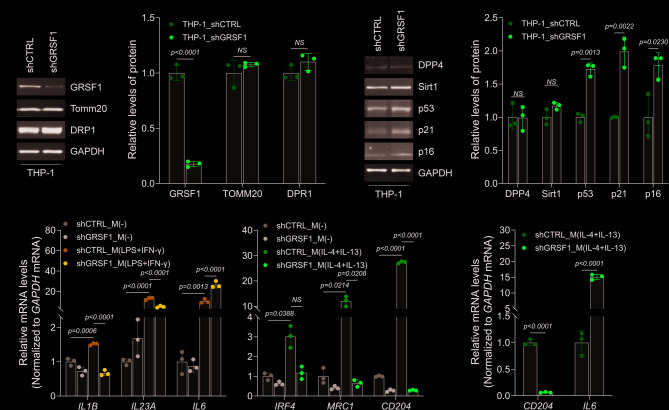
Immunosenescence, the age-associated decline in immune function, is accompanied by altered macrophage phenotypes and increased chronic inflammation. Here, we examined the role of the mitochondrial RNA-binding protein GRSF1 in regulating macrophage-driven inflammation and its impact on neighboring fibroblasts. We found that macrophages differentiated from GRSF1-deficient THP-1 monocytes, particularly M(IL-4 + IL-13) macrophages, displayed elevated IL6 mRNA expression levels and TNF-α secretion, without inducing overt senescence in macrophages themselves. Conditioned media from these macrophages triggered robust senescence-associated transcriptional changes in fibroblasts, including increased expression of IL6, TNF, DPP4, and IL8, as well as elevated SA-β-gal activity. Notably, expression of NF-κB-regulated long noncoding RNAs, such as ANRIL and PACER, was also induced in fibroblasts, suggesting the engagement of an NF-κB-linked inflammatory program. These transcriptional responses were mitigated by red ginseng extract, an anti-inflammatory compound known to suppress TNF-α signaling. Collectively, our findings suggest that GRSF1 depletion in macrophages contributes to a paracrine inflammatory niche that promotes senescence-associated gene expression in surrounding cells.

Keywords: Cell senescence; GRSF1; THP-1 macrophages.


Conflict of interest statement

Declarations. Competing interests: The authors declare no competing interests.

Figures

Fig. 1
Fig. 1
Depleting GRSF1 interferes with macrophage polarization. (A) THP-1 monocytes were transduced with lentiviruses expressing shGRSF1 or shCTRL. Expression levels of GRSF1 and loading controls (TOMM20, DRP1, and GAPDH) were analyzed by Western blot (left). Band intensities were quantified and presented as a bar graph (right). (B) The expression levels of senescence markers (DPP4, Sirt1, p53, p21, and p16) and loading control (GAPDH) were analyzed by Western blot (left). Band intensities were quantified and presented as a bar graph (right). (C) Total RNA was isolated from M(-), M(LPS+IFN-γ), and M(IL-4+IL-13) macrophages differentiated from THP-1 monocytes expressing shGRSF1 or shCTRL. The mRNA levels of M(LPS+IFN-γ)-specific (IL1B, IL23A, and IL6) and M(IL-4+IL-13)-specific markers (IRF4MRC1, and CD204) were determined by RT-qPCR analysis. Relative mRNA expression was normalized to the M(-) group and presented as bar graphs. (D) After isolating total RNA from each of shGRSF1 and shCTRL M(IL-4+IL-13) macrophages, the steady-state levels of CD204 and IL6 mRNAs were quantified by RT-qPCR analysis. The data in (A, B, D) represent the means ± SD of three independent experiments, and statistical significance was analyzed using Student’s t-test. In (C), statistical significance among four groups was assessed by one-way ANOVA, followed by Tukey’s multiple comparison test.
Fig. 2
Fig. 2
GRSF1-deficient macrophages induce the senescence in neighboring fibroblasts.(A) Conditioned media (CM) from M(LPS+IFN-γ) (left) and M(IL-4+IL-13) (right) macrophages expressing shGRSF1 or shCTRL were collected and used to culture pre-senescent WI-38 fibroblasts for 24 h. Serum-free medium containing 0.1% BSA (No Mφ) served as a negative control. Total RNA was isolated from recipient WI-38 fibroblasts, and the mRNA levels of cell cycle inhibitors (CDKN2A and CDKN1A) and senescence markers (IL6, TNF, and DPP4) were measured by RT-qPCR analysis. (B) SA-β-gal activity was analyzed in recipient WI-38 fibroblasts treated with CM from shCTRL of shGRSF1-expressing M(IL-4+IL-13) macrophages (left). SA-β-gal-positive cells were quantified and presented as a bar graph (right). (C) Conditioned media (CM) from GRSF1-deficient or control M(IL-4+IL-13) macrophages were collected, analyzed by antibody array (left), and quantified for released protein levels (right). The data in (A-C) represent the means ± SD from three independent experiments. Statistical significance in (A) was analyzed by one-way ANOVA followed by Tukey’s multiple comparison test, and in (B and C) by Student’s t-test.
Fig. 3
Fig. 3
The altered inflammatory environment modulates NF-κB-dependent transcription. (A) WI-38 fibroblasts were cultured in conditioned media (CM) from GRSF1-deficient (shGRSF1) or control (shCTRL) M(IL-4 + IL-13) macrophages for 24 h, with or without pre-treatment with 800 µg/mL of red ginseng rextract (RGE) for 30 min. Total RNA was isolated from WI-38 fibroblasts, and both mRNA (left) and pre-mRNA (right) levels of IL6, TNF, and DPP4 were measured by RT-qPCR analysis. (B) The levels of NF-κB-related lncRNAs (ANRIL, PACER, NKILA, MIR31HG, and MALAT) were determined by RT-qPCR analysis. (C) Both mRNA and pre-mRNA levels of IL8 were measured by RT-qPCR analysis. (D) Proposed model: Increased IL6 expression in GRSF1-deficient M(IL-4 + IL-13) macrophages promotes an inflammatory environment involving TNF-α. WI-38 fibroblasts exposed to these inflammatory conditions exhibit enhanced transcription of senescence-associated genes and altered expression of NF-κB-dependent transcripts. Figure created with BioRender.com. All the data in (A-C) represent the means ± SD from three independent experiments. Statistical significance was determined by one-way ANOVA followed by Tukey’s multiple comparison test.

Similar articles

References

    1. Ma, S. et al. Caloric restriction reprograms the Single-Cell transcriptional landscape of Rattus Norvegicus aging. Cell180 (5), 984–1001e22 (2020). - PubMed
    1. Walford, R. L. The Immunologic theory of aging. Gerontologist4, 195–197 (1964). - PubMed
    1. Liu, Z. et al. Immunosenescence: molecular mechanisms and diseases. Signal. Transduct. Target. Ther.8 (1), 200 (2023). - PMC - PubMed
    1. Conte, M., Martucci, M., Chiariello, A., Franceschi, C. & Salvioli, S. Mitochondria, Immunosenescence and inflammaging: a role for mitokines? Semin Immunopathol.42 (5), 607–617 (2020). - PMC - PubMed
    1. Ray, D. & Yung, R. Immune senescence, epigenetics and autoimmunity. Clin. Immunol.196, 59–63 (2018). - PMC - PubMed

LinkOut - more resources