

In situ electric field dosimetry analysis for powerline frequency peripheral nerve magnetic stimulation
- PMID: 40813601
- PMCID: PMC12354695
- DOI: 10.1038/s41598-025-12284-0
In situ electric field dosimetry analysis for powerline frequency peripheral nerve magnetic stimulation
Abstract
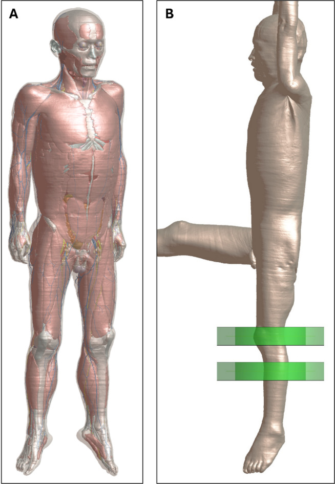
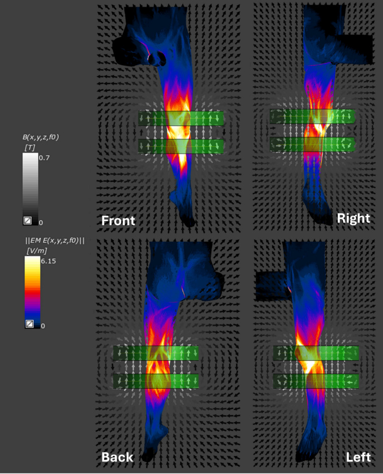
Humans are exposed to environmental 60 Hz magnetic fields (MFs), inducing in our body electric fields (EFs) and currents, potentially stimulating the peripheral nervous system (PNS). Uncertainties exist regarding the 60 Hz MF PNS stimulation threshold. The spatially extended nonlinear node model (SENN) is used to help define international MF exposure guidelines and standards protecting workers and the general public. However, other models exist, particularly the McIntyre-Richardson-Grill (MRG) model, the new gold standard for electrostimulation. This study aims (1) to model a new extremely low frequency MF exposure system for the human leg and (2) to investigate the in situ EFs generated by the system at 60 Hz at the skin level and in the nerves of the leg using a realistic human body model with both the SENN and the MRG models. A Helmholtz like-coil system was designed to generate in situ EFs sufficient for nerve stimulation, modeled using Biot-Savart and Faraday laws. Sim4Life simulations assessed the induced EFs at skin and nerve levels using a detailed human body model and two nerve excitation frameworks: the SENN and MRG models. High EF intensities were observed in four sensory and sensory-motor nerves, with MRG-derived thresholds lower than SENN-derived thresholds. Results also highlight the significance of nerve orientation in EF induction. This study emphasizes the critical role of comprehensive modeling for the design and validation of MF exposure systems and underscores the need for experimental data to refine models, standards, and guidelines.
Keywords: Dosimetry; Extremely low-frequency magnetic fields; MRG; Powerline frequency; SENN.
© 2025. The Author(s).
Conflict of interest statement
Declarations. Competing interests: The authors declare that they have no competing interests. Ethics declarations: This study was approved by Western University’s Ethics Board (protocol REB# 120854). This study is based entirely on a computational dosimetry model and does not involve any human participants, interventions, or biological tissue. Therefore, informed consent and following the declaration of Helsinki were not required for this specific work.
Figures


References
-
- Lambrozo, J. & Souques, M. Electricity and extremely low frequency electric and magnetic fields. In Electromagnetic Fields, Environment and Health (eds Perrin, A. & Souques, M.) 35–50 (Springer, Paris, 2012). 10.1007/978-2-8178-0363-0_4.
-
- International Commission on Non-Ionizing Radiation Protection (ICNIRP). Guidelines for limiting exposure to time-varying electric, magnetic, and electromagnetic fields (up to 300 GHz). Health Phys.74, 494–522 (1998). - PubMed
-
- IEEE International Committee on Electromagnetic Safety on Non-Ionizing Radiation. IEEE Standard for Safety Levels with Respect to Human Exposure to Electromagnetic Fields, 0–3 kHz. IEEE Std C956TM-2002 1–64. 10.1109/IEEESTD.2002.94143 (2002).
MeSH terms
Grants and funding
LinkOut - more resources
Full Text Sources
