. 2025 Aug 14;8(1):1224.
doi: 10.1038/s42003-025-08624-5.

Genetic background and oncogenic driver determines the genomic evolution and transcriptomics of mammary tumor metastasis

Affiliations

Genetic background and oncogenic driver determines the genomic evolution and transcriptomics of mammary tumor metastasis

Christina R Ross et al. Commun Biol. .

Abstract

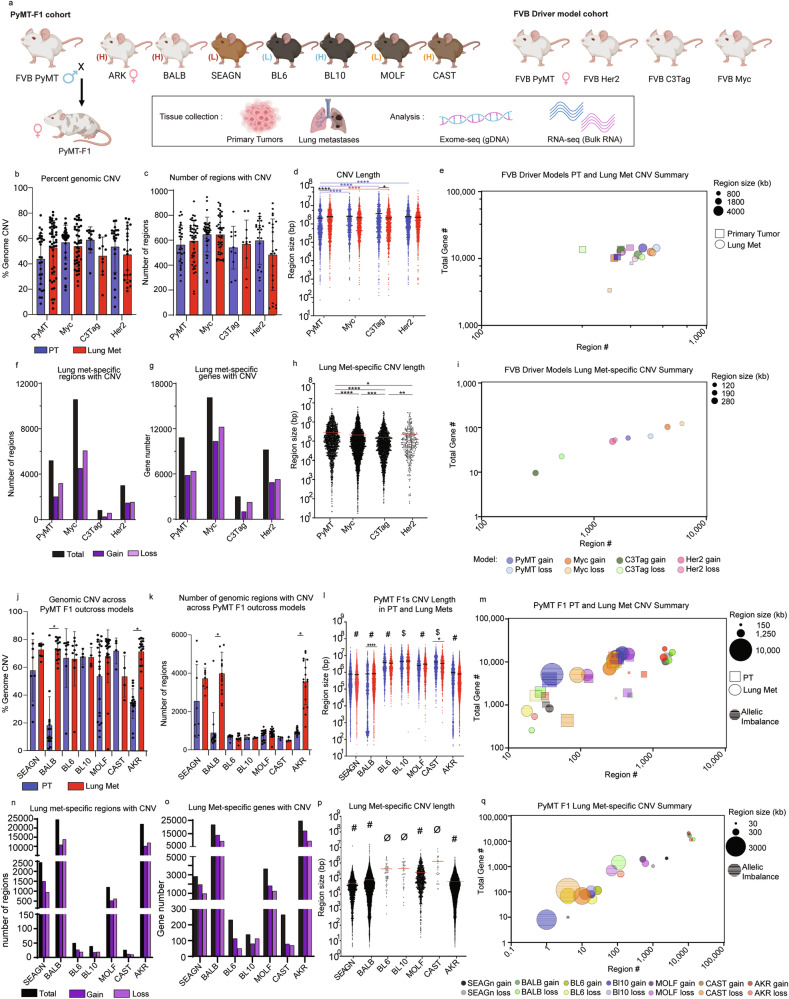
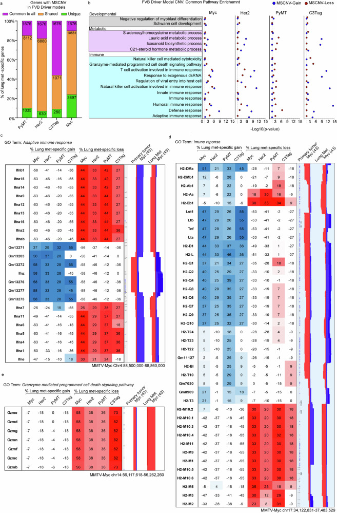
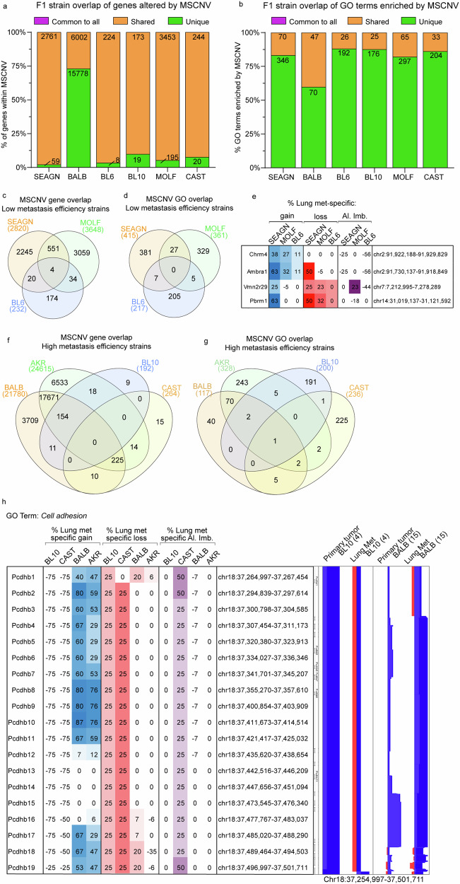
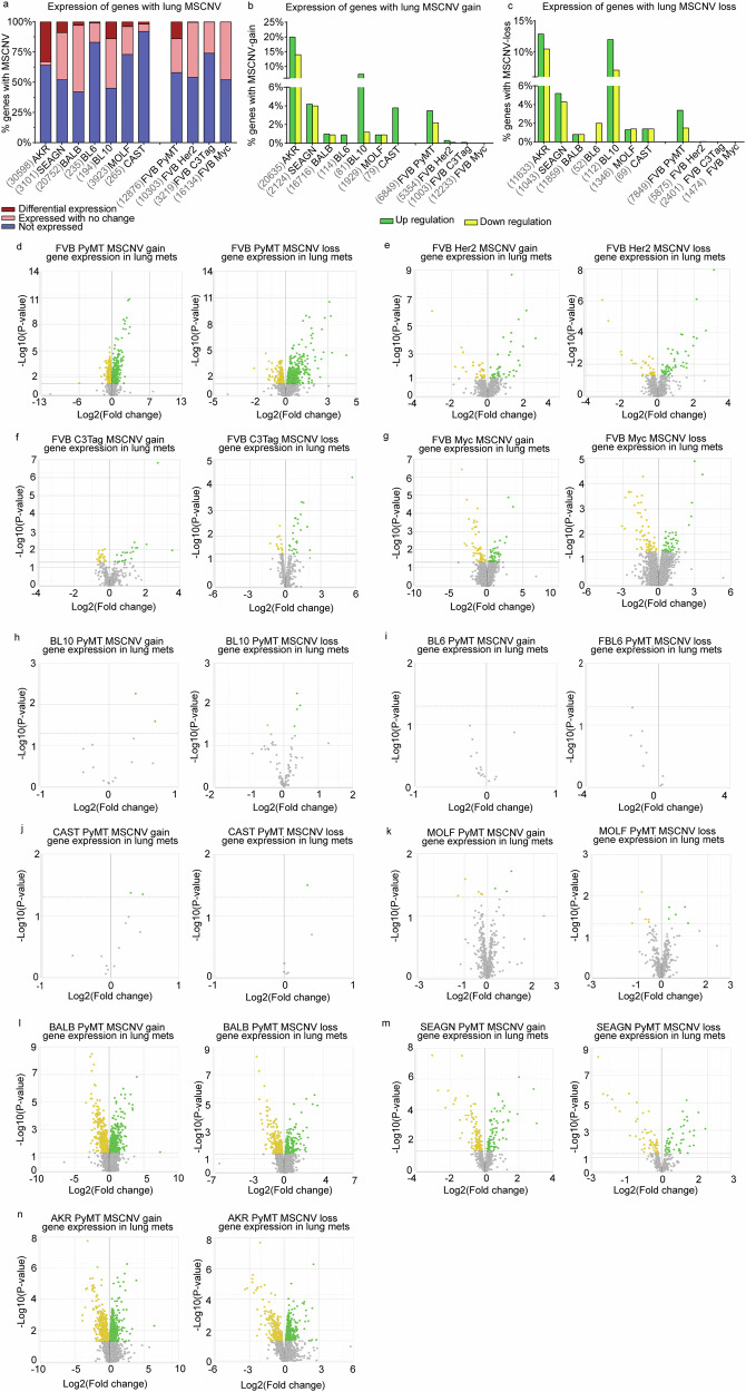
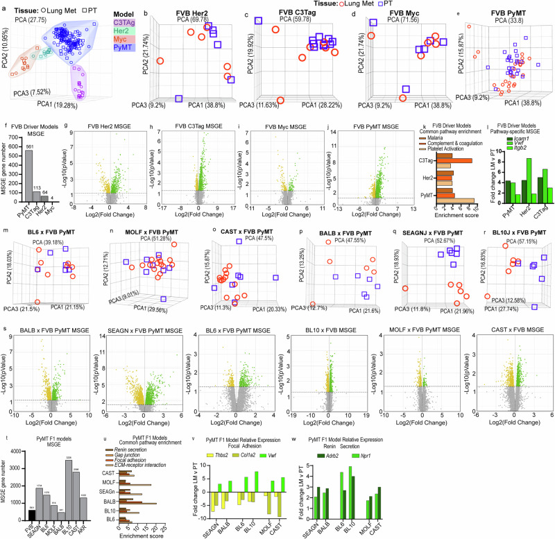
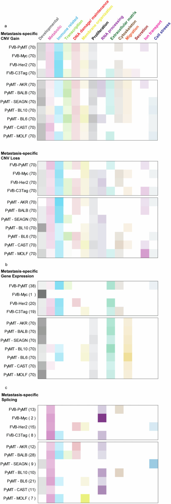
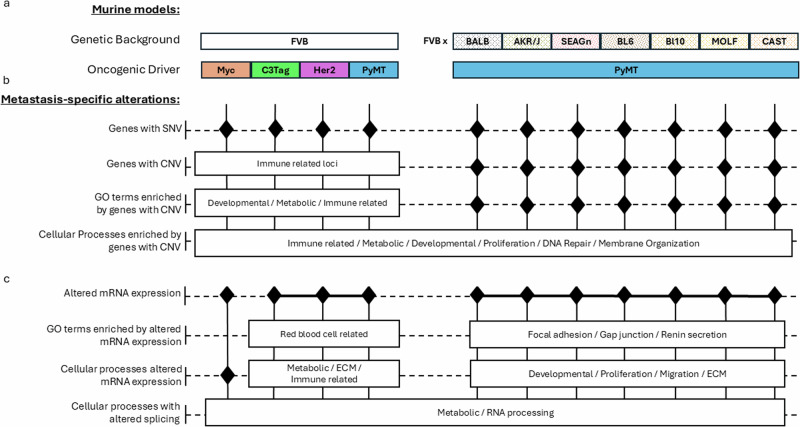
Metastasis remains a major cause of cancer mortality. This study, expanding upon previous findings in the MMTV-PyMT model, investigated four independent mouse models, representing luminal (MMTV-PyMT, MMTV-Myc), HER2-amplified (MMTV-Her2), and triple-negative (C3(1)TAg) breast cancer subtypes. Consistent with previous results, limited evidence for metastasis-associated somatic point mutations was found for all models. We also found that oncogenic drivers significantly influenced the number and size of metastasis-specific copy number variations (MSCNVs), but common driver-independent MSCNVs were rare. Furthermore, analyzing a cohort with varying genetic backgrounds while maintaining a constant oncogenic driver (PyMT) revealed that genetic background profoundly impacts MSCNVs. Transcriptome analysis demonstrated that oncogenic drivers strongly shaped metastasis-specific gene expression (MSGE), with each driver exhibiting distinct expression profiles. In contrast, MSGE in the PyMT-F1 cohort was more variable across strains. Despite the diversity of MSCNV and MSGE, functional analysis revealed that both mechanisms converge on the modulation of key cellular processes, including immune responses, metabolism, and extracellular matrix interactions. These findings emphasize the complex interplay between oncogenic drivers and genetic background in shaping the genomic and transcriptional landscapes of metastatic lesions.


Conflict of interest statement

Competing interests: The authors declare no competing interests.

Figures

Fig. 1
Fig. 1. Model and genetic background shape MSCNV.
a Schematic overview of mouse models, tissues collected, and analyses performed in this study. Histograms showing b the average percentage of the genome and c the average number of regions with CNV with standard deviation (SD), PT (blue), and lung mets (red). d Dot plot of individual CNV region lengths from aggregate data (the average indicated by black line). e Multivariate bubble plot showing average CNV number and aggregate total number of genes with CNV for PT (squares) and lung met (circles) from each model, with bubble size corresponding to CNV region size. Total number (black) of regions (f) and genes (g) with significant CNV gain (dark purple) and loss (light purple) in MSCNV. h Dot plot showing individual CNV region lengths from MSCNV (the average indicated by red line). i Multivariate bubble plot showing average CNV number and aggregate total number of genes with CNV for MSCNV (circles) from each model, with bubble size corresponding to CNV region size. j Histogram showing the average % of the genome and k average number of regions with CNV with SD, PT (blue), and lung mets (red). l Dot plot showing individual CNV region lengths from aggregate data with all animals per model combined (the average indicated by a line). m Multivariate bubble plot showing average CNV number and aggregate total number of genes with CNV for PT (squares) and lung met (circles) from each model, with bubble size corresponding to CNV region size. n Total number (black) of regions and o genes with significant CNV gain (dark purple) and loss (light purple) in Lung mets compared to PT. p Dot plot showing individual CNV region lengths from MSCNV (the average indicated by red line). q Multivariate bubble plot showing average CNV number and aggregate total number of genes with CNV for MSCNV (circles) from each model, with bubble size corresponding to CNV region size. Significance determined by Kruskal–Wallis test (d, h, l, p) or unpaired t test (b, c, j, k). *(p < 0.05), **(p < 0.01), ***(p < 0.001) ****(p < 0.0001), $ = significantly different from 6 groups, # = significantly different from 5 groups, ø = significantly different from 4 groups. Fig. 1a was generated using BioRender.
Fig. 2
Fig. 2. FVB driver models have metastasis-specific regions and gene families with CNV.
a Histogram showing the percent overlap in MSCNV gene lists from each model. Numbers within bars indicate gene number common to all (purple), shared among some (yellow), or unique to each model (green). b DAVID analysis clustering of 1676 MSCNV genes common to all models with enrichment in CNV gain (blue) or loss (red) indicated by −Log10(p value) (dotted line indicates cutoff for significance (≥1.3). Heatmap indicating % lung met-specific gain in blue (% PT gain − % LM gain) and lung met-specific loss in red (% PT loss − % LM loss) for genes within a single genomic region from the c Adaptive immune response, d immune response, and e Granzyme mediated programmed cell death signaling pathway. CNV signal traces from PT and lung mets aligned using Nexus 10 genomic view and the Myc model data set.
Fig. 3
Fig. 3. PyMT F1 models have no common gene sets or ontologies enriched by MSCNV.
Histograms of the percent overlap in gene lists (a) and GO terms (b) enriched by MSCNV in each model. Numbers within bars indicate gene number common to all (purple), shared among some (yellow), or unique to each model (green). Venn diagram analysis of gene sets (c) and GO terms (d) enriched by MSCNV in low metastatic efficiency strains. e Heatmap indicating % lung met-specific gain in blue (% PT gain − % LM gain) and lung met-specific loss in red (% PT loss − % LM loss) for genes with MSCNV in low metastatic efficiency strains. Venn diagram analysis of gene sets (f) and GO terms (g) enriched in MSCNV from high metastatic efficiency strains. h Heatmap indicating % lung met-specific gain in blue (% PT gain − % LM gain), lung met-specific loss in red (% PT loss − % LM loss), and lung met-specific allelic imbalance (Al. Imb.) in purple (% PT Al. Imb. − % LM Al. Imb.) for genes with MSCNV in high metastatic efficiency strains from the Cell adhesion pathway within a single locus. CNV signal traces from PT and Lung mets aligned using Nexus 10 genomic view of the BL10 and BALB model data sets.
Fig. 4
Fig. 4. MSCNV does not significantly impact metastasis-specific gene expression.
a Histogram of the percentage of genes with MSCNV that are not expressed (blue), expressed in lung mets with no change from PT (pink), and differentially expressed in lung mets compared to PTs (red). Numbers listed before each model name represent the MSCNV gene set size. Histograms showing the percentage of genes with MSCNV-gain (b) or MSCNV-loss (c) that had significant up regulation (green) or down regulation (yellow) in lung mets compared to PTs. Paired volcano plots for the MSGE of genes within regions of MSCNV gain or loss for each FVB driver model (dg) and PyMT-F1 model (hn).
Fig. 5
Fig. 5. Metastatic gene expression is shaped by oncogenic driver and genetic background.
a Unsupervised PCA of RNA-seq data from PT and Lung met tissues across all FVB driver and PyMT-F1 mouse models. Unsupervised PCA of individual PT and lung met samples from FVB Her2 (b), FVB C3Tag (c), FVB Myc (d), and FVB PyMT (e). f Histogram of the number of alternatively expressed genes in lung mets vs. PT for each FVB driver model (FDR < 0.05). Volcano plots with significantly up regulated genes in green and down regulated genes in yellow for MSGE in FVB Her2 (g), FVB C3Tag (h), FVB Myc (i), and FVB PyMT (j). k Histogram showing the enrichment score for commonly enriched pathways from MSGE of the C3Tag, Her2, and PyMT models. l Histogram showing the fold change of genes common to pathways enriched by MSGE. Unsupervised PCA of individual PT and lung met samples from BL6 (m), MOLF (n), CAST (o), BALB (p), SEAGN (q), and BL10 (r). s Volcano plots with significantly up regulated genes in green and down regulated genes in yellow for MSGE in the PyMT-F1 models. t Histogram of the number of alternatively expressed genes in lung mets vs. PT for each PyMT-F1 model (FDR < 0.05). u Histogram showing the enrichment score for commonly enriched pathways from MSGE of the F1-PyMT models. Histograms showing the fold change of common MSGE enriched in the Focal Adhesion (v) and Renin Secretion pathways (w).
Fig. 6
Fig. 6. Oncogenic driver and genetic background shape unique MSCNV and MSGE to target common cellular processes.
Categorization of the top 70 (or otherwise indicated) significantly enriched gene ontologies from MSCNV (a), MSGE (b), and lung met-specific alternative splicing (c). Uncategorized ontologies were excluded from the heat maps. Color intensity represents the percent of the analyzed pathways that fell within that category.
Fig. 7
Fig. 7. Host genetic background shapes metastatic SNVs, CNVs, and gene expression more than oncogenic driver.
Schema summarizing a murine model genetic background and oncogenic driver and the unique (black diamonds) or common (white boxes and solid lines) metastasis specific b genomic alterations and c gene expression.

Similar articles

References

    1. Surveillance, Epidemiology, and End Results (SEER) Program (www.seer.cancer.gov) SEER*Stat Database: Mortality - All COD, Aggregated With State, Total U.S. (1969-2023), National Cancer Institute, DCCPS, Surveillance Research Program, released February 2025. Underlying mortality data provided by NCHS (www.cdc.gov/nchs)
    1. Nowell, P. C. The clonal evolution of tumor cell populations. Science194, 23–28 (1976). - PubMed
    1. Patel, S. A., Rodrigues, P., Wesolowski, L. & Vanharanta, S. Genomic control of metastasis. Br. J. Cancer124, 3–12 (2021). - PMC - PubMed
    1. Nguyen, B. et al. Genomic characterization of metastatic patterns from prospective clinical sequencing of 25,000 patients. Cell185, 563–575 e511 (2022). - PMC - PubMed
    1. Dasari, S. et al. Genomic attributes of prostate cancer across primary and metastatic noncastrate and castrate resistant disease states: a next generation sequencing study of 183 patients. Prostate Cancer Prostatic Dis.28, 506–508 (2025). - PubMed