. 2025 May 12;10(8):2800-2804.
doi: 10.1016/j.ekir.2025.05.005. eCollection 2025 Aug.

Non-invasive Diagnosis of Antinephrin-Associated Podocytopathy

Affiliations

Non-invasive Diagnosis of Antinephrin-Associated Podocytopathy

Felicitas E Hengel et al. Kidney Int Rep. .

Abstract

Introduction: Circulating autoantibodies against the podocyte surface protein nephrin have recently been described in patients with podocytopathies, that is, minimal change disease, primary focal segmental glomerulosclerosis, and childhood idiopathic nephrotic syndrome. Their high specificity for podocytopathies in combination with a strong correlation with disease activity hold the potential for a non-invasive diagnosis, but prospective data are lacking.

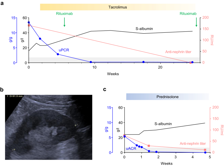
Methods: Here, we describe 3 patients with contraindications or unwillingness for a kidney biopsy, hampering a timely histological diagnosis and choice of appropriate therapy.

Results: In all patients, antinephrin autoantibodies were detected by quantitative immunoprecipitation, prompting the initiation of adequate treatment. These interventions induced a decrease in antinephrin autoantibody levels and clinical remission.

Conclusion: Our study highlights the potential of antinephrin autoantibody measurement for a noninvasive diagnosis of antinephrin-associated podocytopathy.

Keywords: antinephrin autoantibodies; minimal change disease; podocytopathy; serology.


Figures

None
Graphical abstract
Figure 1
Figure 1
Antinephrin autoantibody measurement for noninvasive diagnosis of antinephrin-associated podocytopathy in a patient with severe nephrotic syndrome and thrombotic complications. (a) MR venogram showing no contrast flow in the right transverse sinus (asterisk) indicative of a right transverse sinus thrombosis. (b) CT thorax with contrast shows a filling defect in the segmental branch of the pulmonary artery to the right upper lobe (arrow). (c) CT abdomen and pelvis with contrast in the sagittal plane shows a nonocclusive filling defect through the length of the left common to the external iliac vein indicative of an iliac vein thrombosis (arrows). (d) Serum (S)-albumin levels (black, given as g/l), urinary protein-to-creatinine ratio (uPCR) (blue, given as g/g) and antinephrin autoantibody titers (red, given as relative units RU/ml) as determined by hybrid immunoprecipitation /ELISA; prednisolone treatment period (starting with 60 mg on day 6, rapid taper to 15 mg in week 7) is shown as a blue box, rituximab treatment period (day 16) is shown as a green arrow, and intermittent hemodialysis period (day 11 and day 15) shown as a dashed line. The grey area indicates the normal range of antinephrin antibody levels. CT, Computed tomography; ELISA, enzyme-linked immunosorbent assay; MR, Magnetic resonance.
Figure 2
Figure 2
Antinephrin autoantibody measurement for noninvasive diagnosis of antinephrin-associated podocytopathy in a patient with complicated biopsy conditions and a patient with unwillingness for kidney biopsy. (a) Serum (S)-albumin levels (black, given as g/l) and urinary protein-to-creatinine ratio (uPCR) (blue, given in g/g) and antinephrin autoantibody titers (red, given as relative units RU/ml) as determined by hybrid immunoprecipitation enzyme-linked immunosorbent assay (ELISA); tacrolimus treatment period (starting with 2 mg twice daily in week 2, reduced to 1.5 mg, 1 mg, and 0.5 mg twice daily in week 10, 18, and 26, respectively) is shown as a yellow box and rituximab treatments (1 g as starting dose in week 6, 0.25 g as maintenance therapy in week 26) are shown as green arrows. (b) Renal ultrasound of the patient’s left kidney shows a reduced size in the patient with impaired procedural compliance. (c) S-albumin levels (black, given as g/l) and urinary albumin-to-creatinine ratio (uACR) (blue, given as g/g) and antinephrin autoantibody titers (red, given in relative units RU/ml) as determined by hybrid immunoprecipitation ELISA; prednisolone treatment period (starting with 70 mg daily in week 1, reduced to 20 mg daily in week 5) is shown as a blue box. The gray area indicates the normal range of antinephrin antibody levels.

References

    1. Watts A.J.B., Keller K.H., Lerner G., et al. Discovery of autoantibodies targeting nephrin in minimal change disease supports a novel autoimmune etiology. J Am Soc Nephrol. 2022;33:238–252. doi: 10.1681/ASN.2021060794. - DOI - PMC - PubMed
    1. Hengel F.E., Dehde S., Lassé M., et al. Autoantibodies targeting nephrin in podocytopathies. N Engl J Med. 2024;391:422–433. doi: 10.1056/NEJMoa2314471. - DOI - PubMed
    1. Raglianti V., Angelotti M.L., Cirillo L., et al. Anti-slit diaphragm antibodies on kidney biopsy identify pediatric patients with steroid-resistant nephrotic syndrome responsive to second-line immunosuppressants. Kidney Int. 2024;106:1124–1134. doi: 10.1016/j.kint.2024.09.006. - DOI - PubMed
    1. Hengel F.E., Dehde S., Yilmaz A., et al. Anti-nephrin autoantibodies in steroid-resistant nephrotic syndrome may inform treatment strategy. Kidney Int. 2025;107:951. - PubMed
    1. Hengel F.E., Dehde S., Kretz O., et al. Passive transfer of patient-derived anti-nephrin autoantibodies causes a podocytopathy with minimal change lesions. J Clin Invest. 2025;135 doi: 10.1172/JCI186769. - DOI - PMC - PubMed

LinkOut - more resources