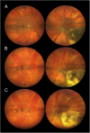
Fundus hypopigmentation and choroidal thinning associated with tebentafusp therapy: report of a case and literature review
- PMID: 40817046
- PMCID: PMC12357441
- DOI: 10.1186/s12886-025-04274-7
Fundus hypopigmentation and choroidal thinning associated with tebentafusp therapy: report of a case and literature review
Abstract
Purpose: To describe a novel case of progressive fundus hypopigmentation and choroidal thinning associated with tebentafusp monotherapy for metastatic uveal melanoma.
Methods: Observational case report and review of the literature.
Case presentation: A 69-year-old male was diagnosed with a choroidal melanoma, measuring 13.3 mm in diameter and 5.8 mm in thickness, in the left eye. Seven years after iodine-125 plaque brachytherapy, systemic imaging identified a solitary liver metastasis, which was laparoscopically resected. About one year later, two new liver metastases were detected. The patient was HLA-A*02:01 positive and started on tebentafusp. Except for transient fever, rash, and pruritus after the first cycles, the therapy was well tolerated. Fourteen months after initiation of tebentafusp, fundoscopy revealed marked hypopigmentation of both fundi and depigmentation of the regressed tumour in left eye. There were no signs of intraocular inflammation in either eye. Upon retrospective review of fundus photographs taken from baseline, the progressive fundus hypopigmentation and depigmentation of the tumour remnants first appeared after the initiation of immunotherapy. A corresponding evaluation of the optical coherence tomography scans of the previously untreated right eye revealed a significant reduction in central choroidal thickness over the same period. Full-field electroretinography demonstrated normal responses in the right eye and attenuated responses in the left eye. Screening for paraneoplastic antibodies was negative. During treatment, he also developed poliosis of the eyebrows and cilia, along with depigmented skin macules and patches. At the last visit, 11 years after the initial diagnosis and 26 months after starting tebentafusp, a repeat CT confirmed stable liver metastases with no new lesions. Both fundi appeared hypopigmented, and best corrected visual acuity was 1.0 in the right eye and hand movements in the left eye.
Conclusions: Tebentafusp therapy can lead to diffuse fundus hypopigmentation and choroidal thinning, similar to what has been reported after immune checkpoint inhibition. The progressive choroidal hypopigmentation, without evidence of associated intraocular inflammation, indicates that glycoprotein 100, the target antigen of tebentafusp, is also expressed by normal choroidal melanocytes.
Keywords: Choroid; Choroidal thinning; Depigmentation; Hypopigmentation; Immune checkpoint inhibitors; Immunotherapy; Metastatic melanoma; Ocular adverse events; Tebentafusp; Uveal melanoma.
© 2025. The Author(s).
Conflict of interest statement
Declarations. Ethics approval and consent to participate: This study followed the tenets of the Declaration of Helsinki. Regional Ethics Committee approval was not required for this case report. Consent for publication: Written informed consent was obtained from the patient for publication of this case report and any accompanying images. Competing interests: The authors declare no competing interests.
Figures


References
-
- Sharma P, Goswami S, Raychaudhuri D, Siddiqui BA, Singh P, Nagarajan A, et al. Immune checkpoint therapy-current perspectives and future directions. Cell. 2023;186(8):1652–69. 10.1016/j.cell.2023.03.006. - PubMed
-
- Larkin J, Chiarion-Sileni V, Gonzalez R, Grob JJ, Rutkowski P, Lao CD, et al. Five-year survival with combined nivolumab and ipilimumab in advanced melanoma. N Engl J Med. 2019;381(16):1535–46. 10.1056/NEJMoa1910836. - PubMed
Publication types
MeSH terms
LinkOut - more resources
Full Text Sources
Medical
Research Materials

