. 2025 Aug 15;17(1):90.
doi: 10.1186/s13073-025-01520-x.

Aging increases susceptibility to liver fibrosis through enhanced NAT10-mediated ac4C modification of TGFβ1 mRNA

Affiliations

Aging increases susceptibility to liver fibrosis through enhanced NAT10-mediated ac4C modification of TGFβ1 mRNA

Xuyun Peng et al. Genome Med. .

Abstract

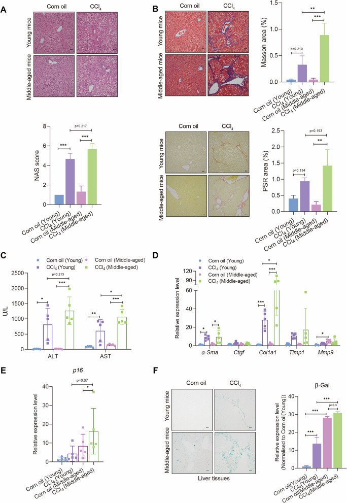
Background: The epidemiological observational studies unveiled that aging is one of the risk factors for liver fibrosis, and the hepatic tissues in the elderly harbor more fibrotic lesions when compared to those in young people. Previous investigations found that TGFβ1 was elevated with aging and promoted liver fibrosis. However, the underlying mechanisms of aging and liver fibrosis remain largely unknown.

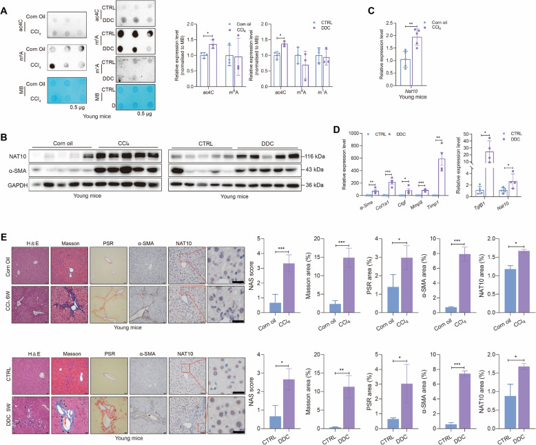
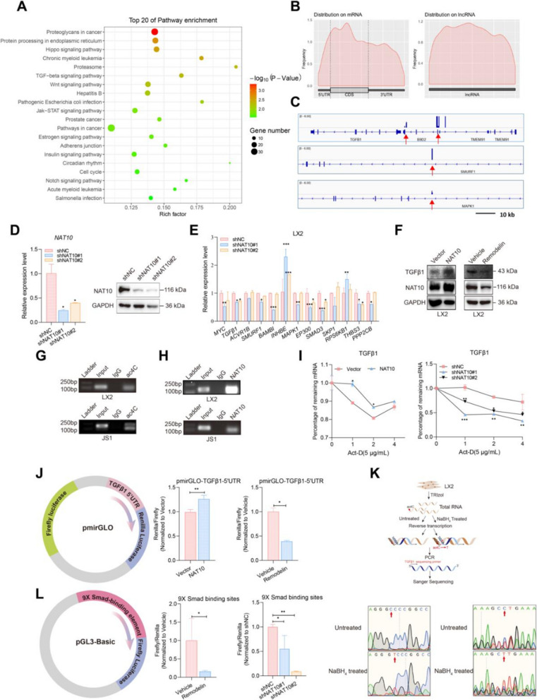
Methods: CCl4-induced liver fibrosis animal models were used in this study. The impact of NAT10 on liver fibrosis and cellular senescence was analyzed by using NAT10 overexpression or knockout hepatic stellate cell lines. The distribution of ac4C RNA modification was monitored by the acRIP-seq. The RNA-protein interaction was examined by the RNA immunoprecipitation.

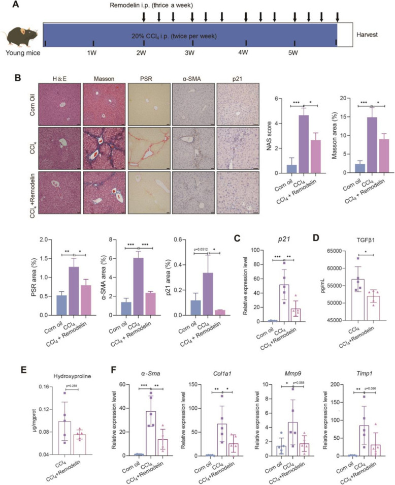
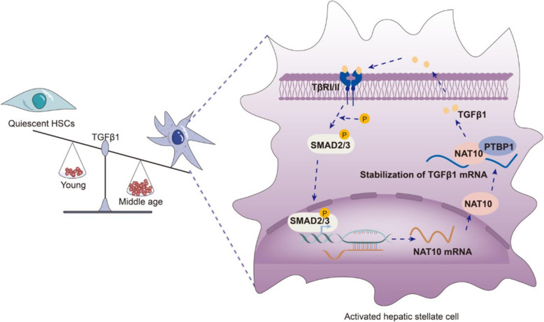
Results: We demonstrated that the middle-aged mice were more susceptible to the CCl4-induced liver fibrosis when compared to the young mice. Then, we found that RNA ac4C-modifying enzyme NAT10 was transcriptionally activated by TGFβ1/SMAD2/3 axis and highly expressed in the aging liver as well as liver fibrosis mouse model. Suppression of NAT10 by its inhibitor Remodelin or specific shRNA attenuated senescence and activation of hepatic stellate cells. Subsequent studies found that NAT10 directly triggered the ac4C RNA modification of TGFβ1 mRNA by physically interacting with the RNA-binding protein PTBP1, enhancing the stabilization of TGFβ1 mRNA and subsequent activation of TGFβ/SMAD signaling pathway. Animal studies demonstrated that inhibition of NAT10 by Remodelin significantly alleviated liver fibrosis and cellular senescence.

Conclusions: Our study identified a previously unknown mechanism of how TGFβ1 drives cellular senescence and liver fibrosis through NAT10-mediated ac4C mRNA modification.

Keywords: Aging; Liver fibrosis; NAT10; RNA modification; TGFβ/SMAD signaling pathway.


Conflict of interest statement

Declarations. Ethics approval: Animals in this study were used in accordance with the Guide for Care and Use of Laboratory Animals of the National Institute of Health. The animal study was approved by the institutional review boards and ethics committees of The Third Affiliated Hospital of Sun Yat-sen University. Consent for publication: Not applicable. Competing interests: The authors declare no competing interests.

Figures

Fig. 1
Fig. 1
Middle-aged mice were more vulnerable to CCl4 insult. Representative photographs of H&E staining (A), Masson staining (B, up), and PSR staining (B, down). Scale bar: 50 µm. C Levels of alanine aminotransferase (ALT) and aspartate aminotransferase (AST) in the serum of mice (n = 5). D qRT-PCR was used to examine the fibrosis-related genes in mice (n = 5). E The RNA expression of p16 in the young and middle-aged liver tissues of corn oil- and CCl4-treated mice (n = 5). F Left: representative photographs of β-Gal staining in the indicated groups. Right: quantification of β-Gal staining. Scale bar: 50 µm (*P < 0.05, **P < 0.01, ***P < 0.001)
Fig. 2
Fig. 2
Increased expression of TGFβ1 and NAT10 were found in the middle-aged mice. A Western blot was used to examine p21 and RNA-modifying enzymes in the liver of mice (n = 5). B Left: TGFβ1 levels were monitored by ELISA in the serum of mice. Right: qRT-PCR was used to examine the expression of TGFβ1 in liver tissues of the indicated groups (n = 5). Western blot (C) and qRT-PCR (D) were conducted to examine the expression of NAT10 in mouse primary HSCs, LX2, and JS1 after treatment with TGFβ1. E Dot blot of ac4C in LX2 and JS1 cells treated with or without TGFβ1. The values were normalized to methylene blue staining (*P < 0.05, **P < 0.01, ***P < 0.001)
Fig. 3
Fig. 3
NAT10 and ac4C RNA modification were elevated in the mice with liver fibrosis. A Left panel: dot blot detecting ac4C levels in the indicated groups. Right panel: quantification of dot blot results. The values were normalized to the methylene blue staining. B Western blot was used to detect Nat10 in CCl4- and DDC-treated mice model (n = 5). C qRT-PCR was used to detect Nat10 in liver tissues of the indicated groups (n = 5). D qRT-PCR for Tgfβ1, Nat10, and fibrotic genes in the liver tissues (corn oil: n = 5; DDC: n = 4). E Representative photographs of H&E staining, Masson staining, PSR staining, and IHC staining. Scale bar: 50 µm. Scale bar of a zoom-in Nat10-staining region: 25 µm (*P < 0.05, **P < 0.01, ***P < 0.001)
Fig. 4
Fig. 4
SMAD2 and SMAD3 directly potentiated the RNA transcription of NAT10. A The visual genome graphs of SMAD2 and SMAD3 binding sites within NAT10 gene promoter. B Schematic overview of the ChIP primer design and ChIP assay by using SMAD2 and SMAD3 antibody. The PCR product size of human NAT10 gene locus is 232 bp, and the PCR product size of mouse Nat10 gene locus is 185 bp. C Overexpression of SMAD2 and SMAD3 elevated the protein level of NAT10. D Left panel: schematic diagram of luciferase plasmid construction. Right panel: luciferase reporter assay of HEK293T cells co-transfected with Renilla and pGL4 construct harboring the NAT10 promoter. E The LX2 and JS1 cells were treated with SB431542 or LY364947 plus TGFβ1 for 24 h. Western blots were performed to examine the expression of NAT10 and α-SMA. F The HEK293T cells were transfected with pGL4 construct harboring the NAT10 promoter and then treated with SB431542 or LY364947 plus TGFβ1 for 24 h. Luciferase activity was measured and values were referred to Renilla values and normalized to untreated pGL4-NAT10 promoter (*P < 0.05, **P < 0.01, ***P < 0.001)
Fig. 5
Fig. 5
NAT10 increased the RNA ac4C modification and stability of TGFβ1 mRNA. A Top 20 pathways with acetylated mRNAs. B The genome-wide distribution of ac4C modification in mRNA and lncRNA. C The visual genome of mRNAs with ac4C modification. D qRT-PCR was conducted to evaluate the expression of NAT10 after silencing of NAT10. E The RNA levels of acetylated mRNA transcripts of TGFβ signaling pathway were examined after knock down of NAT10. F Western blot was used to detect TGFβ1 and NAT10 in LX2 cells. Nucleic acid electrophoresis for RIP assay by using antibody against ac4C (G) and NAT10 (H). The PCR product size of human TGFβ1 mRNA is 151 bp and the PCR product size of mouse TGFβ1 mRNA is 133 bp. I qRT-PCR was conducted to examine the RNA stability of TGFβ1 after overexpression or silence of NAT10 in LX2. Cells were treated with Act-D and samples were collected at the indicated time. J Left panel: schematic diagram of luciferase plasmid construction. Middle panel: luciferase reporter assay of NAT10-overexpressing HEK293T cells transfected with pmirGLO constructs with the TGFβ1 5′UTR. Right panel: luciferase reporter assay of HEK293T cells transfected with pmirGLO constructs harboring the TGFβ1 5′UTR. Twenty-four hours after transfection, cells were treated with or without Remodelin. Luciferase assay was conducted. K Top: schematic diagram of ac4C modification sites in TGFβ1 mRNA. Bottom: Sanger sequencing results. C to T mutation indicates an ac4C modification site. L Left: schematic diagram of luciferase plasmid construction. Right: luciferase reporter assay of HEK293T cells co-transfected with Renilla and pGL3-based constructs harboring the 9 × Smad binding sites. Luciferase assays were conducted after overexpression of or knockdown of NAT10 (*P < 0.05, **P < 0.01, ***P < 0.001)
Fig. 6
Fig. 6
PTBP1 recognized acetylated TGFβ1 mRNA. A NAT10 physically interacted with various RNA-binding proteins. B and C NAT10 physically interacted with RNA-binding protein PTBP1. D Immunofluorescence staining of NAT10 (red) with PTBP1 (green) in LX2 cells. Scale bar: 10 μm. E Schematic diagram of dot blot by using PTBP1 antibody in the RIP experiment. F Dot blot detecting ac4C modification in LX2 cells. G RIP assay was used to validate the mutual interaction between TGFβ1 mRNA and PTBP1 in LX2 and JS1 cells. H Cells were treated with or without siPTBP1 for 72 h. qRT-PCR was conducted to examine the RNA expression of PTBP1 and TGFβ1 mRNA in LX2 cells. I qRT-PCR was used to examine the half-life of TGFβ1 in LX2 cells after silencing of PTBP1. J Luciferase reporter assay of HEK293T cells co-transfected with pmirGLO constructs of the TGFβ1 5′UTR and PTBP1 plasmid (left) or siRNA against PTBP1 (right). K NAT10-overexpressing LX2 cells were treated with siRNA against PTBP1. qRT-PCR was conducted to examine the expression of TGFβ1 (*P < 0.05, **P < 0.01, ***P < 0.001)
Fig. 7
Fig. 7
NAT10 inhibitor Remodelin alleviated CCl4-mediated liver fibrosis. A Schematic of the animal experiment design. B Representative photographs of H&E staining, Masson staining, PSR staining, and IHC staining. Scale bar: 50 µm. C qRT-PCR was conducted to examine the expression of p21 after treatment of Remodelin. D ELISA assay was conducted to examine the levels of TGFβ1 after treatment of Remodelin. E ELISA assay was conducted to examine the levels of hydroxyproline after treatment of Remodelin. F qRT-PCR was conducted to examine the expression of fibrosis-related genes in the liver tissues (*P < 0.05, **P < 0.01, ***P < 0.001)
Fig. 8
Fig. 8
RNA modification drives age-related liver fibrosis. A hypothetical model was used to illustrate the underlying mechanisms. Middle-aged mice exhibit higher levels of TGFβ1. TGFβ/SMAD signaling pathway directly potentiates the transcription of NAT10, and NAT10 subsequently increases the ac4C RNA modification of TGFβ1 mRNA. The RNA-binding protein PTBP1 then recognizes this acetylated (ac4C-modified) mRNA, leading to the stabilization of TGFβ1 mRNA and reactivation of the TGFβ/SMAD signaling pathway

References

    1. Sheedfar F, Di Biase S, Koonen D, Vinciguerra M. Liver diseases and aging: friends or foes? Aging Cell. 2013;12:950–4. - PubMed
    1. Bawa FNC, Zhang Y. Retinoic acid signaling in fatty liver disease. Liver Res. 2023;7:189–95. - PMC - PubMed
    1. Delire B, Lebrun V, Selvais C, Henriet P, Bertrand A, Horsmans Y, Leclercq IA. Aging enhances liver fibrotic response in mice through hampering extracellular matrix remodeling. Aging (Albany NY). 2016;9:98–113. - PMC - PubMed
    1. Zhong HH, Hu SJ, Yu B, Jiang SS, Zhang J, Luo D, Yang MW, Su WY, Shao YL, Deng HL, et al. Apoptosis in the aging liver. Oncotarget. 2017;8:102640–52. - PMC - PubMed
    1. Caballeria L, Pera G, Arteaga I, Rodriguez L, Aluma A, Morillas RM, de la Ossa N, Diaz A, Exposito C, Miranda D, et al. High prevalence of liver fibrosis among European adults with unknown liver disease: a population-based study. Clin Gastroenterol Hepatol. 2018;16:1138-1145 e1135. - PubMed

MeSH terms

LinkOut - more resources