

Anti-MDA5 antibody IgG1 subtype is associated with rapidly progressive interstitial lung disease in anti-MDA5-positive dermatomyositis
- PMID: 40817077
- PMCID: PMC12357345
- DOI: 10.1186/s13023-025-03921-y
Anti-MDA5 antibody IgG1 subtype is associated with rapidly progressive interstitial lung disease in anti-MDA5-positive dermatomyositis
Abstract
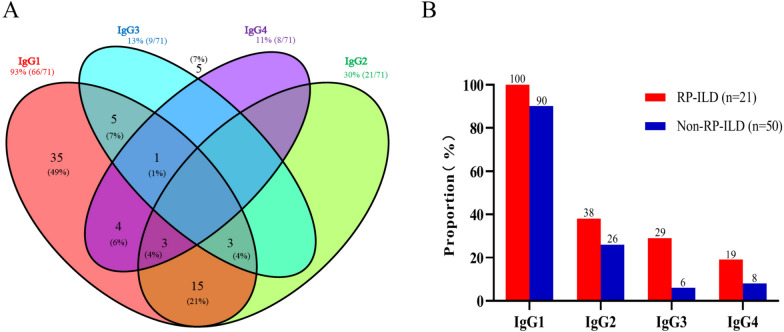
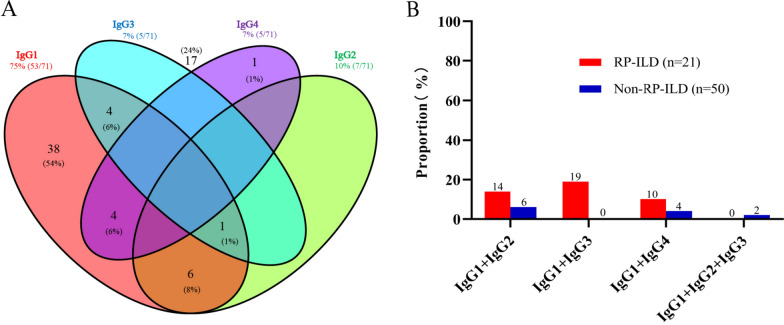
Background: Rapidly progressive interstitial lung disease (RP-ILD) is a severe, often fatal complication in patients with anti-melanoma differentiation-associated gene 5 antibody-positive dermatomyositis (MDA5+ DM). Early prediction of RP-ILD still remains challenging. We aimed to explore the link between anti-MDA5 IgG subtypes and ILD prognosis in individuals with MDA5+ DM.
Methods: In a retrospective study involving 71 MDA5+ DM-ILD patients, initial serum titers of anti-MDA5 IgG subtypes were measured using indirect immunofluorescence. We then analyzed the associations between these IgG subclasses and the development of RP-ILD.
Result: Of the 71 patients, 30% developed RP-ILD. RP-ILD patients had a shorter disease course and a higher mortality rate than non-RP-ILD patients (both P < 0.001). A notable link was found between RP-ILD and anti-MDA5 IgG1 (P < 0.05), with 100% of RP-ILD patients showing IgG1 titers ≥ 1:100. Additionally, IgG3 positivity was more prevalent in RP-ILD (P < 0.05). Multivariate logistic regression analysis identified high titers of anti-MDA5 IgG1 and a high neutrophil-lymphocyte ratio (NLRhigh≥5.22) as independent risk factors for RP-ILD (P = 0.020, 0.017, respectively). The combination of anti-MDA5 IgG1 ≥ 1:100 with an NLR ≥ 5.22 improved the predictive accuracy for RP-ILD, yielding an AUC of 0.80.
Conclusions: Elevated anti-MDA5 IgG1 titers are strongly related to RP-ILD in MDA5+ DM and function as an important marker for early detection of individuals at high risk. Combining anti-MDA5 IgG1 levels with NLR further enhances predictive accuracy for RP-ILD, offering a practical approach for clinical monitoring and early intervention.
Keywords: Anti-MDA5 antibody; IgG1; Indirect immunofluorescence; Rapidly progressive interstitial lung disease.
© 2025. The Author(s).
Conflict of interest statement
Declarations. Conflict of interest: The authors report no conflicts of interest. Ethical approval and consent to participate: The Ethics Committee of the First Affiliated Hospital of Nanjing Medical University approved the study (ID: 2020-SR-265), and all participants gave informed consent. Consent for publication: Not applicable.
Figures

Similar articles
-
[A study of differences in the course of anti-MDA5-positive dermatomyositis and early diagnosis of rapidly progressive interstitial lung disease].Zhonghua Jie He He Hu Xi Za Zhi. 2025 Aug 12;48(8):726-731. doi: 10.3760/cma.j.cn112147-20241229-00761. Zhonghua Jie He He Hu Xi Za Zhi. 2025. PMID: 40764133 Chinese.
-
Mortality Risk Prediction in Patients With Antimelanoma Differentiation-Associated, Gene 5 Antibody-Positive, Dermatomyositis-Associated Interstitial Lung Disease: Algorithm Development and Validation.J Med Internet Res. 2025 Feb 5;27:e62836. doi: 10.2196/62836. J Med Internet Res. 2025. PMID: 39908093 Free PMC article.
-
Diagnostic significance of carcinoembryonic antigen and anti-MDA5 antibodies in polymyositis/dermatomyositis-associated rapidly progressive interstitial lung disease.Med Clin (Barc). 2025 Sep;165(3):107048. doi: 10.1016/j.medcli.2025.107048. Epub 2025 Jun 13. Med Clin (Barc). 2025. PMID: 40516211 English, Spanish.
-
Management of MDA-5 antibody positive clinically amyopathic dermatomyositis associated interstitial lung disease: A systematic review.Semin Arthritis Rheum. 2022 Apr;53:151959. doi: 10.1016/j.semarthrit.2022.151959. Epub 2022 Jan 31. Semin Arthritis Rheum. 2022. PMID: 35134633
-
Risk factors for mortality in anti-MDA5 antibody-positive dermatomyositis with interstitial lung disease: a systematic review and meta-analysis.Front Immunol. 2025 Jul 17;16:1628748. doi: 10.3389/fimmu.2025.1628748. eCollection 2025. Front Immunol. 2025. PMID: 40746563 Free PMC article.
References
-
- Moghadam-Kia S, Oddis CV, Aggarwal R. Anti-MDA5 antibody spectrum in Western World. Curr Rheumatol Rep. 2018;20(12):78. - PubMed
-
- Shi J, Li S, Yang H, Zhang Y, Peng Q, Lu X, et al. Clinical profiles and prognosis of patients with distinct antisynthetase autoantibodies. J Rheumatol. 2017;44(7):1051–7. - PubMed
-
- Gono T, Kuwana M. Inflammatory myopathies: choosing the right biomarkers to predict ILD in myositis. Nat Rev Rheumatol. 2016;12(9):504–6. - PubMed
-
- Hoshino K, Muro Y, Sugiura K, Tomita Y, Nakashima R, Mimori T. Anti-MDA5 and anti-TIF1-gamma antibodies have clinical significance for patients with dermatomyositis. Rheumatology (Oxford). 2010;49(9):1726–33. - PubMed
-
- Nakashima R, Imura Y, Kobayashi S, Yukawa N, Yoshifuji H, Nojima T, et al. The RIG-I-like receptor IFIH1/MDA5 is a dermatomyositis-specific autoantigen identified by the anti-CADM-140 antibody. Rheumatology (Oxford). 2010;49(3):433–40. - PubMed
MeSH terms
Substances
Grants and funding
LinkOut - more resources
Full Text Sources
Medical
