. 2025 Aug 15;16(1):7604.
doi: 10.1038/s41467-025-62972-8.

RKIP regulates bone marrow macrophage differentiation to mediate osteoclastogenesis and H-type vessel formation

Affiliations

RKIP regulates bone marrow macrophage differentiation to mediate osteoclastogenesis and H-type vessel formation

Zeyu Zheng et al. Nat Commun. .

Abstract

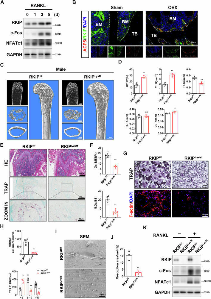
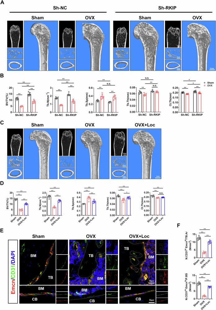
As central cells involved in osteoimmunology, bone niche macrophages possess diverse functions, and their differentiation fate regulates bone homeostasis. Elucidation of the underlying mechanism involved in macrophage differentiation is important for developing new therapeutic targets for osteoporosis. Here, we show that knocking out Raf kinase inhibitor protein (RKIP), either globally or in macrophages, results in dramatically increased bone mass in mice due to synergistic inhibition of bone resorption and promotion of bone formation. Mechanistically, RKIP knockout inhibits differentiation of macrophages into osteoclasts and promotes their differentiation towards pro-angiogenic subclusters, which enhances formation of H-type vessels. RKIP enhances osteoclastogenesis by interacting with ARHGAP to suppress CDC42 inactivation. Intranuclear RKIP suppresses angiogenic genes expression by bridging the association between HIF-1α and VHL to reduce the protein stability of HIF-1α in macrophages. Furthermore, RKIP deletion or inhibitor rescues ovariectomy (OVX)-induced bone loss in vivo. Collectively, this study provides insights into the different roles of extranuclear or intranuclear RKIP in regulating differentiation of bone niche macrophages and could inform potential therapies for bone homeostasis-related diseases.


Conflict of interest statement

Competing interests: The authors declare no competing interests.

Figures

Fig. 1
Fig. 1. Global knockout of RKIP inhibits bone resorption and promotes bone formation.
A Micro-CT and three-dimensional (3D) reconstruction images of the distal femur of 3-month-old male RKIP+/+ or RKIP-/- mice are shown. B Quantification of bone volume/tissue volume (BV/TV), trabecular number (Tb.N), trabecular separation (Tb.Sp), trabecular thickness (Tb.Th), and cortical thickness (Ct.Th) (n = 5). C H&E staining and TRAP staining of 3-month-old male RKIP+/+ or RKIP-/- mice femurs sections. D Quantification of osteoclast number per bone surface (N.Oc/BS) and osteoclast surface per bone surface (Oc.S/BS) (n = 6). E Calcein double staining and Von Kossa staining of 3-month-old male RKIP+/+ or RKIP-/- mice femurs sections. F Quantification of mineral apposition rate (MAR) and bone formation rate per bone surface (BFR/BS) (n = 6). G OCN (green) and DAPI (blue) immunofluorescence of 3-month-old male RKIP+/+ or RKIP-/- mice femurs sections; The arrow indicates the cells which express OCN. H Quantification of OCN+ cell number per bone perimeter (N.OCN+/B.Pm) (n = 6). I Serum C-terminal telopeptide of type I collagen (CTX-1) and Procollagen I N-Terminal Propeptide (PINP) concentration measured by ELISA from RKIP+/+ or RKIP-/- male mice (n = 6). Data are presented as mean ± SD. *P < 0.05, **P < 0.01 for a comparison with the control group or as indicated. n value means the number of repetitions in each independent experiment.
Fig. 2
Fig. 2. Conditional knockout of RKIP in macrophages impedes osteoclastogenesis in vivo and in vitro.
A Western blots of RKIP, c-Fos and NFATc1 protein levels in BMMs under the treatment of M-CSF and RANKL for indicated time. B RKIP (green), ACP5 (red) and DAPI (blue) immunofluorescence staining of femur sections from sham or OVX mice; The arrow indicates the cells which express both RKIP and ACP5. C Micro-CT and three-dimensional (3D) reconstruction images of the distal femur of 3-month-old male RKIPf/f or LysM-Cre-RKIPf/f mice are shown. D Quantification of BV/TV, Tb.N, Tb.Sp, Tb.Th, and Ct.Th (n = 5). E H&E staining and TRAP staining of 3-month-old male RKIPf/f or LysM-Cre-RKIPf/f mice femurs sections. F Quantification of N.Oc/BS and Oc.S/BS (n = 6). G TRAP staining and F-actin immunofluorescence staining to detect osteoclastogenesis of BMMs from RKIPf/f or LysM-Cre-RKIPf/f male mice. H Quantification of size and nuclei numbers of TRAP-positive multinuclear cells (n = 6). I Scanning electron microscopy (SEM) images of bone resorption pits of osteoclast precursors from RKIPf/f or LysM-Cre-RKIPf/f male mice. J Quantification of resorbed area/well (n = 3). K Western blots of RKIP, c-Fos and NFATc1 protein levels in BMMs from RKIPf/f or LysM-Cre-RKIPf/f male mice with or without the treatment of RANKL. Data are presented as mean ± SD. *P < 0.05, **P < 0.01 for a comparison with the control group or as indicated. n value means the number of repetitions in each independent experiment.
Fig. 3
Fig. 3. RKIP enhances osteoclastogenesis by interacting with ARHGAP to suppress CDC42 inactivation.
A GO enrichment analysis of RNA-seq analysis of BMMs from RKIP+/+ and RKIP-/- mice. B CDC42 peptides identified through mass spectrometry are shown. C Western blots of CDC42-GTP and total CDC42 protein levels in BMMs from RKIP+/+ or RKIP-/- male mice with RANKL treatment for indicated time. D RKIP (green), CDC42 (red) and DAPI (blue) immunofluorescence staining of BMMs from RKIP+/+ or RKIP-/- male mice with or without RANKL treatment. E Various truncated fragments of RKIP (R1, 1–90; R2, 91–140; R3, 141–187). R1–R3 and full length of CDC42 plasmid were co-transfected in HEK-293T cells. The interactions were detected through the indicated IP and IB analyses. F Various truncated fragments of CDC42 (C1, 1–47; C2, 48–104; C3, 105–191). C1–C3 and full length of RKIP plasmid were co-transfected in HEK-293T cells. The interactions were detected through the indicated IP and IB analyses. G Overall structure of CDC42 (pink) and RKIP (purple) and key residues of their interaction are shown. H HEK-293T cells were transfected with CDC42-Flag or CDC42-Flag and RKIP-Myc; The interactions were detected through the indicated IP and IB analyses. I HEK-293T cells were transfected with ARHGAP-HA and CDC42-Flag, or ARHGAP-HA, CDC42-Flag and RKIP-Myc; The interactions were detected through the indicated IP and IB analyses. J HEK-293T cells were transfected with RKIP-Myc, ARHGAP-HA or RKIP-Myc and ARHGAP-HA; The interactions were detected through the indicated IP and IB analyses. K The illustration of RKIP inhibiting CDC42 inactivation by interacting with ARHGAP.
Fig. 4
Fig. 4. RKIP regulates macrophage differentiation fate and influences H-type vessel formation.
A The illustration of scRNA-seq on cells isolated from the bone marrow and metaphysis. B UMAP dimensionality reduction plot for unsupervised clustering of macrophage subtypes. C Percentage distribution of macrophage subclusters. D Dot plot of the top 5 cluster-specific marker genes of macrophage subclusters. E The box plot of AUCell scores of the pro-angiogenic gene set for each subcluster. F Percentage distribution of non-pro-angiogenic and pro-angiogenic macrophage subcluster in RKIPf/f or LysM-Cre-RKIPf/f male mice. G GO enrichment analysis of pro-angiogenic macrophage subcluster. H The mRNA level of VEGFA-VEGFD and PDGFA-PDGFD in BMMs from RKIP+/+ or RKIP-/- male mice (n = 6). I F-actin immunofluorescence staining of matrigel tube formation assay using conditioned medium (CM) of BMMs from RKIP+/+ or RKIP-/- male mice. J Quantification of cumulative tube length (n = 4). K CD31 (green), Emcn (red) and DAPI (blue) immunofluorescence staining of femur sections from RKIPf/f or LysM-Cre-RKIPf/f male mice. L Quantification of the number of CD31hiEmcnhi cell per trabecular area (N. CD31hiEmcnhi/TB.Ar) and the number of CD31hiEmcnhi per periosteal bone surface (N. CD31hiEmcnhi/P.BS) (n = 6). Data are presented as mean ± SD. *P < 0.05, **P < 0.01 for a comparison with the control group or as indicated. n value means the number of repetitions in each independent experiment.
Fig. 5
Fig. 5. Intranuclear RKIP modulates angiogenesis by regulating the protein stability of HIF-1α in macrophages.
A Western blots of HIF-1α, HIF-2α and RKIP protein levels in BMMs from RKIP+/+ or RKIP-/- male mice under hypoxia culture for indicated time. B RKIP (green), HIF-1α (red) and DAPI (blue) immunofluorescence staining of BMMs from RKIP+/+ or RKIP-/- male mice under hypoxia culture for 24 h. C Western blots of HIF-1α and RKIP protein levels in BMMs following the adenovirus transduction of RKIP or the empty vector with the treatment of MG132 or chloroquine (CQ) under hypoxia culture for 48 h. D Western blots of the ubiquitination of HIF-1α in BMMs from RKIP+/+ or RKIP-/- male mice under hypoxia culture for 24 h, which were immunoprecipitated with anti-HIF1α. E BMMs were cultured under normoxia or hypoxia for 48 h; The interactions were detected through the indicated IP and IB analyses. F Western blots of HIF-1α, VHL, and RKIP in cytoplasmic and nuclear of BMMs from RKIP+/+ or RKIP-/- male mice under hypoxia culture for 48 h. G The illustration and images of proximity ligation assays (PLA) of indicated proteins in BMMs under hypoxia culture for 48 h. H Putative murine, rat, human, and canine RKIP NLS sequences, predicted from analysis with the PredictNLS program. I Flag(green), HIF-1α (red) and DAPI (blue) immunofluorescence staining of HEK-293T cells transfected with RKIP-Flag or RKIP-NLS-Del-Flag plasmid, which were under hypoxia culture for 24 h. J Western blots of HIF-1α, VHL, and Flag in cytoplasmic and nuclear of HEK-293T cells transfected with RKIP-Flag or RKIP-NLS-Del-Flag plasmid, which were under hypoxia culture for 48 h. K RKIP(green), HIF-1α (red) and DAPI (blue) immunofluorescence staining of BMMs transfected with NC or KPNB1 siRNA, which were under hypoxia culture for 24 h.
Fig. 6
Fig. 6. Knockdown of RKIP and RKIP inhibitor prevent OVX-induced bone loss.
A Micro-CT and three-dimensional reconstructed images of distal femurs from intramedullary injection of Sh-NC or Sh-RKIP adeno-associated virus (AAV) mice that underwent either sham or OVX operation. B Quantification of BV/TV, Tb.N, Tb.Sp, Tb.Th, and Ct.Th (n = 6). C Micro-CT and three-dimensional reconstructed images of distal femurs from sham and OVX mice injected with saline or Locostatin (1 mg/kg) for 2 months. D Quantification of BV/TV, Tb.N, Tb.Sp, Tb.Th, and Ct.Th (n = 6). E CD31 (green), Emcn (red) and DAPI (blue) immunofluorescence staining of femur sections from sham and OVX mice injected with saline or Locostatin (1 mg/kg). F Quantification of N. CD31hiEmcnhi/TB.Ar and N. CD31hiEmcnhi/P.BS (n = 6). Data are presented as mean ± SD. *P < 0.05, **P < 0.01 for a comparison with the control group or as indicated. n value means the number of repetitions in each independent experiment.
Fig. 7
Fig. 7. RKIP regulates osteoclast differentiation in the human.
A RKIP (green), ACP5 (red) and DAPI (blue) immunofluorescence staining of femur sections from normal or osteoporosis patients; The arrow indicates the cells which express both RKIP and ACP5. B Quantification of the number of ACP5+RKIP+ cells per trabecular perimeter (N. ACP5+RKIP+/TB. Pm) (n = 6). C Western blots of RKIP, c-Fos protein levels in PBMCs from normal or osteoporosis patients. D Quantification and normalization of the gray levels of RKIP and c-Fos proteins to that of GAPDH using Image J (n = 3). E The mRNA level of RKIP in PBMCs from normal or osteoporosis patients (n = 3). F Western blots of RKIP, c-Fos, and NFATc1 protein levels in PBMCs with h-M-CSF and h-RANKL treatment for indicated time. G TRAP staining to detect osteoclastogenesis of PBMCs with various concentrations of Locostatin treatment. H Quantification of size and nuclei numbers of TRAP-positive multinuclear cells (n = 6). I The mRNA level of ACP5, CTSK, NFATc1 in PBMCs with vehicle or Locostatin treatment in the presence or absence of h-RANKL for 10 days (n = 6). J Western blots of RKIP, c-Fos, and NFATc1 protein levels in PBMCs with vehicle or Locostatin treatment in the presence or absence of h-RANKL for 10 days. K TRAP staining to detect osteoclastogenesis of PBMCs with various concentrations of Didymin treatment. L Quantification of size and nuclei numbers of TRAP-positive multinuclear cells (n = 6). M The mRNA level of ACP5, CTSK, NFATc1 in PBMCs with vehicle or Didymin treatment in the presence or absence of h-RANKL for 10 days (n = 6). N Western blots of RKIP, c-Fos, and NFATc1 protein levels in PBMCs with vehicle or Didymin treatment in the presence or absence of h-RANKL for 10 days. Data are presented as mean ± SD. *P < 0.05, **P < 0.01 for a comparison with the control group or as indicated. n value means the number of repetitions in each independent experiment.
Fig. 8
Fig. 8. The illustration of RKIP regulating bone homeostasis by mediating the differentiation fate of bone niche macrophages.
Schematic illustration showing the different roles of extranuclear or intranuclear RKIP in differentiation fate regulation of bone niche macrophages. RKIP is a critical molecule regulating the differentiation fate of bone niche macrophage via the RKIP/ARHGAP/CDC42 and RKIP/VHL/HIF-1α pathways.

Similar articles

References

    1. Clarke, B. Normal bone anatomy and physiology. Clin. J. Am. Soc. Nephrol.3, S131–S139 (2008). - PMC - PubMed
    1. Zaidi, M. Skeletal remodeling in health and disease. Nat. Med.13, 791–801 (2007). - PubMed
    1. Zaidi, M., Yuen, T., Sun, L. & Rosen, C. J. Regulation of Skeletal Homeostasis. Endocr. Rev.39, 701–718 (2018). - PMC - PubMed
    1. Riddle, R. C. & Clemens, T. L. Bone cell bioenergetics and skeletal energy homeostasis. Physiol. Rev.97, 667–698 (2017). - PMC - PubMed
    1. Kurihara, N., Chenu, C., Miller, M., Civin, C. & Roodman, G. D. Identification of committed mononuclear precursors for osteoclast-like cells formed in long term human marrow cultures. Endocrinology126, 2733–2741 (1990). - PubMed

MeSH terms

Substances

LinkOut - more resources