. 2025 Aug 15;16(1):7629.
doi: 10.1038/s41467-025-62813-8.

Design and nonviral delivery of live attenuated vaccine to prevent chronic hepatitis C virus-like infection

Affiliations

Design and nonviral delivery of live attenuated vaccine to prevent chronic hepatitis C virus-like infection

Sheetal Trivedi et al. Nat Commun. .

Abstract

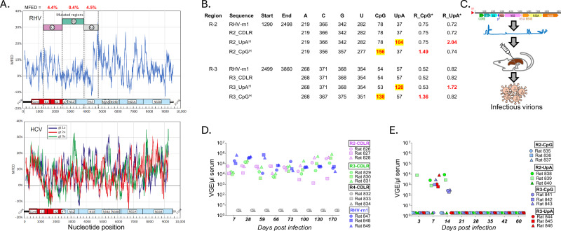
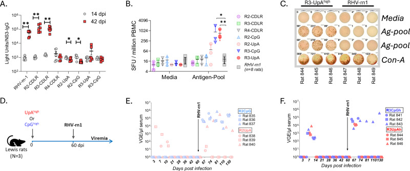
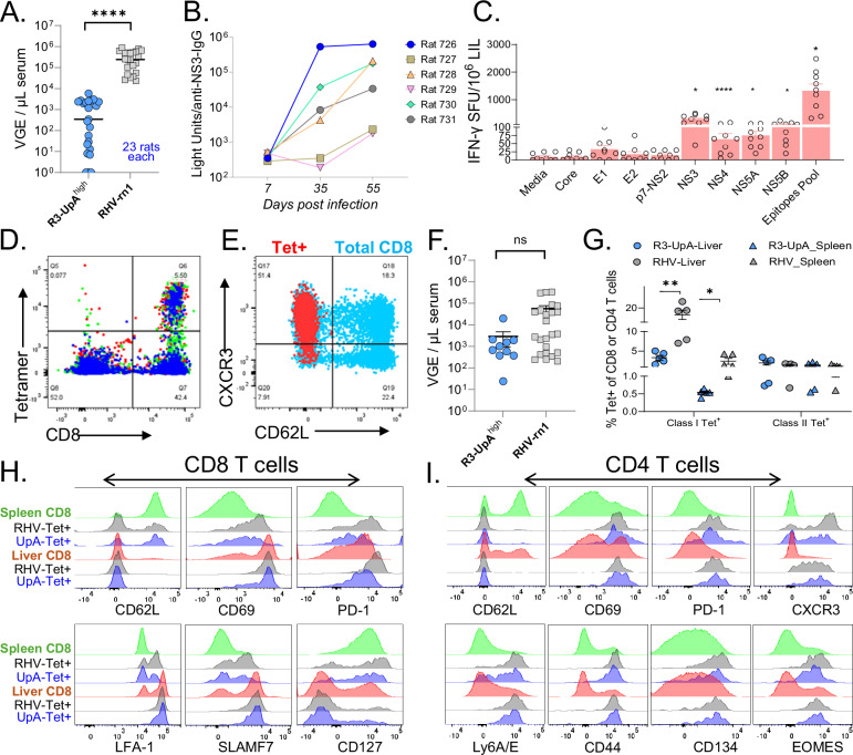
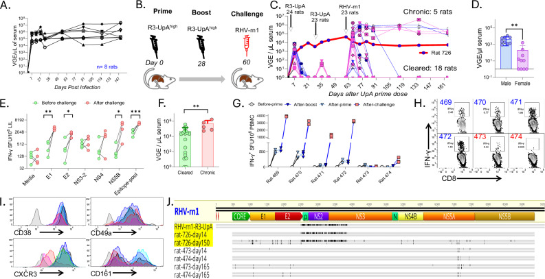
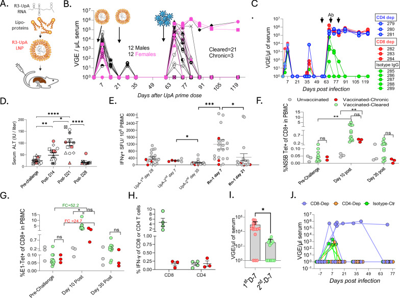
An effective vaccine for the hepatitis C virus (HCV) remains an unmet medical need. There is no animal model for assessing HCV vaccines; however, rodent hepacivirus (RHV) infection in laboratory rats recapitulates the lifelong chronic hepatotropic infection and immune evasion of HCV. Here, we designed a live-attenuated vaccine (LAV) for RHV and determined its immunogenicity and efficacy for preventing chronic infection. The LAV strains are generated by synonymous mutagenesis to increase the frequencies of naturally suppressed dinucleotides, UpA or CpG, in genomic regions that lack extensive RNA secondary structures. Rats vaccinated using LAV containing infectious virions (LAV-IV), or lipid nanoparticle-encapsulated viral RNA (LNP-vRNA) developed short-term viremia and robust T cell responses. After challenge with RHV-rn1, while all unvaccinated rats developed chronic infection, 75% and 85% of rats vaccinated with LAV-IV and LAV-vRNA cleared the infection. Clearance of RHV-rn1 was associated with expansion of memory T cells, transient rise in serum ALT, and, more importantly, enhanced protection against reinfection. In conclusion, we identified a genomic region of hepacivirus that can be synonymously mutated to attenuate its persistence, and vaccines based on these modified genomes protect against chronic hepacivirus infection, a strategy with an apparent translational path toward HCV immunization.


Conflict of interest statement

Competing interests: The authors declare no competing interests.

Figures

Fig. 1
Fig. 1. Design of RHV mutants evaluated as LAV vaccine candidates and their infection outcomes in rats.
A The minimum folding energy differences (MFED) in the RHV-rn1 genome were plotted as the blue line in the upper panel and for different HCV genotypes in the lower panel. MFED values were calculated for consecutive 240 base fragments, incrementing by 15 bases across the genome. MFED values were calculated by subtraction of the MFE of the native sequence from the mean value of the same sequence scrambled in base order while maintaining native dinucleotide frequencies. R2-4 depicts the genomic regions selected for mutagenesis, and their MFED values are shown in red fonts. B Table shows mononucleotide and dinucleotide compositions of wild-type (RHV-rn1), CDLR, and UpA- and CpG-maximized sequences in R-2 and R-3 regions. The yellow highlighted numbers indicate the final number of dinucleotides in the R-2 and R-3 regions of the UpA and CpG mutants. *Ratio of observed frequencies of CpG or UpA frequencies to expected values based on mononucleotide composition. C Schematics of in vivo rescue of infectious virus from mutated genomes using intrahepatic injection of transcribed RNA in rats. Each rat was injected with 10 μg viral RNA in PBS. D Course of viremia of CDLR mutants and wild-type RHV-rn1. Note that the CDLR-4 mutant failed to produce consistent and high-titer viremia observed in R2 and R-3 CDLR mutants. E Course of viremia in rats injected with R-2 and R-3 variants with elevated UpA or CpG dinucleotide frequencies. Source data are provided as a Source Data file.
Fig. 2
Fig. 2. Nature of immunity and protection conferred by RHV-rn1 and mutant viruses.
A Anti-NS3 IgG titers in the serum of rats infected with RHV-rn1 and different mutants (n = 6). Data are presented as individual values and mean ± SEM. Two-tailed paired t-test was used for analysis, and p-values <  0.01 or 0.05 were considered statistically significant; p-values between day14 and day42 for RHV-rn-1 (**, 0.002), R2-CDLR (**, 0.01), R3-CDLR (**, 0.002), R2-UpA (*, 0.034), and R2-CpG (*, 0.035). Sequences of R2 and R3 regions of RHV-rn1 and mutants are available as Supplementary Information S3. B Spot forming units (SFU) indicating the IFN-γ secreting T cells in ELISPOT assay in PBMC stimulated with a pool of peptides representing the RHV-rn1 T cell epitopes. PBMC were collected on 28-31 dpi. SFU in the antigen pool of mutants was compared to RHV-rn1 (n = 8 rats) using a two-tailed unpaired t-test, and p-values for R3-CpG (*, 0.0285) and R3-UpA (**, 0.001) as compared to RHV-rn1 in Antigen-Pool. Data points are presented as individual values and error bar at mean ± SD. C Image of ELIPOST assay showing the SFU counts in rats infected with R3-UpAhigh and RHV-rn1 at 28 dpi. D Outline of vaccination and challenge studies. E, F Rats that cleared the R-2 and R-3 mutants were challenged with wild-type RHV-rn1 and followed for viremia for >2 months post-infection. Source data are provided as a Source Data file.
Fig. 3
Fig. 3. Nature of viremia, antibody and antigen-specific T cells induced by R3-UpA mutant in rats and mice.
A Viremia in R3-UpAhigh and RHV-rn1 infected rats (23 rats in each group) on 7 dpi. RHV-rn1 viremia data were compiled from our other studies using the same viral stock and from 8 rats used as controls in this study. Two-sided unpaired t-test was performed; p-value (****,  <0.0001). B Serum anti-NS3 IgG antibody titers after R3-UpAhigh infection in six vaccinated rats. C Specificities of antigen-specific T cells isolated from the liver of R3-UpAhigh infected rats (n = 3) at 14 dpi, three animals with their 3 replicates were shown as dots. Unpaired t-test and p-values between Media and NS3 (*, 0.0269), NS4 (***, 0.0006), NS5A (*, 0.0403), NS5B (*, 0.0429), Epitopes-Pool (*, 0.0358) respectively. Data points are presented as individual values and error bar at mean ± SEM. D Rat MHC class I tetramer incorporating an NS5B peptide was used to determine the frequencies of virus-specific CD8 T cells in the livers of R3-UpA vaccinated rats euthanized on 14 dpi. Representative data from three rats were shown using different colors. E Representative data showing the difference in phenotype of vaccine-primed memory CD8 T cells specific for NS5B peptide (red) and total CD8 (blue) T cells in the liver of vaccinated rats. F Viremia in R3-UpAhigh (n = 10) and RHV-rn1 infected mice on day 7 pi. Two-tailed unpaired t-test with p-value = (ns,0.1222). Data points were shown with mean ± SEM. G Frequencies of mouse MHC class I and class II specific CD8 and CD4 T cells in the liver (circle) and spleen (triangle) in R3-UpA and RHV-rn1 cleared mice (n = 5) on 45 dpi. Two-tailed unpaired t-test for Class I Tet+ response between R3-UpA Liver and RHV -Liver (**, 0.0084) and between R3-UpA Spleen and RHV-Spleen (*, 0.0356). Data points are presented as individual values and error bars at mean ± SEM. H Comparative analysis of total CD8 T cells in the liver and spleen with antigen-specific memory CD8 T cells in R3-UpA and RHV mice on 45 dpi. I Comparative analysis of total CD4 T cells in the liver and spleen with antigen-specific memory CD4 T cells in R3-UpA and RHV mice on 45 dpi. H and I use the same color scheme. Source data are provided as a Source Data file.
Fig. 4
Fig. 4. Infection, immunity, and protection of LAV-IV vaccine.
A Course of viremia in 8 RHV-rn1 infected naïve rats. B Experimental outline and timeline of vaccination and challenge study. C Course of viremia after R3-UpAhigh prime and booster vaccination and RHV-rn1 challenge infection in 12 male (blue) and 12 female (pink) rats. In vivo rescued R3-UpAhigh virus was used for vaccination. Rat-726 (male) developed persistent viremia of the R3-UpA virus, shown as a blue circle and red connecting lines. D Comparison of R3-UpA viremia after prime dose in male (n = 9) and female (n = 10) rats at day 7 pi. Two-tailed unpaired t-test between Male and Female group, p-value = (**, 0.0024). Data points are presented as individual values and error bars at mean ± SD. E Comparison of virus-specific T cell frequencies targeting different viral proteins before and after 14 days of RHV-rn1 challenge in 4 vaccinated rats. The frequencies of IFN-γ secreting T cells, shown as SFU, in ELISPOT assay in LILs stimulated with a pool of peptides representing the RHV-rn1 T cell epitopes. The mean value of SFU counts in triplicate wells is shown. Two-tailed paired t-test- before and after challenge for each peptide pool. P-values for E1 (**, 0.006), E2 (*, 0.0106), NS5B (*, 0.036) and Epitope-Pool (***, 0.0001). F Viremia on day 7 after RHV-rn1 challenge in rats that subsequently cleared the infection (n = 18) or remained chronic (n = 5). Two-tailed unpaired t-test; p-value (**, 0.0066). Data points are presented as individual values and error bars at mean ± SD. G Frequencies of virus-specific IFN-γ + T cells in PBMC of rats, before and after RHV-rn1 challenge, showing their significant expansion after challenge, on 9 dpi. Rat 473 and 474 developed chronic RHV-rn1 infection. H Frequencies of CD8 T cells producing IFN-γ after antigen stimulation in the liver of cleared (blue font) and chronic (red font) rats after 100 dpi. I Surface expression of various CD8 T cell markers shown as Fluorescence minus one (FMO) control (grey) and rat MHC class I Tetramer+ CD8 T cells in the liver of cleared (shades of blue) and chronic (shades of red) rats. J Sequence alignments of RHV-rn1 and R3-UpA genomes recovered from rats that developed chronic infection. The parallel grey lines show nucleotide (upper) and amino acid (lower) mutations (black marks) compared to the RHV-rn-1 genome. Alignment was generated using a consensus sequence derived from >200 X coverage of each nucleotide position, and the recognized base was present in >75% of sequence reads. The sequences are submitted to GenBank (PV639513-PV639519). Source data are provided as a Source Data file.
Fig. 5
Fig. 5. Infection, immunity, and protection of LNP-vRNA vaccine.
A Composition of R3-UpA vRNA-LNP vaccine. B Timeline of vaccination and challenge studies, and course of viremia in vaccinated rats. C Vaccinated rats were transiently depleted for CD8α + (n = 3) or CD4 (n = 3) T cells by antibody (Ab) or isotype control (n = 5) before challenge with RHV-rn1 to assess the role of T cell subsets in vaccine-induced immunity. D Serial analysis of serum ALT values in 12 vaccinated rats before and after RHV-rn1 challenge. Rats that developed chronic infection are shown in red-filled shapes. Two-tailed unpaired t-test and p-value for Pre-Challenge vs Post D14 (**,0.0047), Pre-Challenge vs Post-D21 (****, <0.0001), Post-D14 vs Post-D21, (*,0.0242) and Post-D21 vs Post-D28 (****, < 0.0001). Data points are presented as individual values and error bars at mean ± SEM. E Serial analysis of virus-specific T cells in PBMC of rats using IFN-γ ELISPOT assay. The frequencies of IFN-γ secreting T cells, shown as SFU, in ELISPOT assay in LILs stimulated with a pool of peptides representing the RHV T cell epitopes. The mean value of SFU counts in triplicate wells is shown. The number of animals whose PBMC samples were available for analysis is shown in parentheses; UpA 1st day28 (n = 21), UpA 2nd day7 (n = 6), UpA 2nd day35 (n = 21), Rn-1 day 7 (n = 20), and Rn-1 day21 (n = 20). Two-tailed unpaired t-test and p-value between UpA 2nd day 7 vs UpA 2nd day 35 (*, 0.0284); UpA-2nd day 35 vs Rn-1 day 7 (***, 0.0006); Rn-1 day 7 vs Rn-1 day 21 (*, 0.0327). F, G Serial analysis of the frequencies of NS5B2511- and E1191-specific CD8 T cells in the PBMC of unvaccinated (grey) and vaccinated rats that cleared (green) or developed chronic the RHV-rn1 challenge infection (red). FC=folds change. Two-tailed unpaired t-test and p-values between pre-challenge and day 10 post for cleared (**, 0.0081); and between unvaccinated and chronic (**, 0.0046). In Fig. 5F, p-values for Pre-challenge, Day 10 post, and Day 35 post (cleared vs chronic) were ns (0.5562, 0.8372, and 0.1492, respectively). In Fig. 5G p-values for Pre-challenge, Day 10 post, and Day 35 post (cleared vs chronic) were ns (0.8372, 0.1740, and 0.1581, respectively). H Frequencies of cells producing IFN-γ in intracellular cytokine production assay (ICS) of liver infiltrating leukocytes isolated from vaccinated rats with cleared (n = 4, green) and chronic (n = 3, red) RHV-rn1 infection. For ICS, cells were stimulated for 5-h with pools of peptides representing RHV T cell epitopes at 10 µg/mL concentration. Data points are presented as individual values and error bars at mean ± SEM. P-values for % IFN-γ of CD8 and CD4 between cleared and chronic were ns (0.0665,0.8703, respectively). I Viremia titers on day 7 after the first (n = 14) and second (n = 12) RHV-rn-1 challenge infection. Two-tailed unpaired t-test was performed, and a significant p-value was shown as *(0.0371). Data points are presented as individual values and error bars at mean ± SD. J Protective immunity against reinfection in rats that cleared the primary RHV-rn1 challenge infection. Viremia after RHV-rn1 reinfection in IgG-isotype control (green, n = 7), anti-CD8α (blue, n = 4), and anti-CD4 (brown, n = 4) cell-depletion antibody-treated rats. Source data are provided as a Source Data file.

Similar articles

References

    1. Houghton, M. The long and winding road leading to the identification of the hepatitis C virus. J. Hepatol.51, 939–948 (2009). - PubMed
    1. Ghany, M. G. et al. The 2020 Nobel Prize for medicine or physiology for the discovery of Hepatitis C Virus: A triumph of curiosity and persistence. Hepatology74, 2813–2823 (2021). - PMC - PubMed
    1. Liang, T. J., Feld, J. J., Cox, A. L. & Rice, C. M. Controlled human infection model - fast track to HCV Vaccine?. N. Engl. J. Med.385, 1235–1240 (2021). - PMC - PubMed
    1. Hartlage, A. S. & Kapoor, A. Hepatitis C Virus vaccine research: time to put up or shut up. Viruses13, 10.3390/v13081596 (2021). - PMC - PubMed
    1. Bailey, J. R., Barnes, E. & Cox, A. L. Approaches, progress, and challenges to Hepatitis C vaccine development. Gastroenterology156, 418–430 (2019). - PMC - PubMed

LinkOut - more resources