. 2025 Aug 15;16(1):7620.
doi: 10.1038/s41467-025-62775-x.

Functionally deficient UBOX5 variants and primary angle-closure glaucoma

Zheng Li #  1 Wee Ling Chng #  1   2 Zhehao Liu #  1   2 Tan Do #  3 Masakazu Nakano #  4 Li Jia Chen #  5 Yunhua Loo  2   6 Anita S Y Chan  2   6 Fotis Topouzis  7 Monisha E Nongpiur  2   6 Mineo Ozaki  2   6   8 Satoko Nakano  9 Toshiaki Kubota  9 Shamira A Perera  2   6 Rahat Husain  2   6 Tina T L Wong  2   6 Ching-Yu Cheng  2   6 Ching Lin Ho  2   6 Khaled Abu-Amero  10 Hon-Tym Wong  11 Mônica Barbosa de Melo  12 Nguyen Do Thi Ngoc Hien  3 Nguyen Van Trinh  3 Nguyen Thi Thanh Huong  3 Yaakub Azhany  13 Rodolfo Perez-Grossmann  14 Poemen Pm Chan  5 Kelsey V Stuart  15 Mahantesh I Biradar  15 Anita Szabo  15 Eleftherios Anastasopoulos  7 Dimitrios A Giannoulis  7 Panagiota Ntonti  7 Evangelia Papakonstantinou  7 Alexandros Lambropoulos  16 Anthoula Chatzikyriakidou  16 Vassilis Kilintzis  7 Humaira Ayub  17 Shazia Micheal  18 Yee Yee Aung  19 Edgar U Leuenberger  20   21 Antonio Fea  22 Naing Naing Mon  2 Amihan Anajao  23 Xuezhi Bi  2   23 Yee Jiun Kok  23 Rachel S Chong  2   6 Pui-Yi Boey  2   6 Darrell Zi Jing Tan  1 Wendy Wan Ling Sin  2 Balram Chowbay  24 Chaw Chaw Khaing  25 Yin Mon Aung  26   27 Rigo Daniel Reyes  28 Evangelia S Panagiotou  7 Dimitrios G Mikropoulos  7 Irini C Voudouragkaki  7 Georgios D Panos  7   29 Zhicheng Xie  1 Xiao Yin Chen  1 Yi Ting Lim  1 Wee Yang Meah  1 Ying Shi Lee  6 Candice Ee Hua Ho  6 Pearlyn Mei Xin Yeo  2 Yoko Ikeda  30 Yuichi Tokuda  4 Masami Tanaka  4 Natsue Omi  4 Morio Ueno  30 José P C de Vasconcellos  31 Vital P Costa  31 Ricardo Y Abe  31 Bruno B de Souza  12 Guillermo B Fong  32 Vania V Castro  33 Ricardo Fujita  34 Maria L Guevara-Fujita  34 Farah Akhtar  35 Mahmood Ali  35 Mary Ann T Catacutan  20 Irene R Felarca  20 Chona S Liao  20 Carlo Lavia  22 Hlaing May Than  36 Khin Thida Oo  26 Phyu P Soe-Kyaw  26 Paolo Frezzotti  37 Francesca Pasutto  38 Raquel Quino  28   39 Zaw Minn-Din  26 Nay Lin Oo  25 Laura Dallorto  22 Saw Htoo Set  19 Vi Huyen Doan  40 Raheel Qamar  41   42 Jamil Miguel Neto  31 Saleh Al-Obeidan  43 Clement C Tham  5 Kazuhiko Mori  30 Chie Sotozono  30 Shigeru Kinoshita  44 Anastasios G Konstas  7 Ahmad Tajudin Liza-Sharmini  13 Juan C Zenteno  45   46 Nhu Hon Do  3 Paul J Foster  15 Kei Tashiro  4 Chi Pui Pang  5 Anthony P Khawaja  15 Tin Aung  2   6 Zhenxun Wang  47   48   49 Chiea Chuen Khor  50
Affiliations

Functionally deficient UBOX5 variants and primary angle-closure glaucoma

Zheng Li et al. Nat Commun. .

Abstract

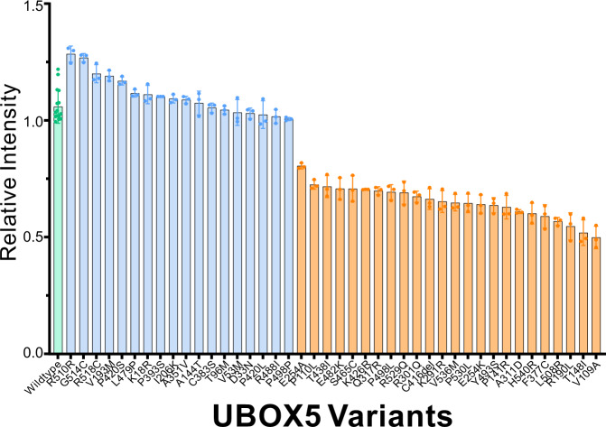
Primary angle-closure glaucoma is a major cause of irreversible blindness worldwide afflicting >20 million people. Through whole exome sequencing, we analysed the association between gene-based burden of rare, protein-altering genetic variants and disease risk in 4,667 affected individuals and 5,473 unaffected controls. We tested genes surpassing exome-wide significance (P < 2.5 × 10⁻⁶) for replication in a further 2,519 cases and 472,189 controls. We observed carriers of rare, protein-altering variants at UBOX5 (observed in 154 out of 7,186 affected individuals [2.1%] and in 3,975 out of 477,197 unaffected controls [0.83%]) to be associated with 2.13-fold increased risk of PACG (95%ci, 1.69 - 2.69; P = 1.25 × 10⁻¹⁰). We performed substrate trapping assays coupled with mass spectrometry and observed Binding Immunoglobulin Protein (BIP) as a key substrate for UBOX5. Biological assays showed UBOX5 acts by ubiquitinating BIP. We evaluated the functional status of 35 UBOX5 variants and observed that functionally deficient variants were enriched in affected individuals compared to controls. We validated this finding in an independent collection where 3 persons carrying functionally deficient variants were observed out of 208 cases (1.4%), whereas none were observed in 600 controls. Our findings suggest the UBOX5-BIP signalling pathway might be involved in biology of primary angle-closure glaucoma.


Conflict of interest statement

Competing interests: A.P.K has acted as a paid consultant or lecturer to Abbvie, Aerie, Allergan, Google Health, Heidelberg Engineering, Novartis, Reichert, Santen, Thea and Topcon. The remaining authors declare no competing interests.

Figures

Fig. 1
Fig. 1. UBOX5 is associated with increased risk of primary angle-closure glaucoma (PACG).
Cochran Mantel-Haenszel stratified meta-analysis was used to calculate an overall odds ratio for the study cohorts in the discovery (a) and validation (b) stage. Data are presented as Odds Ratios with accompanying 95 percent confidence intervals. P-values are two-sided, with no adjustments for multiple comparisons. A Forest plots for the discovery exome sequencing stage (comprising 4,667 persons with PACG and 5,473 unaffected individuals enrolled from 4 sites). Primary analysis describing the association between carriage of rare, protein-altering variants at UBOX5 and risk of PACG are shown together with post-hoc analyses stratifying by sex, as well as by presence of acute primary angle closure. B Forest plot from the primary analysis of the validation stage (comprising 2,519 affected individuals and 471,724 unaffected individuals enrolled from 10 sites).
Fig. 2
Fig. 2. UBOX5 has E3 ubiquitin ligase activity.
a A UBOX5 chimeric construct (UBOX5-UBD) where the UBOX5 open reading frame was fused by a flexible linker to a FLAG-tagged ubiquitin binding domain was generated. FLAG-tagged UBOX5 (3 lanes on the right) or FLAG-tagged UBOX5-UBD construct (4 lanes on the left) and HA-tagged ubiquitin was co-transfected into HEK293 cells. After the first immunoprecipitation of lysates by anti-FLAG antibody, 20% of eluates was kept for analysis. Eluate from the UBOX5-UBD was further immunoprecipitated with anti-HA antibody to enrich for ubiquitinated proteins. To serve as antibody specificity control (negative controls), lysates were mock immunoprecipitated with mouse immunoglobulin. Immunoblotting of BIP was performed on inputs and eluates as indicated. Bands corresponding to ubiquitinated BIP and ubiquitinated UBOX5-UBD chimeric protein are indicated by vertical lines, while the unmodified proteins are indicated by arrows on the right. Positions of the molecular weight markers are indicated by arrows on the left. IB: FLAG and IB:HA are shown in Supplementary Fig. 13. The experiment was repeated independently 3 times. Source data are provided as a Source data file. b MYC-tagged BIP, empty vector, UBOX5, or HA-tagged ubiquitin was co-transfected into HEK293 cells in the indicated combinations. 24 h later, cells were treated with 0.7 uM Tharpsigargin for 16 h, and then further treated with MG132 (a proteasomal inhibitor) for 6 h as indicated. Cells were then harvested and a MYC immunoprecipitation was performed on the input lysates. Eluates were immunoblotted with antibodies against HA to assess the extent of BIP ubiquitination. The membrane was then stripped and a MYC immunoblot was performed to assess immunoprecipitation efficiency. Ubiquitination of BIP was only observed when UBOX5 was expressed. The degree of ubiquitination of BIP did not appear to differ with the addition of MG132, a proteasomal inhibitor, suggesting that the ubiquitinated BIP was not degraded by the proteasome pathway. The experiment was repeated independently 3 times. Source data are provided as a Source data file. (c) UBOX5 or its empty vector contains a GFP open reading frame, which allows for assessment of transfection efficiency by assessing GFP abundance in input lysates. Human UBOX5 immunoblots were used to verify expression of UBOX5. GAPDH was used as loading control. The experiment was repeated independently 3 times. Source data are provided as a Source data file.
Fig. 3
Fig. 3. The biological properties of UBOX5.
a, b UBOX5 and BIP are both induced by ER stress. a NIH3T3 cells were treated with the indicated ER stress inducers tunicamycin (Tu) and thapsigargin (Tg). (Upper) Endogenous UBOX5 mRNA abundance was quantified by qPCR in biological triplicates, normalised against mouse beta-actin transcript. Relative fold change of transcript is reported against control DMSO treatment. Error bars represent standard deviation. (b, Top) Protein abundance of endogenous mouse  UBOX5 in ER-stressed NIH3T3 cells. Beta-actin was used as loading control. Of note, UBOX5 mRNA and protein is induced in response to ER stress. (b, Bottom): Densitometric quantitation of UBOX5 bands. UBOX5 band intensity of each sample is normalised to corresponding beta-actin intensity. The amount of TG or TU used is indicated on the x-axis. P-values were generated from two-sided Welch’s t-test. Error bars represent standard deviation. Source data are provided as a Source data file. c Cellular Localization of UBOX5: Immunoblots of HEK293 cells transiently transfected with UBOX5 expression plasmid and treated with 0.7 uM Thapsigargin for 16 h. Cellular fractions are indicated above. Whole cell lysates were fractionated into cytoplasmic, endoplastic reticulum (ER) fractions and nuclear fractions by stepwise centrifugation. Indicated antibodies are shown. Positions of molecular weight markers are indicated on the left with arrows. GFP is used as transfection control, Calnexin is used as fractionation control for ER and nuclear fraction; Histone H2B is used as fractionation control for nuclear fraction. GAPDH is used as fractionation control for cytoplasmic and nuclear fractions. The experiment was repeated independently 3 times. Source data are provided as a Source data file. d, e The ability of UBOX5 to ubiquitinate BIP is not dependent on cellular stress. d MYC-tagged BIP, empty vector, UBOX5, or HA-tagged ubiquitin was co-transfected into HEK293 cells in the indicated combinations. 24 hours later, cells were treated with 0.7 μM Thapsigargin (TG) or DMSO for 16 h. TG is a known inducer of endoplasmic reticulum stress. Cells were then harvested and a MYC immunoprecipitation was performed on the input lysates. Eluates were immunoblotted with antibodies against HA to assess the extent of BIP ubiquitination. e UBOX5 or its empty vector contains a GFP open reading frame, which allows for assessment of transfection efficiency by assessing GFP abundance in input lysates. GAPDH was used as loading control.
Fig. 4
Fig. 4. Gel quantitation data measuring the functional activity of UBOX5 protein-altering variants to ubiquitinate BIP.
Data are presented in biological triplicates. Intensity of the HA band was quantified and then normalized to the MYC signal of the same elute sample in the same gel. MYC signal was obtained after stripping the initial HA immunoblot and re-probing with MYC antibody. Wild-type UBOX5 (green bar) was measured independently 15 times to provide a confidence estimate of wild-type UBOX5 activity, so that functionally deficient UBOX5 variants (defined as <80% activity of wild-type UBOX5) can be more confidently identified. Variants in blue bars represent normally functioning UBOX5 alleles. A total of 42 UBOX5 variants were tested, comprising 35 variants detected in the initial study, and 7 variants in the validation study from Italy and Pakistan. Representative Western Blot gel photographs are presented as Supplementary Fig. 7. Error bars represent standard deviation. Source data are provided as a Source data file.
Fig. 5
Fig. 5. UBOX5 affects BIP half-life.
a Wild-type UBOX5 increases the half-life of BIP in the presence of thapsigargin-induced ER stress. Top left panel: Pulse chase of BIP with or without expression of UBOX5 in HEK293 cells. HEK 293 cells were transfected with UBOX5 or empty vector. 24 h later, cells were treated with a pulse of 0.25 μM Tharpsigargin for 2 hours, and cycloheximde (150 mM) was added. Cells were collected at indicated time points for immunoblotting. GAPDH was used as loading control. Positions of molecular weight standards are indicated on the left. Top right panel: The same experiment repeated with Tharpsigargin substituted with DMSO carrier control. Bottom panels: Quantitation of BIP band intensities normalized to GAPDH intensity for every indicated time point. Separate graphs were shown for cells treated with Tharpsigargin (Left) or DMSO control (Right). Intensities are shown as fold changes compared to normalized BIP intensity at t = 0 of pulse chase. Data are presented as mean values +/- standard deviation. Error bars indicate standard deviation; 3 biological replicates were used for quantitation. Source data are provided as a Source data file. b Variant UBOX5 and their effects on the half-life of BIP. Top: Pulse chase of BIP in the presence of wildtype UBOX5 or variant UBOX5 (D33N, K291R, R301Q, and S465C) in HEK293 cells. HEK 293 cells were transfected with wildtype UBOX or indicated variants. 24 h later, cells were treated with a pulse of 0.25 μM Tharpsigargin for 2 h, and cycloheximde (150 mM) was added at time = 0. Cells were then subsequently collected at indicated time points (in hours) for immunoblotting. GAPDH was used as loading control. UBOX5 expression was verified as indicated. GFP, expressed from a separate locus in the vector used, was also used to verify success of transfection. Bottom: Densitometric quantitation of BIP band intensities in cells expressing indicated UBOX5 variants, normalized to GAPDH intensity for every time point on the same blot. Intensities are shown as fold changes compared to normalized BIP intensity at t = 0 of pulse chase. Three biological replicates were analyzed. Error bars represent standard deviation. Source data are provided as a Source data file.

References

    1. Zhang, N., Wang, J., Chen, B., Li, Y. & Jiang, B. Prevalence of primary angle closure glaucoma in the last 20 years: a meta-analysis and systematic review. Front Med. (Lausanne)7, 624179 (2020). - PMC - PubMed
    1. Quigley, H. A. & Broman, A. T. The number of people with glaucoma worldwide in 2010 and 2020. Br. J. Ophthalmol.90, 262–267 (2006). - PMC - PubMed
    1. George, R., Panda, S. & Vijaya, L. Blindness in glaucoma: primary open-angle glaucoma versus primary angle-closure glaucoma-a meta-analysis. Eye36, 2099–2105 (2022). - PMC - PubMed
    1. Ahram, D. F., Alward, W. L. & Kuehn, M. H. The genetic mechanisms of primary angle closure glaucoma. Eye29, 1251–1259 (2015). - PMC - PubMed
    1. Waseem, N. H. et al. Mutations in SPATA13/ASEF2 cause primary angle closure glaucoma. PLoS Genet.16, e1008721 (2020). - PMC - PubMed

LinkOut - more resources