Clinical Trial
. 2025 Sep;31(9):2927-2935.
doi: 10.1038/s41591-025-03822-4. Epub 2025 Aug 15.

Dual-vector rAAVrh8 gene therapy for GM2 gangliosidosis: a phase 1/2 trial

Affiliations
Clinical Trial

Dual-vector rAAVrh8 gene therapy for GM2 gangliosidosis: a phase 1/2 trial

Florian Eichler et al. Nat Med. 2025 Sep.

Abstract

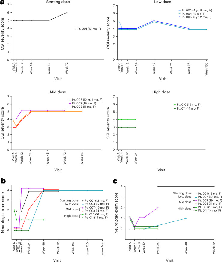
The dual rAAVrh8-HEXA and rAAVrh8-HEXB vector can restore central nervous system hexosaminidase (Hex) enzyme activity, decrease GM2 levels in cerebrospinal fluid and rescue phenotypic consequences of GM2 gangliosidosis, Tay-Sachs and Sandhoff diseases in animal models following simultaneous bi-thalamic (BiT) injections. Following up on an n = 2 expanded access trial, we initiated a phase 1/2, single-dose, dose-escalation of combined BiT, intra-cisterna magna and intrathecal infusion in children with Tay-Sachs and Sandhoff diseases (six infantile, three juvenile). The BiT injection volume and vector dose were doubled between four cohorts, with the lowest dose matching the earlier expanded access trial. Cerebrospinal fluid HexA enzyme activity, serum total Hex activity and GM2 levels showed a dose-dependent biochemical correction of the disease. Serum Hex activity surpassed 40 nmol h-1 ml-1, two times the lower limit of normal, and neuroimaging demonstrated increased fiber tracts. Correction was greatest at 12 weeks, but in decline by 24 weeks postdosing. Infantile patients experienced global clinical stabilization and prolonged oral feeding without aspiration until 3-3.5 years. Seizures had a later onset, were less frequent, less severe and more responsive to anti-convulsant medication. Adverse events were rare in infantile patients, but worsening dystonia was observed in juvenile patients, who were excluded from ongoing enrollment. ClinicalTrials.gov registration: NCT04669535 and NCT06614569 .


Conflict of interest statement

Competing interests: M.S.-E. may be entitled to receive licensing revenue from the patents covering the technologies used in this study. The remaining authors declare no competing interests.

Figures

Fig. 1
Fig. 1. Neuroimaging after BiT injection.
a, Immediate postoperation (postop) MRI depicting BiT injection of AAVrh8-HEXA and AAVrh8-HEXB (1,250 μl and 4.1 × 1013 vector genomes (vg) per thalamus). MRI without contrast showing T2-FLAIR hyperintensity in the thalamus at the injection site. The hyperintensity decreases over time (at 3 and 6 months). There is no evidence of hemorrhage or other major complication associated with the injection. b, Two patients demonstrated an increase in cortical T2 hypointensity, suggestive of myelin production, over the course of three months postinfusion. The effect was observed in the frontoparietal cortex, but not in the temporal cortex. c, DTI for five patients taken at baseline and 24 weeks after treatment showing neural tract fiber growth (green) and loss (red). d, Quantification of percent change in tract fiber numbers and volume from DTI data at 12 and 24 weeks posttreatment. DWI, diffusion-weighted imaging; F, female; mo, months, Pt., patient; SWI, susceptibility-weighted imaging.
Fig. 2
Fig. 2. Liver enzymes and specific gamma interferon ELISpots for capsid and transgene epitope libraries.
Normal ranges are indicated for liver enzymes in dotted lines. Positive and negative ELISpot responses are denoted by red and blue shading, respectively. Complete immune response data are presented in the companion publication. ALT, alanine transaminase; AST, aspartate aminotransferase; GGT, gamma-glutamyl transferase; IFNɣ, interferon-ɣ; M, male; NA, not applicable (no sample collected); yr, years.
Fig. 3
Fig. 3. Bioactivity of rAAVrh8-HexA and rAAVrh8-HexB in CNS and systemically.
a, HexA enzyme activity in CSF collected at the indicated time points. Analysis for HexA enzyme was only interpretable in patients with TSD. b, Total serum Hex levels collected at the indicated time points. The dashed line indicates normal Hex levels. c, C20:0 GM2 quantification by mass spectrometry. We note that juvenile patients have lower levels of GM2 as predicted by their disease subtype. d, CSF HexA activity levels from a generally correlated with GM2 20:0 levels.
Fig. 4
Fig. 4. Clinical assessment of patients over time.
a, CGI severity scores at each time point. Lower scores indicate better neurological status. b,c, Infantile patients’ neurologic exam scores for seizure severity (b) and sucking or swallowing (c). A higher score indicates worsened severity.
Extended Data Fig. 1
Extended Data Fig. 1. Diagram of study design.
The study was designed to test the safety and bioactivity of a single two-part delivery of rAAVrh8-HexA/HexB (bilateral thalamic plus CSF) with the dose escalation based on the volume of vector delivered by convection-enhanced delivery to the thalamus on each side. The Starting Dose was equivalent to that in the previously published Expanded Access study. The dose per thalamus ranged from 5.9E + 12 to 4.1E + 13 vg/thalamus and the total dose ranged from 1.42E + 14 to 3.6E + 14 vg/patient.
Extended Data Fig. 2
Extended Data Fig. 2. Thalamic infusion dose escalation ranged from starting dose (180 mcl) to high dose (1250 mcl).
Infusion accuracy was seen in thalami bilaterally as displayed with T2/Flair hyperintensity. There was no significant bleeding, as demonstrated by susceptibility-weighted imaging (SWI). There was no significant stroke, as demonstrated by DWI imaging, except for one clinically insignificant caudate area of ischemia immediately post-operation from cannula placement (bottom right).
Extended Data Fig. 3
Extended Data Fig. 3. Volumetric measurements.
Volumetric measurements are shown for the brain and ventricles in GM2 infantile patients (Patients 001, 004, 007, 008, 010, 011) in the first column.
Extended Data Fig. 4
Extended Data Fig. 4. DTI measurements.
DTI measurements of the lentiform nucleus are shown for the infantile patients (Patients 001, 004, 007, 008, 010, 011).
Extended Data Fig. 5
Extended Data Fig. 5. Cerebellar cortex volumes.
Cerebellar cortex volumes of GM2 patients at the time of treatment and subsequently compared to natural history data of GM2 patients.
Extended Data Fig. 6
Extended Data Fig. 6. Neurologic and developmental outcomes.
Clinical Global Impression severity and change from baseline are shown for each patient.

References

    1. Bley, A. E. et al. Natural history of infantile GM2 gangliosidosis. Pediatrics128, e1233–e1241 (2011). - PMC - PubMed
    1. Regier, D. S., Proia, R. L., D’Azzo, A. & Tifft, C. J. The GM1 and GM2 gangliosidoses: natural history and progress toward therapy. Pediatr. Endocrinol. Rev.13, 663–673 (2016). - PMC - PubMed
    1. Day, J. W. et al. Onasemnogene abeparvovec gene therapy for symptomatic infantile-onset spinal muscular atrophy in patients with two copies of SMN2 (STR1VE): an open-label, single-arm, multicentre, phase 3 trial. Lancet Neurol.20, 284–293 (2021). - PubMed
    1. Hwu, W.-L. et al. Gene therapy for aromatic l-amino acid decarboxylase deficiency. Sci. Transl. Med.4, 134ra61 (2012). - PubMed
    1. Pearson, T. S. et al. Gene therapy for aromatic L-amino acid decarboxylase deficiency by MR-guided direct delivery of AAV2-AADC to midbrain dopaminergic neurons. Nat. Commun.12, 4251 (2021). - PMC - PubMed

Publication types

Substances

Associated data