. 2025 Aug 16:271678X251369258.
doi: 10.1177/0271678X251369258. Online ahead of print.

Abnormal perinidal cerebral metabolism is associated with symptoms in brain arteriovenous malformation: New insights by a novel approach using oxygen-15 labelled tracers and PET

Affiliations

Abnormal perinidal cerebral metabolism is associated with symptoms in brain arteriovenous malformation: New insights by a novel approach using oxygen-15 labelled tracers and PET

Atsushi Hashio et al. J Cereb Blood Flow Metab. .

Abstract

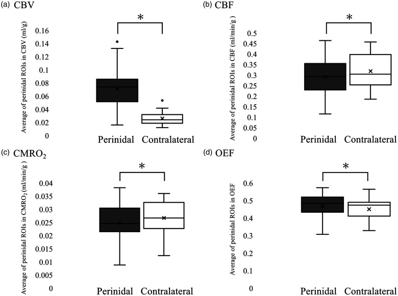
The mechanisms underlying neurological symptoms, including epilepsy, in unruptured brain arteriovenous malformations (bAVM) remain to be elucidated. The dual-tracer basis function method (DBFM) with single-scan dual-tracer (15O2 and C15O2) approach is a novel PET imaging technique that can evaluate the hemodynamics and metabolism of brain tissue adjacent to the nidus without the influence of abundant vascular radioactivity. We aimed to clarify the relationship between neurological symptoms and hemodynamic and metabolic abnormalities using the DBFM. Cerebral blood volume (CBV), cerebral blood flow (CBF), cerebral metabolic rate of oxygen (CMRO2), and oxygen extraction fraction (OEF) were compared between symptomatic and asymptomatic bAVMs and before and after treatment. Among 46 patients with unruptured bAVMs who underwent DBFM, 26 cases were included. While CBV and OEF increased in the perinidus (p < 0.01), CBF and CMRO2 decreased (p < 0.01). Symptoms were significantly associated with higher OEF (symptomatic vs. asymptomatic, ratio of perinidal to contralateral, n = 13 vs. n = 13, 1.07 ± 0.030 vs. 1.01 ± 0.080, p = 0.017). All elevated OEFs in symptomatic bAVMs with therapeutic symptom resolved in post-therapeutic PET imaging. DBFM revealed a higher OEF was associated with symptomatic bAVM. The DBFM potentially aids in predicting post-therapeutic symptom improvement.

Keywords: Brain arteriovenous malformation; dual-tracer basis function method; epilepsy; oxygen extraction fraction; positron emission tomography.


Conflict of interest statement

The author(s) declared no potential conflicts of interest with respect to the research, authorship, and/or publication of this article.

Figures

Figure 1.
Figure 1.
Region of interest (ROI) setting in magnetic resonance angiography (MRA). (a–c) An example of the ROI setting in MRA is shown in a case with left basal ganglia brain arteriovenous malformations (bAVM) (a: upper end of the nidus; (b) a slice with the largest diameter of the nidus; c: the lower end of the nidus). Circular ROIs, 2 cm2, are placed adjacent to the nidus––one in the slice at the upper end of the nidus and one on the opposite side (a), three in the slice at the maximum diameter of the nidus and three on the opposite side (b), one in the slice at the lower end of the nidus and one on the opposite side (c).
Figure 2.
Figure 2.
Comparison of the parameters between the ipsilateral and contralateral brain tissues. Comparison of the region of interest (ROI)-p and the ROI-c for each parameter––cerebral blood volume (CBV, a), cerebral blood flow (CBF, b), cerebral metabolic rate of oxygen (CMRO2, c), oxygen extraction fraction (OEF, d)––are shown. The average ROI adjacent to the nidus was defined as ROI-p, and the average ROI contralateral to the nidus was defined as ROI-c. Asterisks indicate statistically significant differences (p < 0.05).
Figure 3.
Figure 3.
Representative cases of symptomatic and asymptomatic brain arteriovenous malformations (bAVMs). Magnetic resonance angiography (MRA) and positron emission tomography (PET) qualitative images of symptomatic (a−e) and asymptomatic patients (f−j) are shown. (a) An MRA shows symptomatic bAVM in the right parietal lobe. (b−e) PET images of cerebral blood volume (CBV, b), cerebral blood flow (CBF, c), cerebral metabolic rate of oxygen (CMRO2, d), and oxygen extraction fraction (OEF, e) are shown. The red arrow indicates elevated OEF levels in the perinidal brain tissue. (f) An MRA reveals an asymptomatic bAVM in the left occipital lobe. (g−j) PET images of CBV (g), CBF (H), CMRO2 (i), and OEF (j) are shown. For quantification, please refer to Method.
Figure 4.
Figure 4.
Comparison of the region of interest (ROI)-p/c ratio between the asymptomatic and symptomatic bAVMs. A comparison of the ratio of ROI-p to ROI-c between the asymptomatic (white) and symptomatic (black) groups for each parameter is shown. (a) cerebral blood volume (CBV); (b) cerebral blood flow (CBF); (c) cerebral metabolic rate of oxygen (CMRO2); and (d) oxygen extraction fraction (OEF). The average of the ROI adjacent to the nidus is defined as ROI-p, and the average is contralateral to the nidus as ROI-c. The asterisk indicates a significant statistical difference of p < 0.05.
Figure 5.
Figure 5.
A representative case of symptomatic brain arteriovenous malformations (bAVMs) treated by gamma knife surgery (GKS). Magnetic resonance angiography (MRA) and positron emission tomography (PET) qualitative images pre-GKS (a−e) and post-GKS (f−j) are shown. (a) An MRA reveals bAVM in the left temporal lobe. (b–e) PET images of cerebral blood volume (CBV, b), cerebral blood flow (CBF, c), cerebral metabolic rate of oxygen (CMRO2, d), and oxygen extraction fraction (OEF, e) before GKS are shown. The red arrow indicates elevated OEF levels in the perinidal brain tissue. f: Post-GKS MRA image is shown. (g–j) PET images of CBV (g), CBF (h), CMRO2 (i), and OEF (j) after GKS are shown. The white arrow indicates normalized OEF in the perinidal brain tissue.
Figure 6.
Figure 6.
Change in the region of interest (ROI)-p/c before and after gamma knife surgery (GKS) in patients with nidal occlusion. (a–d) Changes in the ratio of ROI-p to ROI-c before and after GKS in cerebral blood volume (CBV, a), cerebral blood flow (CBF, b), cerebral metabolic rate of oxygen (CMRO2, c), and oxygen extraction fraction (OEF, d). Symptomatic: solid lines; asymptomatic: dotted lines. The average of the ROI adjacent to the nidus is defined as ROI-p and the average of the ROI contralateral to the nidus as ROI-c.

References

    1. Lawton MT, Rutledge WC, Kim H, et al. Brain arteriovenous malformations. Nat Rev Dis Primers 2015; 1: 15008. - PubMed
    1. Choi JH, Mast H, Hartmann A, et al. Clinical and morphological determinants of focal neurological deficits in patients with unruptured brain arteriovenous malformation. J Neurol Sci 2009; 287: 126–130. - PMC - PubMed
    1. Okabe T, Meyer JS, Okayasu H, et al. Xenon-enhanced CT CBF measurements in cerebral AVM’s before and after excision. Contribution to pathogenesis and treatment. J Neurosurg 1983; 59: 21–31. - PubMed
    1. Homan RW, Devous MD, Stokely EM, et al. Quantification of intracerebral steal in patients with arteriovenous malformation. Arch Neurol 1986; 43: 779–785. - PubMed
    1. Kaminaga T, Hayashida K, Iwama T, et al. Hemodynamic changes around cerebral arteriovenous malformation before and after embolization measured with PET. J Neuroradiol 1999; 26: 236–241. - PubMed

LinkOut - more resources