Review
. 2025 Aug 26;44(8):116153.
doi: 10.1016/j.celrep.2025.116153. Epub 2025 Aug 14.

From thought to action: The organization of spinal projecting neurons

Affiliations
Review

From thought to action: The organization of spinal projecting neurons

Carla C Winter et al. Cell Rep. .

Abstract

Spinal projecting neurons (SPNs) are specialized neurons with cell bodies residing in the brain and axons extending into the spinal cord, providing a direct communication pathway that enables top-down control of nearly every bodily function. Disruptions to these pathways contribute to a wide range of neurological disorders, including developmental, degenerative, and traumatic pathologies. Advances in retrograde labeling, activity monitoring, and circuit manipulation have enabled increasingly precise and comprehensive characterizations of SPNs. Here, we provide a historical overview of brain-spinal cord connectivity research, followed by an in-depth synthesis of the current knowledge of SPN anatomical connections, molecular identities, and functional properties. We then propose a conceptual framework in which distinct SPN modules coordinately regulate motor, autonomic, and sensory processes to support bodily readiness and drive behavioral action. Beyond revealing the organizational logic of SPNs, these insights provide a foundation for designing therapies to restore brain-spinal cord communication following injury or disease.

Keywords: CP: Neuroscience; descending control; spinal cord; spinal cord injury.


Conflict of interest statement

Declaration of interests Z.H. is a co-founder of Rugen and Myrobalan and an advisor of Axonis.

Figures

Figure 1.
Figure 1.. Anatomy of brain-body communication
The brain commands the body via three primary routes. Left: the hypothalamic-pituitary-peripheral gland axis is a neuroendocrine system that allows the brain to directly influence, and be influenced by, peripheral targets. The hypothalamus, a part of the central nervous system, produces and releases hormones (e.g., CRH and TRH) into the pituitary portal circulation, which stimulate the release of hormones (e.g., ACTH and TSH) from the anterior pituitary gland. Anterior pituitary hormones are released into the systemic circulation and travel through the body’s circulation to impact distant organs (e.g., adrenal and thyroid). The hormones (e.g., cortisol and thyroid hormones) released by the peripheral glands can influence the hypothalamus and pituitary, creating a feedback loop that maintains appropriate hormone balances via bi-directional communication between the brain and the body. Arrows represent stimulation, and lines terminating in a vertical bar represent feedback inhibition. Right: the brain also directly influences bodily functions via the efferent somatic and autonomic motor pathways. The somatic motor pathway relays command signals from the brain to skeletal muscle. Here, spinal projecting neurons relay information from the brain, either via mono-synaptic or poly-synaptic connections, to somatic motor neurons throughout the spinal cord. The somatic motor neurons then relay cholinergic commands to peripheral skeletal muscle targets. The autonomic nervous system is broadly divided into two main pathways: the sympathetic and parasympathetic systems, which govern ‘‘fight or flight’’ or ‘‘rest and digest’’ bodily functions, respectively. The sympathetic nervous system is controlled by descending commands from spinal projecting neurons, which innervate SPGNs in the thoracolumbar spinal cord. These SPGNs send short cholinergic axons to synapse onto the sympathetic ganglia close to the central nervous system. The post-ganglionic neurons send long noradrenergic axons to peripheral targets. Some SPGNs directly innervate the adrenal medulla. The parasympathetic nervous system is controlled by descending signals that innervate PSPGNs in the brainstem or the sacral spinal cord. Both the brainstem and sacral PSPGNs send cholinergic axons to synapse with post-ganglionic neurons located in the ganglia close to their target organs, which in turn release ACh to modulate the function of the target organs. PSPGNs residing in the brainstem command peripheral targets via the motor component of the vagus nerve. CRH, corticotrophin releasing hormone; TRH, thyroid releasing hormone; ACTH adrenocorticotrophic hormone; TSH, thyroid-stimulating hormone; ACh, acetylcholine; Epi, epinephrine; Norepi, norepinephrine; SPGN, sympathetic preganglionic neuron; PSPGN, parasympathetic preganglionic neuron.
Figure 2.
Figure 2.. Approaches to defining spinal projecting neuron populations
Historical and more modern approaches have enabled the characterization of SPNs across several domains. Top: first and foremost, SPNs can be defined by their connectome. Specifically, identifying their points of origin, spinal terminations, and axonal collateralization can offer a significant foundation for their functional roles. Their points of origin are throughout the brain, with most abundant populations in the CTX, HY, MB, CB, and HB. Their spinal terminations are defined by which rostral-caudal segment and gray matter zone their axons terminate in the spinal cord. Rostral-caudal segments are broadly divided into cervical, thoracic, lumbar, and sacral levels. Within each segment, the spinal cord gray matter is divided into three main gray zones (i.e., dorsal, intermediate, and ventral), which can be further subdivided into 10 Rexed laminae. SPNs may also be characterized by their axon collaterals, with many populations projecting to multiple target regions, enabling simultaneous control over multiple downstream circuits. Middle: SPNs may also be classified according to their molecular makeup. Single-nucleus transcriptomics has enabled high-throughput molecular characterization of brain-wide SPNs (detailed further in Figure 3). Images adapted from Winter et al., 2023. Bottom: finally, many efforts have sought to classify SPNs based on their functional outputs. Neural activity is primarily measured using in vivo Ca2+ imaging and intracellular electrophysiology. Behavioral analyses assess the contribution of SPNs to sensory (e.g., von Frey mechanical sensitivity test), motor, and autonomic systems. SPN, spinal projecting neuron; CTX, cortex; MB, midbrain; CB, cerebellum; HY, hypothalamus; Med, medulla; HB, hindbrain; C, cervical; T, thoracic; L, lumbar; S, sacral; IML, intermediolateral nucleus; IMM, intermediomedial nucleus; cc, central canal; Str, striatum; APN, anterior pretectal area; Pn, pontine nuclei; GiV, gigantocellular reticular nucleus ventral part; CSN, corticospinal neuron; RuSN, rubrospinal neuron; CbSN, cerebellospinal neuron; ReSN, reticulospinal neuron; Ca2+, calcium.
Figure 3.
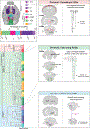
Figure 3.. A three-division framework for spinal projecting neuron organization
Synthesizing insights into the anatomical and transcriptomic identities of SPN populations yields a three-division framework for SPNs and informs how these neurons lead to diverse functional outputs. Left, top: adeno-associated virus-mediated retrograde labeling and whole-brain imaging of SPNs reveal that they arise throughout the brain, with most abundant concentrations in the CTX (M1M2S1, RFA, and S2), MED, PONS, MB, HY, and CB. The bar plot indicates the percentage contribution of each of the anatomically defined populations, rounded to the nearest digit. Left, bottom: single-nucleus transcriptomics of ∼65,000 retrogradely labeled SPNs across the whole brain revealed a hierarchical organization with 3 major divisions, 13 subclasses, and 76 cell types. The color blocks shading the taxonomy tree indicate division. The nodes at the end of the dendrogram indicate type, and the bar plots at far right indicate subclass. Right, top: division 1 of the SPN taxonomy corresponds to SPN types 1–7. Anatomically, MERFISH localizes these neurons to the L5 CTX, RN, and DCN (note: CSNs are in more restricted areas of L5 than indicated in the MERFISH results). These CSNs, RuSNs, and CbSNs each arise from discrete points of origin and have well-defined, somatotopically organized spinal projections. Molecularly, they are marked by relatively simple transcriptomes with discrete molecular markers and exclusively glutamatergic identity. These features suggest that they are suited for relaying commands to specific spinal circuits for ‘‘point-to-point’’ control of specific functions. Right, middle: division 2 corresponds to SPN types 8–56. MERFISH localizes these to sparse locations throughout the reticular formation. They comprise numerous transcriptionally defined cell types, have exceptionally complex transcriptomic expression patterns, and have glutamatergic, GABAergic, and glycinergic cell types. These anatomically and molecularly complex ReSNs with broad spinal termination patterns suggest that they are suited for relaying commands related to activities of the entire spinal cord, such as posture and autonomic functions. Right, bottom: division 3 corresponds to SPN types 57–76. MERFISH localizes these neurons to the hypothalamus (PVN and LHA), A11, EW, LC, and Ra. Molecularly, these neurons have unique neuro-modulatory properties, expressing slow-acting neurotransmitters (e.g., noradrenaline and serotonin) and/or neuropeptides. The release of these neuromodulators may allow these neurons to serve as a ‘‘gain setting’’ mechanism to amplify and/or prolong neural responses. For example, noradrenergic LC projections to the dorsal horn have been shown to decrease pain transmission. Images are adapted from Winter et al., 2023. RFA, rostral forelimb area; M1, primary motor cortex; M2; secondary motor cortex; S1, primary somatosensory cortex; S2, secondary somatosensory cortex; CB, cerebellum; MB, midbrain, MED, medulla; HY, hypothalamus; CSN, corticospinal neuron; RuSN, rubrospinal neuron; CbSN, cerebellospinal neuron; ReSN, reticulospinal neuron; L5, layer 5; CTX, cortex; RN, red nucleus; DCN, deep cerebellar nuclei; FL, forelimb; HL, hindlimb; C, cervical; T, thoracic; L, lumbar; S, sacral; RF, reticular formation; PVN, paraventricular nucleus of the hypothalamus; A11, cell group A11; LHA, lateral hypothalamic area; EW, Edinger-Westphal nucleus; LC, locus coeruleus; Ra, raphe nucleus; SPN, spinal projecting neuron.
Figure 4.
Figure 4.. SPN modules control complex behaviors
Spinal projecting neurons command complex bodily functions. Conceptually, these complex behaviors are the result of ‘‘modules’’ of motor, autonomic, and sensory functions, which, when executed together, generate unique behavioral outputs. These modules can be categorized into those supporting ‘‘readiness’’ and ‘‘action.’’ Top: ‘‘readiness’’ modules refer to a combination of motor, autonomic, and sensory states that prepares an organism to be able to perform a subsequent action. For example, an organism at rest has baseline muscle tone, sympathetic tone, and sensory sensitivity that prepares it to be able to perform a subsequent action. SPN populations (such as the rVMM, Zhang et al., Cell 2024) may control all three components of this readiness module. This readiness module is similar to an orchestra, with the conductor commanding the instrumentalists to be alert for the conductor’s subsequent cues to produce music. Bottom: ‘‘action’’ modules comprise a more complex set of motor, autonomic, and sensory functions that together generate complex behaviors like locomotion or handling. Action modules are likely the result of combinatorial recruitment of many different SPN populations throughout the brain. This is like a conductor who cues unique combinations of instrumentalists to produce distinct symphonies. SPN, spinal projecting neuron; rVMM, rostral ventro-medial medulla; FL, forelimb; HL, hindlimb; HR, heart rate; BP, blood pressure; RR, respiratory rate.

References

    1. Zheng B, and Tuszynski MH (2023). Regulation of axonal regeneration after mammalian spinal cord injury. Nat. Rev. Mol. Cell Biol 24, 396–413. 10.1038/S41580-022-00562-Y. - DOI - PubMed
    1. Lemon RN (2008). Descending Pathways in Motor Control. Annu. Rev. Neurosci 31, 195–218. 10.1146/annurev.neuro.31.060407.125547. - DOI - PubMed
    1. Blackstone C (2018). Converging cellular themes for the hereditary spastic paraplegias. Curr. Opin. Neurobiol 51, 139–146. 10.1016/j.conb.2018.04.025. - DOI - PMC - PubMed
    1. Ozdinler PH, Gautam M, Gozutok O, Konrad C, Manfredi G, Area Gomez E, Mitsumoto H, Erb ML, Tian Z, and Haase G (2020). Better understanding the neurobiology of primary lateral sclerosis. Amyotroph. Lateral Scler. Frontotemporal Degener. 21, 35–46. 10.1080/21678421.2020.1837175. - DOI - PMC - PubMed
    1. Dervishi I, and Ozdinler PH (2018). Incorporating upper motor neuron health in ALS drug discovery. Drug Discov. Today 23, 696–703. 10.1016/j.drudis.2018.01.027. - DOI - PMC - PubMed

LinkOut - more resources