. 2025 Aug 16;15(1):288.
doi: 10.1038/s41398-025-03506-0.

Neural transmission in the wired brain, new insights into an encoding-decoding-based neuronal communication model

Affiliations

Neural transmission in the wired brain, new insights into an encoding-decoding-based neuronal communication model

Sivan Kinreich. Transl Psychiatry. .

Abstract

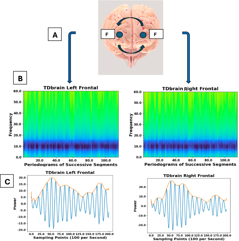
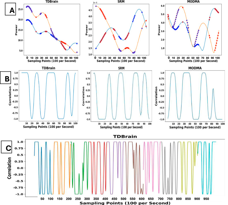
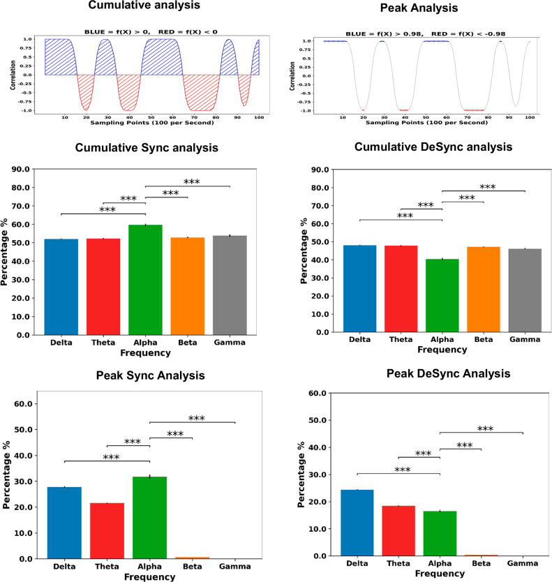
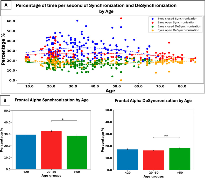
Brain activity is known to be rife with oscillatory activity in different frequencies, which are suggested to be associated with intra-brain communication. However, the specific role of frequencies in neuronal information transfer is still an open question. To this end, we utilized EEG resting state recordings from 5 public datasets. Overall, data from 1668 participants, including people with MDD, ADHD, OCD, Parkinson's, Schizophrenia, and healthy controls aged 5-89, were part of the study. We conducted a running window of Spearman correlation between the two frontal hemispheres' Alpha envelopes. The results of this analysis revealed a unique pattern of correlation states alternating between fully synchronized and desynchronized several times per second, likely due to the interference pattern between two signals of slightly different frequencies, also named "Beating". Subsequent analysis showed this unique pattern in every pair of ipsilateral/contralateral, across frequencies, either in eyes closed or open, and across all ages, underscoring its inherent significance. Biomarker analysis revealed significantly lower synchronization and higher desynchronization for people older than 50 compared to younger ones and lower ADHD desynchronization compared to age-matched controls. Importantly, we propose a new brain communication model in which frequency modulation creates a binary message encoded and decoded by brain regions for information transfer. We suggest that the binary-like pattern allows the neural information to be coded according to certain physiological and biological rules known to both the sender and recipient. This digital-like scheme has the potential to be exploited in brain-computer interaction and applied technologies such as robotics.


Conflict of interest statement

Competing interests: The author declares no competing interests. Ethics approval and consent to participate: All methods were performed in accordance with the relevant guidelines and regulations. Our study utilizes data from five publicly available databases that do not contain identifiable patient-level data, and as such, informed consent from individual participants was not required.

Figures

Fig. 1
Fig. 1. One participant’s example illustrates signal extraction.
A. Location and names of frontal electrodes in the cap. B. Spectrogram of one TDbrain participant, right and left frontal lobes. 2-minute signal, after filtering for Alpha. The 8–12 Hz power is visible in both the left and right lobes. C. A two-second example of the envelope of alpha power in the left/right frontal lobe (color orange). The envelope signal of the two alpha lobes was used later in Spearman correlation analysis to examine the communication between the frontal contralateral and ipsilateral lobes.
Fig. 2
Fig. 2. The expression of beating from the alpha signal of two frontal electrodes.
A. An illustration of the Beating phenomenon in red and blue shows that the two sine waves are at slightly different frequencies. In green is the envelope of their sum. B. An illustration of frontal F3, F4 electrodes, where the envelope of the alpha signal is extracted. C. Correlational analysis of the envelopes of the two Alpha signals resulted in the alternating state of full synchronization and desynchronization. The alternating states resemble a binary code of 0 and 1 s as illustrated over the time points captured by the red arrow.
Fig. 3
Fig. 3. Three participants, each from a different dataset (TDbrain, SRM, MODMA), to illustrate brain communication via signal correlation.
Spearman Correlation between two Alpha frontal lobes, each from the contralateral hemisphere. A. One second of the alpha envelope from the left hemisphere (light blue) and the right hemisphere (orange) are presented to illustrate times of correlation (synchronization, bold blue dots) and anti-correlation (desynchronization, bold red dots) in three participants, each from a different dataset. B. The same one-second from the same three participants, 100 correlation values per second between the two frontal alpha envelopes. C. 10 consecutive seconds from one TDbrain participant. In each second (colored in a different color), there are 100 correlation values. From the one-second example for the participants in the middle panel and the consecutive seconds in the bottom panel, it is evident that the communication between the frontal hemispheres goes in and out of sync several times per second.
Fig. 4
Fig. 4. Peak analysis synchronization time across frequencies.
Upper panel. Left, the Illustration of f(x) > 0 and f(x) < 0 (Cumulative analysis) and on the right, the Illustration of f(x) > 0.98 and f(x)<−0.98 (Peak analysis). The middle panel (Cumulative analysis) shows significantly higher values of alpha synchrony vs. the lower bands, delta (p < 0.001) and theta (p < 0.001) and the higher bands, that is, beta(p < 0.001) and gamma (p < 0.001). Desynchronization was also significantly different between Alpha and the lower bands, delta (p < 0.001) and theta (p < 0.001), where the lower bands showed a higher percentage time of desynchronization. The lower panel (Peak analysis) shows similar results with full synchronization and desynchronization, where the distinction between Alpha and the other frequencies is clearer, while the higher bands show almost no synchronization/desynchronization at all. The error bars are standard error. ***p < 0.01.
Fig. 5
Fig. 5. Age analysis.
A. The percentage of synchronization across the lifespan for all participants. B. The sample was stratified by age. The age analysis was conducted only with the eyes-closed group because there were not enough participants in the eyes-open group for age stratification. Frontal alpha synchronization was significantly higher and desynchronization was significantly lower in the 20–50 age group compared to the older group. The error bars are standard error. *p < 0.05, **p < 0.01.
Fig. 6
Fig. 6. An example of data transmission between a sender and a receiver.
A. The start and end of transmission are indicated by the dotted green line (signal), the sampling edge (clock) is indicated in orange, and the shifting edge (clock) is indicated in blue. The clock determines the packets. With the constraint of the clock, the signal is translated into a binary zero or one. B. Encoding and decoding information between brain regions based on a binary code generated by frequency modulation.

Similar articles

References

    1. Koch C, Massimini M, Boly M, Tononi G. Neural correlates of consciousness: progress and problems. Nat Rev Neurosci. 2016;17:307–21. - PubMed
    1. Pascual-Marqui RD, Biscay RJ, Bosch-Bayard J, Lehmann D, Kochi K, Kinoshita T, et al. Assessing direct paths of intracortical causal information flow of oscillatory activity with the isolated effective coherence (iCoh). Front Hum Neurosci. 2014;8:448. - PMC - PubMed
    1. Fries P. Rhythms for cognition: communication through coherence. Neuron. 2015;88:220–35. - PMC - PubMed
    1. Mushtaq F, Welke D, Gallagher A, Pavlov YG, Kouara L, Bosch-Bayard J, et al. One hundred years of EEG for brain and behaviour research. Nat Hum Behav. 2024;8:1437–43. - PubMed
    1. Chiarion G, Sparacino L, Antonacci Y, Faes L, Mesin L. Connectivity analysis in EEG data: a tutorial review of the state of the art and emerging trends. Bioengineering. 2023;10:372. - PMC - PubMed

MeSH terms

LinkOut - more resources