. 2025 Dec;32(1):2545519.
doi: 10.1080/10717544.2025.2545519. Epub 2025 Aug 17.

Albumin-based nanoparticles encapsulating SN-38 demonstrate superior antitumor efficacy compared to irinotecan

Affiliations

Albumin-based nanoparticles encapsulating SN-38 demonstrate superior antitumor efficacy compared to irinotecan

Guojun Xiong et al. Drug Deliv. 2025 Dec.

Abstract

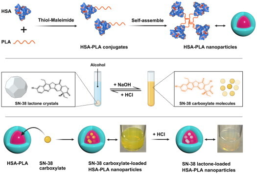
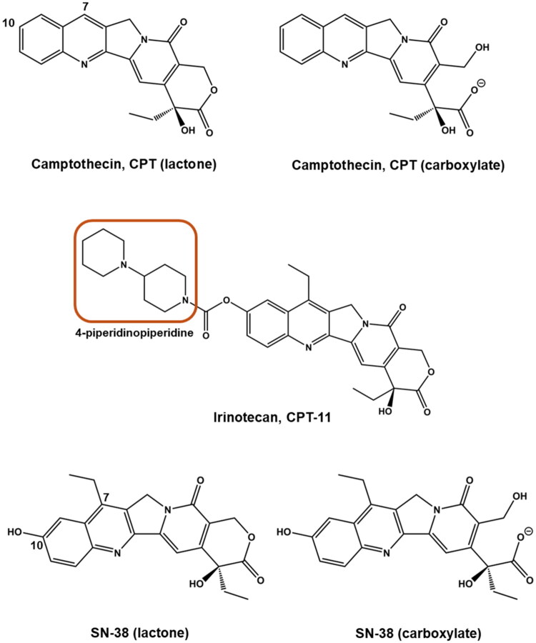
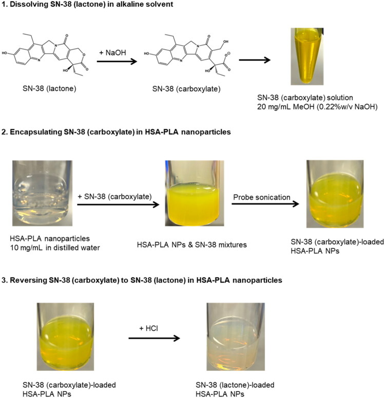
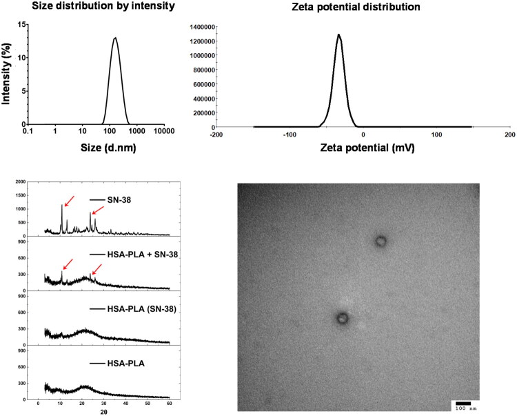
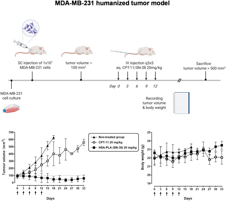
Efficient formulation of SN-38 for broad-spectrum chemotherapy remains an unmet medical need. The limited solubility of SN-38 in both aqueous and organic solvents poses a major challenge for formulation development. As a result, the predominant strategy, polymer-SN-38 drug conjugates, often involves complex synthetic procedures and low drug loading (1-5% w/w). Such limitations hinder their large-scale production and clinical translation. In this study, we developed an encapsulation strategy that utilizes the reversible lactone-carboxylate equilibrium of SN-38 to simplify the formulation process and achieve enhanced drug loading. The major issue of SN-38 solubility in organic solvents was effectively addressed by sodium hydroxide (NaOH)-induced conversion of the lactone to the carboxylate form. We have demonstrated that SN-38 carboxylate, once encapsulated within human serum albumin-polylactic acid (HSA-PLA) nanoparticles, retains its reversibility and can be converted back to the active lactone form simply by the addition of hydrochloric acid (HCl). The drug loading capacity of SN-38 in the HSA-PLA nanoparticles was increased to 19% w/w. In vitro cytotoxicity assays confirmed that HSA-PLA (SN-38) nanoparticles exhibited significantly lower IC50 values (0.5-194 nM) across multiple cancer cell lines compared to the clinical standard, irinotecan (CPT-11), indicating superior potency under physiological conditions. In vivo studies in 4T1 and MDA-MB-231 tumor-bearing mice further validated the enhanced therapeutic efficacy of this formulation. Overall, this study presents a promising alternative strategy for SN-38 delivery via encapsulation rather than polymer-drug conjugation, significantly simplifying the formulation process and enhancing the translational potential of SN-38 for broad chemotherapeutic applications.

Keywords: HSA–PLA; Human serum albumin; Protein drug delivery system; SN-38 loaded nanoparticles; lactone–carboxylate equilibrium.


Conflict of interest statement

Guojun Xiong, Andreas G. Schätzlein, and Ijeoma F. Uchegbu are inventors of a PCT patent (WO2025083423) filed by the UCLB. This work has no conflict of interest with Nanomerics Ltd.

Figures

None
Graphical abstract
Figure 1.
Figure 1.
Chemical structures of camptothecin (CPT) and its derivatives. The structural forms of camptothecin (CPT) and its derivatives, including irinotecan (CPT-11) and SN-38. Camptothecin exists in two forms: lactone (active) and carboxylate (inactive). Similarly, SN-38, the active metabolite of irinotecan, also exhibits a lactone–carboxylate equilibrium. The structure of irinotecan (CPT-11) includes a 4-piperidinopiperidine moiety (highlighted), which forms salts with hydrochloride to enhance water solubility. The 7-ethyl and 10-hydroxy groups on SN-38 inhibit the transformation to its carboxylate form under physiological conditions when compared with CPT.
Figure 2.
Figure 2.
Schematic representation of SN-38 encapsulation in HSA–PLA nanoparticles and lactone–carboxylate conversion. (1) SN-38 (lactone) is dissolved in an alkaline solvent (0.22% w/v NaOH in methanol), transforming into SN-38 (carboxylate). (2) The SN-38 (carboxylate) form is encapsulated into HSA–PLA nanoparticles through probe sonication, resulting in SN-38 (carboxylate)-loaded nanoparticles. (3) The SN-38 (carboxylate) in HSA–PLA nanoparticles is converted back to the lactone form by acidification with HCl, ensuring the active form of SN-38 is retained for drug delivery applications.
Figure 3.
Figure 3.
Characterization of SN-38-loaded HSA–PLA nanoparticles. Top left: the size distribution by intensity shows a monodisperse population of nanoparticles with a narrow size range. Top right: zeta potential measurement showing a single negative peak, further confirming the surface uniformity and good colloidal stability of the nanoparticles. Bottom left: X-ray diffraction (XRD) patterns of free SN-38, physical mixture (HSA–PLA + SN-38), SN-38-loaded HSA–PLA nanoparticles and blank HSA–PLA nanoparticles. The disappearance of SN-38 crystalline peaks after encapsulation indicates successful drug incorporation and conversion into an amorphous form. Bottom right: transmission electron microscopy (TEM) image confirms the spherical morphology of the nanoparticles with a uniform size distribution (scale bar: 100 nm).
Figure 4.
Figure 4.
Internalization of HSA–PLA nanoparticles via macropinocytosis in 4T1 cells. Schematic (A) illustrates the uptake mechanism of HSA–PLA nanoparticles (green) into 4T1 cells through macropinocytosis, where they become entrapped within macropinosomes (labeled with dextran). Confocal microscopy images (B) show 4T1 cells incubated with FITC-labeled HSA–PLA nanoparticles (green) and Texas Red–dextran (red), a marker for macropinosomes. Cell nuclei were stained with Hoechst 33342 (blue). Scale bars = 20 µm. Panel (C) displays a zoomed-in confocal image of the white-boxed region. Co-localization of HSA–PLA nanoparticles with macropinosomes is indicated by yellow signals in the merged image (white arrows), confirming macropinocytosis as a major pathway for nanoparticle uptake.
Figure 5.
Figure 5.
Apoptosis analysis of MDA-MB-231 cells after treatment with CPT-11 and HSA–PLA (SN-38) nanoparticles. (A) Schematic illustration of apoptosis detection using FITC-annexin V and propidium iodide (PI). In healthy cells (left), the phosphatidylserine (PS) remains on the inner leaflet of the plasma membrane. In early apoptotic cells (middle), PS is translocated to the outer membrane, allowing annexin V binding, while the membrane remains intact and impermeable to PI. In late apoptotic or necrotic cells (right), both annexin V and PI can bind due to compromised membrane integrity. (B) Method validation for apoptosis detection using flow cytometry. The first dot plot (from left to right) shows the unstained control (Annexin V−/PI−). The second plot represents cells stained with Annexin V-FITC only. The third plot shows cells stained with PI only. The final plot displays the distribution of cells stained with both dyes. (C) Dot plots (from left to right) represent the untreated cell control, cells treated with 200 nM CPT-11 for 48 hours, cells treated with the vehicle (HSA–PLA), and cells treated with HSA–PLA (SN-38) at 200 nM for 48 hours. Cells treated with HSA–PLA (SN-38) showed a significant increase in the proportion of apoptotic cells.
Figure 6.
Figure 6.
Cytotoxicity of CPT-11 and HSA–PLA (SN-38) nanoparticles in various cancer cell lines. Dose–response curves show the cytotoxic effects of CPT-11 (blue) and HSA–PLA (SN-38) nanoparticles (red) on different cancer cell lines (A431, A549, BT549, MIA PaCa-2, HT29, A2780, and PC3) after 48 hours of treatment. Cell viability (%) was assessed using a WST-1 assay, and drug concentrations are expressed as the logarithm of active pharmaceutical ingredient (API) concentrations (µg/mL). HSA–PLA (SN-38) nanoparticles exhibit a greater cytotoxic effect compared to CPT-11 across all tested cell lines, indicating enhanced potency. Error bars represent standard deviations from triplicate experiments.
Figure 7.
Figure 7.
In vivo antitumor study in 4T1 tumor bearing models. The top illustration provides a brief overview of the in vivo study design. Created with biorennder.com. The bottom left panel shows tumor volume growth over time in mice treated with HSA–PLA nanoparticles (control), CPT-11 (40 mg/kg), and HSA–PLA (SN-38) nanoparticles (40 mg/kg). The HSA–PLA (SN-38) nanoparticles exhibited a significantly greater tumor suppression effect compared to CPT-11 and the control group (****p < .0001). Data are shown as mean ± standard deviation (SD), n = 5. The bottom right panel presents body weight measurements over the study period, demonstrating no significant weight loss in any treatment group, indicating the safety of the formulations. Arrows indicate the time of drug administration. Data are presented as mean ± SD, n = 5.
Figure 8.
Figure 8.
Tumor growth inhibition and body weight monitoring in the MDA-MB-231 humanized tumor model. (Top) A schematic overview of the anticancer study conducted in the humanized tumor model (MDA-MB-231). Created with biorennder.com. (Bottom left) Tumor volume progression over 33 days in mice treated with HSA–PLA (SN-38) nanoparticles (20 mg/kg), CPT-11 (20 mg/kg), or left untreated. The HSA–PLA (SN-38) group exhibited a significant reduction in tumor growth compared to the non-treated and CPT-11-treated groups. Data are presented as mean ± SD, n = 5. (Bottom right) Body weight measurements over the same period, showing no significant weight loss across all groups, indicating good tolerability of the treatments. Arrows indicate the time points of drug administration. Data are presented as mean ± SD, n = 5.

Similar articles

References

    1. Burke TG, Mi Z.. 1993. Ethyl substitution at the 7 position extends the half-life of 10-hydroxycamptothecin in the presence of human serum albumin. J Med Chem. 36(17):2580–2582. doi: 10.1021/jm00069a020. - DOI - PubMed
    1. Chu S, Wang AL, Bhattacharya A, et al. 2022. Protein based biomaterials for therapeutic and diagnostic applications. Prog Biomed Eng. 4(1):012003. doi: 10.1088/2516-1091/ac2841. - DOI - PMC - PubMed
    1. Hong S, Choi DW, Kim HN, et al. 2020. Protein-based nanoparticles as drug delivery systems. Pharmaceutics. 12(7):604. doi: 10.3390/pharmaceutics12070604. - DOI - PMC - PubMed
    1. Innocenti F, Kroetz DL, Schuetz E, et al. 2009. Comprehensive pharmacogenetic analysis of irinotecan neutropenia and pharmacokinetics. J Clin Oncol. 27(16):2604–2614. doi: 10.1200/JCO.2008.20.6300. - DOI - PMC - PubMed
    1. Jiang M, Li W, Liang J, et al. 2024. Developing a palladium(II) agent to overcome multidrug resistance and metastasis of liver tumor by targeted multiacting on tumor cell, inactivating cancer-associated fibroblast and activating immune response. J Med Chem. 67(18):16296–16310. doi: 10.1021/acs.jmedchem.4c01175. - DOI - PubMed

LinkOut - more resources