

Therapeutic potential of greenly synthesized selenium nanoparticles against experimental cyclosporiasis
- PMID: 40820021
- PMCID: PMC12358599
- DOI: 10.1038/s41598-025-15238-8
Therapeutic potential of greenly synthesized selenium nanoparticles against experimental cyclosporiasis
Abstract
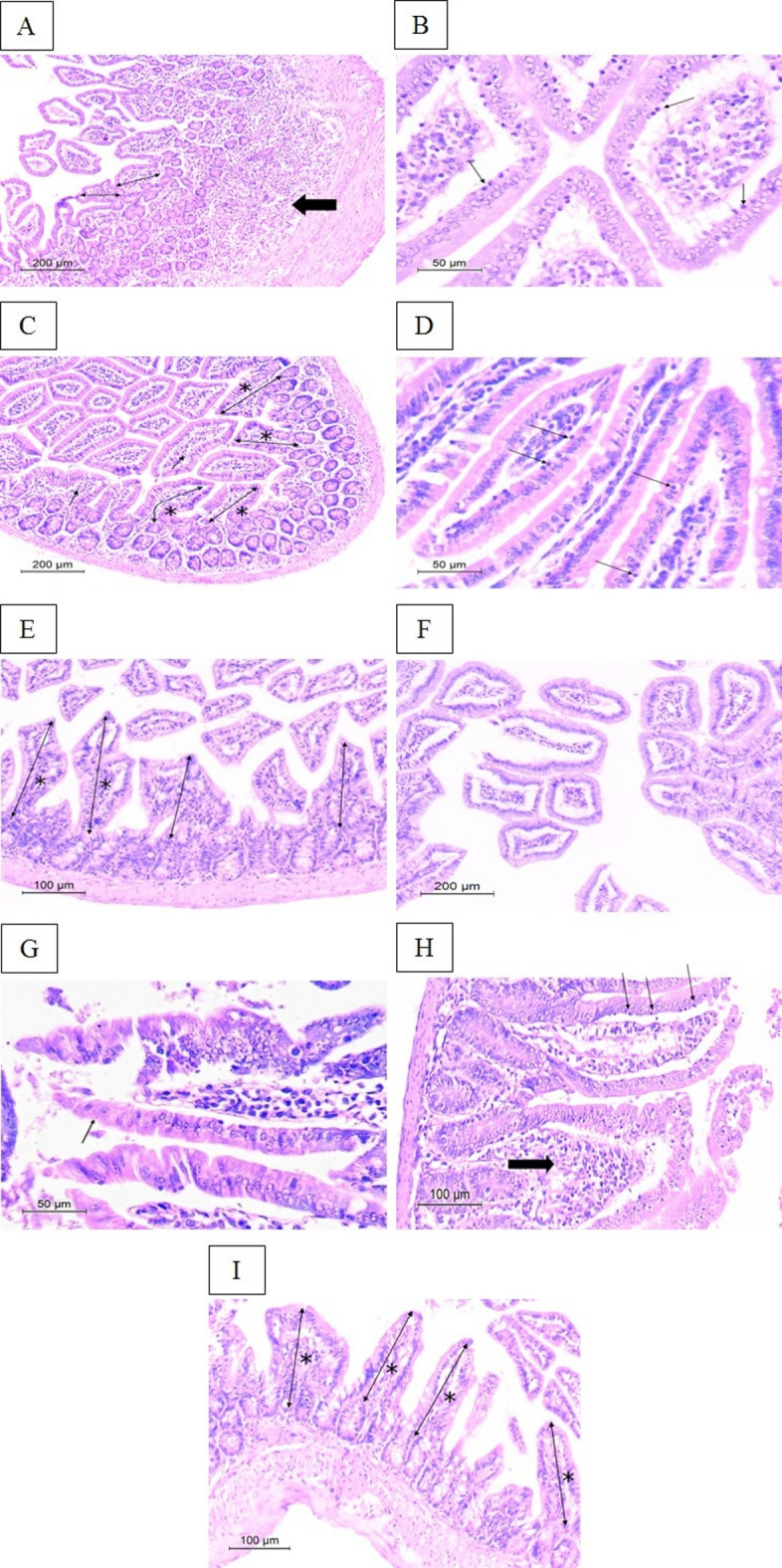
Cyclospora cayetanensis (C. cayetanensis), an opportunistic intracellular coccidian, is responsible for chronic debilitating diarrheal outbreaks possessing life-threatening penalties, especially in immunocompromised patients. The solemn therapeutic agents against cyclosporiasis are limited by their grave effects and high recurrence rate. The current study aimed to utilize greenly synthesized selenium nanoparticles (SeNPs) and evaluate their therapeutic efficacy on cyclosporiasis in immunosuppressed murine models. They were biosynthesized proficiently using Alcaligenes faecalis and characterized by assorted analytical techniques. After molecular confirmation of the parasite strain, immunosuppressed mice were infected with 10,000 C. cayetanensis sporulated oocysts. The anti-Cyclospora activity of seven-day oral treatment of 10 mg/kg of SeNPs was assessed through parasitological, ultrastructural, histopathological, and biochemical studies. The in vivo safety of SeNPs was assessed clinically, biochemically, and histopathologically. Parasitologically, SeNPs recorded the highest statistically significant decrease in the fecal oocyst burden (97.96%R) on the 30th day post-infection (PI). Scanning electron microscopic examination revealed remarkably deformed SeNPs-treated oocysts. SeNPs-treated mice exhibited impressive amelioration in intestinal architecture and inflammation, protracted to the 30th day PI. Colorimetric analysis revealed that SeNPs have recorded the highest serum reduced glutathione (GSH) level (300% increase) that swiftly repressed malondialdehyde (MDA) by 63.46%R. The present work has shed the first light on biogenic SeNPs as a safe, promising, proficient antioxidant nanotherapeutic for the treatment of experimental cyclosporiasis.
Keywords: Cyclospora cayetanensis; Anti-inflammatory; Antioxidant; Green synthesis; Scanning electron microscopy; Selenium nanoparticles.
© 2025. The Author(s).
Conflict of interest statement
Declarations. Competing interests: The authors declare no competing interests. Ethics approval: The present experimental study was conducted according to the Egyptian national regulations for laboratory animal experimentation. The study protocol was approved by the Ethics Committee of the Faculty of Medicine, Alexandria University, Egypt, under the approval number: 0306386. The authors complied with ARRIVE guidelines in all methods in the experiment conducted in the current study. Consent for publication: We declare that all authors have read and approved the manuscript for submission. We affirm that the manuscript is original and has not been published previously nor under consideration for publication elsewhere.
Figures


Similar articles
-
Size reduction of selenium nanoparticles synthesized from yeast beta glucan using cold atmospheric plasma.Sci Rep. 2025 Jul 16;15(1):25875. doi: 10.1038/s41598-025-09192-8. Sci Rep. 2025. PMID: 40670484 Free PMC article.
-
Silver nanoparticles as a therapeutic agent in experimental cyclosporiasis.Exp Parasitol. 2019 Dec;207:107772. doi: 10.1016/j.exppara.2019.107772. Epub 2019 Oct 11. Exp Parasitol. 2019. PMID: 31610183
-
Nutritional conditions affecting of selenium nanoparticles synthesized by Fusarium oxysporum (CCASU-2023-F9), and their biological activities against mycotoxin-producing fungi isolated from animal feed.Braz J Microbiol. 2024 Dec;55(4):3465-3476. doi: 10.1007/s42770-024-01494-9. Epub 2024 Sep 6. Braz J Microbiol. 2024. PMID: 39240496 Free PMC article.
-
Worldwide Epidemiology of Cyclospora cayetanensis in HIV/AIDS Patients: A Systematic Review and Meta-Analysis.Acta Parasitol. 2025 Jul 21;70(4):163. doi: 10.1007/s11686-025-01099-8. Acta Parasitol. 2025. PMID: 40690092 Review.
-
A Snapshot of Selenium-enclosed Nanoparticles for the Management of Cancer.Curr Pharm Des. 2024;30(11):841-858. doi: 10.2174/0113816128297329240305071103. Curr Pharm Des. 2024. PMID: 38462835 Review.
References
-
- Totton, S. C., O’Connor, A. M., Naganathan, T., Martinez, B. A. F. & Sargeant, J. M. A review of Cyclospora cayetanensis in animals. Zoonoses Public. Health. 68, 861–867. 10.1111/zph.12872 (2021). - PubMed
MeSH terms
Substances
LinkOut - more resources
Full Text Sources
Research Materials
Miscellaneous
