. 2025 Aug 17;15(1):30153.
doi: 10.1038/s41598-025-15891-z.

Modulation of retinal inflammation delays degeneration in a mouse model of geographic atrophy

Affiliations

Modulation of retinal inflammation delays degeneration in a mouse model of geographic atrophy

Raela B Ridley et al. Sci Rep. .

Abstract

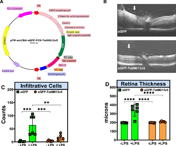
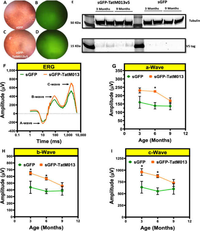
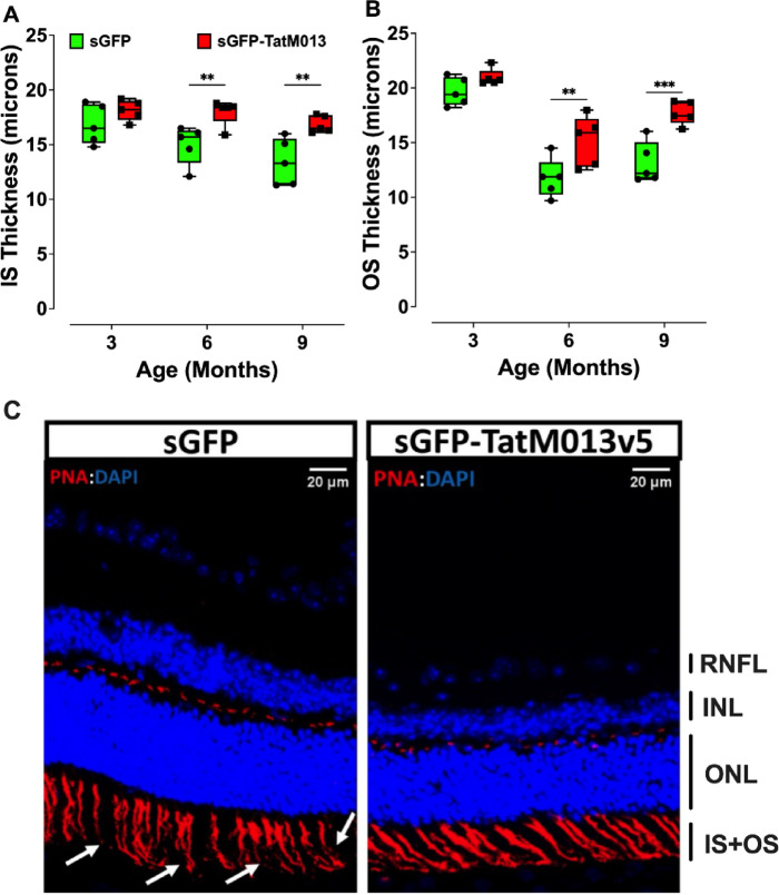
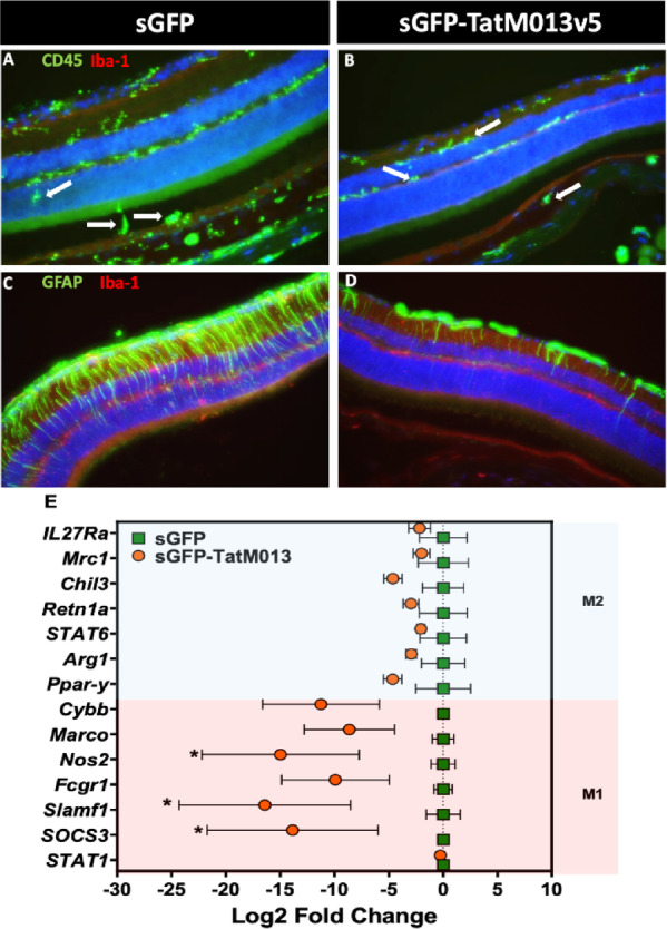
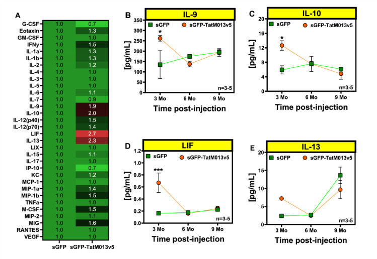
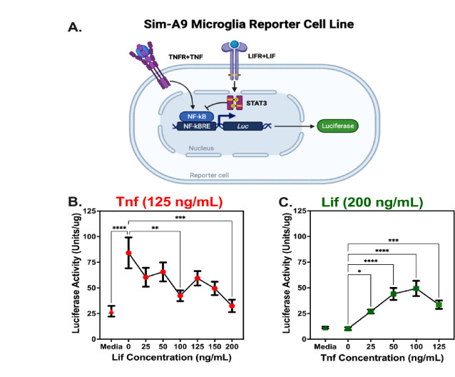
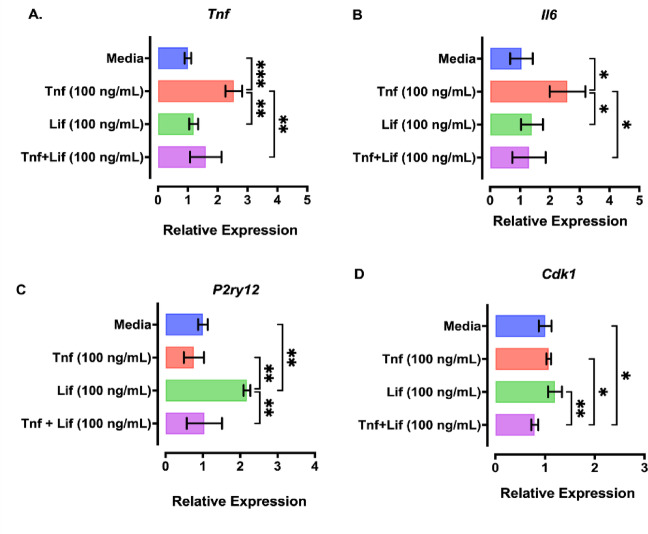
Geographic atrophy, the advanced form of age-related macular degeneration (AMD), is associated with increased oxidative stress and chronic inflammation. Pro-inflammatory genes, like TNF-α and IL-1β, are under the regulation of the transcription factor p65/RelA. We have previously shown that adeno-associated virus (AAV) delivery of the RelA inhibitory gene M013 blocks retinal inflammation in uveitis models. In this study, we evaluated the effects of RelA inhibition in an oxidative stress-driven geographic atrophy mouse model. We injected Sod2RPEcKO mice with rAAV, delivering either secreted GFP (sGFP control) or sGFP fused to a cell-penetrating version of the tagged M013 (sGFP-TatM013v5). Over nine months, we measured retinal function, structure, and morphological changes using electroretinography, optical coherence tomography, and fundoscopy. We quantified changes in inflammatory markers using multiplex ELISA, RT-qPCR, and immunofluorescence staining of the retinal tissue. Finally, we generated an NF-kB-luciferase reporter microglia cell line to study the impact of immune signaling changes on microglia. Mice injected with the rAAV delivering M013 had transient protection of their retinal function at 3 months. Based on ERG evaluations, the intravitreal injection of rAAV delivering sGFP-TatM013v5 significantly delayed the loss of retinal function. Furthermore, the rAAV-mediated expression of the sGFP-TatM013v5 protected photoreceptors' outer and inner segments based on OCT and immunofluorescence analysis. Analysis of postmortem tissues showed decreased migration of immune cells towards the RPE. Retinas injected with the sGFP-M013v5 vector showed increased levels of IL-9, IL10 and LIF. Finally, adding LIF to our NF-kB reporter cell line showed decreased TNF-induced reporter expression and modulation of microglia-specific genes. Our results indicate that modulating retinal inflammation could significantly slow the degeneration associated with geographic atrophy. Specifically, inhibiting the RelA protein in the retina may offer protective effects against retinal degeneration. Additionally, we demonstrated that LIF can counteract the influence of TNF on microglial gene expression. Future research will explore the dynamic interactions between RelA and other transcription factors and the NF-kB signaling pathway in the retina as they relate to retinal diseases.

Keywords: Age-related macular degeneration; Myxoma virus; NF-kB; Retinal inflammation.


Conflict of interest statement

Declarations. Ethics approval and consent to participate: The University of Florida Institutional Animal Care and Use (IACUC) Board approved all the animal experiments conducted for this research (Protocol 201709854). All procedures adhered to ARVO guidelines for the use of animals in biomedical research. The study is reported in accordance with ARRIVE guidelines. Competing interests: The authors declare no competing interests.

Figures

Fig. 1
Fig. 1
TatM013v5 decreases ocular inflammation in the EIU mouse model. (A) Genetic map of the pTR-smCBA-sGFP-FCS-TatM013v5 vector. The control vector has the same sequence except with the TatM013v5 insert. (B) Representative SD-OCT B-scans of mice treated with AAV that delivered either sGFP or sGFP-TatM013v5, twenty-four hours after endotoxin (LPS) injection. (C) Two-masked individuals quantified infiltrative cells in the vitreous humor using Image J software. LPS injection only significantly increased infiltrative cells in sGFP-treated eyes. (D) Total retinal thickness measurements were done before and after the LPS injection. LPS injection caused a significant increase in retina thickness only in sGFP-treated eyes. Values represent average ± standard deviation (n = 5–6 mice). ** = adjusted p-value ≤ 0.01, *** = adj. p-val ≤ 0.001, **** = adj. p-val ≤ 0.0001 (pTR – plasmid with terminal repeats, smCBA – small chicken-beta actin promoter, sGFP – secretable green fluorescent protein, FCS – furin cleavage site, SD-OCT – spectral domain optical coherence tomography, LPS – lipopolysaccharide).
Fig. 2
Fig. 2
Retinal delivery of TatM013v5 protects the retina function of the RPESod2cKO mouse model. (AD) Two-month-old mice of the correct genotype were injected intravitreally with 1010 vgc of AAV2quad(Y-F) + T491V, delivering either sGFP (A,B) or sGFP-TatM013v5 (C,D). One month later, fluorescent fundoscopy using a Micron III camera confirmed transgene expression. Representative images demonstrate the presence of GFP fluorescence in the retina of either sGFP or sGFP-TatM013-treated eyes. (E) Western blot showing the retinal expression of TatM013v5 in sGFP-TatM013v5-treated mice. Mice were evaluated with ERG at 3-, 6-, and 9-months of age. (F) Representative ERG recording at six months of age. The average amplitudes of their (G) a-wave, (H) b-wave, and (I) c-wave at each time point are graphed as a function of time in months. Eyes treated with the AAV vector delivering sGFP-TatM013 had significantly higher amplitudes when compared to sGFP-treated eyes. Values are reported as average ± standard deviation. (n = 5 mice per group). * = adj. p-val ≤ 0.05 (ERG – electroretinogram).
Fig. 3
Fig. 3
Effects of TatM013v5 on photoreceptor’s inner and outer segments of the RPESod2cKO mouse model. Two hundred and fifty B-scans of mice retinas were acquired using a Bioptigen spectral-domain optical coherence tomography. We averaged every 10 B-scans to obtain 25 high-resolution images. Bioptigen Diver auto-segmentation software was used to measure the thickness of each retina layer. The thickness of the (A) inner segments (IS), and (B) outer segments (OS) of retinas treated with either sGFP (green) or sGFP-TatM013v5 (red) were plotted as a function of the animal age. (C) Retina cross sections were stained with peanut agglutinin (PNA) to detect the structure of IS and OS. sGFP-TatM013v5 treated retinas had more intact IS and OS based on the PNA staining (red channel). Values were reported as average +/- standard deviation (n = 5 mice). * = adj. p-val ≤ 0.05, *** = adj. p-val. ≤ 0.001.
Fig. 4
Fig. 4
TatM013v5 retinal expression decreases reactive gliosis and retinal inflammation. Cryosections from a 3-month-old Sod2RPEcKO mouse model were stained with antibodies against CD45 and Iba-1 or GFAP and Iba-1. (A) CD45-positive cells were detected through all the retinal layers in retinas treated with the sGFP vector, especially in the RPE layer (white arrows). (B) In contrast, retinas treated with the sGFP-TatM013v5 vector showed CD45 positive cells only between the RNFL and the IPL or below the RPE layer (white arrows). (C) Increased expression of GFAP through all retinal layers was observed in sGFP-treated retinas, indicating Müller glial activation. (D) The GFAP staining pattern in sGFP-TatM013v5 treated retinas was mainly limited to the RNFL, showing astrocyte expression and minimal staining in Müller glial cells. (E) Total RNA was isolated from retinas treated with an AAV vector, delivering either sGFP or sGFP-TatM013v5. Gene expression of M1-associated genes (e.g., STAT1 and Nos2) and M2-associated genes (e.g., PPar-γ and Arg1) were quantified using RT-qPCR. Beta Actin was used as the reference housekeeping gene for all samples, and the ΔΔCt method was used to determine the fold-change. Values are plotted as the Log2 of the fold-change (average ± standard deviation) to better illustrate the decrease in M1-gene expression induced by the expression of sGFP-TatM013v5. No significant changes were identified within the M2 genes. (n = 3 retina samples). * = adjusted p-value ≤ 0.05 (CD45 – cluster of differentiation 45, Iba-1 – ionized calcium-binding adaptor molecule 1, GFAP – glial fibrillary acidic protein, RNFL – retina nerve fiber layer, IPL – inner plexiform layer, RPE – retinal pigmented epithelium, RT-qPCR – reverse-transcribed quantitative polymerase chain reaction).
Fig. 5
Fig. 5
Retinal expression of TatM013v5 increases retinal concentration of IL-10, IL-9, and LIF. Retinal protein extracts from RPESod2cKO-treated mice were evaluated by multiplex ELISA using a pre-mixed kit for 32 cytokines and chemokines. Four of these analytes were significantly different, at least at a one-time point in the assay. (A) At three months of age, the anti-inflammatory cytokine IL-10 was increased in the sGFP-TatM013v5-treated retinas compared to sGFP-treated retinas (values relative to sGFP). (B) IL-9 was increased dramatically in sGFP-TatM013v5-treated retinas at three months of age. Likewise, IL-10 (C) and LIF (D) were increased substantially in sGFP-TatM013v5-treated retinas at three months. Finally, the cytokine IL-13 concentration increased in the sGFP-treated retinas by nine months and not in the sGFP-TatM013v5 (E). Values represent average ± standard deviation (n = 3–5 retina lysates). * = adj. p-val ≤ 0.05 (IL-10 – interleukin 10, IL-9 – interleukin 9, LIF – leukemia inhibitory factor, ELISA – enzyme-linked immunosorbent assay).
Fig. 6
Fig. 6
LIF antagonizes pro-inflammatory signals in microglia. (A) Sim-A9 cells expressing the luciferase gene under the control of the NF-kBRE were incubated with or without either (B) Tnf or (C) Lif and with different concentrations of Lif (B) or Tnf (C) for 24 h. Values represent average ± SEM, compared by one-way ANOVA followed by Holm-Sidak test for multiple comparisons. *= adj. p-value≤ 0.05, **= adj. p-val 0.01, ***= adj. p-val 0.001, ****= adj p-val 0.0001. (n = 3 independent experiments, 15 biological replicates) (NF-kBRE – nuclear factor kappa-light-chain-enhancer of activated B cells response element, TNF – tumor necrosis factor).
Fig. 7
Fig. 7
Tnf and Lif regulate microglia pro-inflammatory, homeostatic, and proliferative genes in vitro. We stimulated Sim A9 microglia cells with Tnf, Lif, or Tnf in the presence of Lif for 24 h. RT-qPCR for inflammatory genes (A,B), homeostasis (C) and proliferation (D) were used to quantify gene expression changes. Values represent average ± StDev. Values were compared with a one-way ANOVA followed by Holm-Sidak test for multiple comparisons. *= adj p-val≤ 0.05, **= adj p-val≤ 0.01, ***= adj. p-val≤ 0.001 (n = 3 independent experiments).

Update of

Similar articles

References

    1. Jonas, J. B., Cheung, C. M. G. & Panda-Jonas, S. Updates on the epidemiology of age-related macular degeneration. Asia Pac. J. Ophthalmol.6, 493–497 (2017). - PubMed
    1. Crabb, J. W. The proteomics of Drusen. Cold Spring Harb Perspect. Med.4, a017194 (2014). - PMC - PubMed
    1. Winkler, T. W. et al. Genome-wide association meta-analysis for early age-related macular degeneration highlights novel loci and insights for advanced disease. BMC Med. Genomics. 13, 120 (2020). - PMC - PubMed
    1. Lorés-Motta, L. et al. Common haplotypes at the CFH locus and low-frequency variants in CFHR2 and CFHR5 associate with systemic FHR concentrations and age-related macular degeneration. Am. J. Hum. Genet.108, 1367–1384 (2021). - PMC - PubMed
    1. Mao, H. et al. Mitochondrial oxidative stress in the retinal pigment epithelium leads to localized retinal degeneration. Invest. Ophthalmol. Vis. Sci.55, 4613–4627 (2014). - PMC - PubMed

LinkOut - more resources