. 2025 Sep 30;152(13):939-956.
doi: 10.1161/CIRCULATIONAHA.125.074845. Epub 2025 Aug 18.

An Aberrant Resurgence of Endogenous Retroviruses Prompts Myocarditis and Heart Failure

Affiliations

An Aberrant Resurgence of Endogenous Retroviruses Prompts Myocarditis and Heart Failure

Junhao Xiong et al. Circulation. .

Abstract

Background: Endogenous retroviruses (ERVs) occupy >8% of the human genome. Aberrant resurgence of ERVs has been implicated recently in several critical pathologies. However, the possible incidence and role of ERV resurgence in heart failure (HF), a leading cause of global morbidity and mortality, remain unexplored.

Methods: We established a total RNA sequencing analyzing pipeline to assess the ERV occurrence in human and murine HF models. We generated 2 myocardium-specific mouse lines by crossing Myh6-MerCreMer (Myosin heavy chain 6 promoter driving MerCreMer recombinase) with TRIM28f/f and SETDB1f/f mice to identify the molecular regulators of ERV resurgence and the downstream pathways in the heart. We evaluated ERV expression by total RNA sequencing, reverse transcription-quantitative polymerase chain reaction and RNA fluorescence in situ hybridization. We restrained ERV activation by overexpressing TRIM28 (tripartite motif-containing 28) using adeno-associated virus serotype 9. The therapeutic potential of the ERV-mediated inflammatory pathway was tested in a myocardial ischemia/reperfusion model.

Results: ERVs, particularly class I ERVs, were prominently activated in multiple cross-species models of HF. Depletion of TRIM28, an epigenetic repressor, attenuated the epigenetic surveillance of trimethylation at lysine 9 of histone H3 and N6-methyladenosine, leading to the activation of ERVs in the failing heart. This ERV activation stimulated the antiviral innate immune pathways of TLR7/9 (Toll-like receptor 7/9) and NF-κB and lead to myocarditis and acute HF. Furthermore, restraining ERV activation and ERV-mediated innate immune responses by either adeno-associated virus serotype 9-mediated TRIM28 expression or a small-molecule TLR7/9 inhibitor improved heart function and alleviated HF in an ischemia/reperfusion model.

Conclusions: ERV resurgence is a specific molecular trait of HF, driven by TRIM28 depletion in cardiomyocytes. ERV resurgence activates the innate immune TLR7/9-NF-κB pathway and induces myocarditis and HF. Inception of ERVs and the ERV-mediated immune pathway confers cardiac protection. These results identify TRIM28-ERV-TLR7/9-NF-κB as a target for therapeutic management of myocarditis and HF.

Keywords: NF-κB; TRIM28; endogenous retrovirus; heart failure; myocarditis.


Conflict of interest statement

None.

Figures
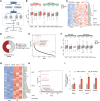
Figure 1.
Figure 1.
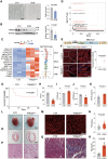
A retroviral provirus revives in heart failure. A, The scheme for the discovery of heart failure (HF)–associated retrotransposon elements (RTEs) in cross-species HF models by Figdraw. RTE sequences were annotated using Repeat Masker across multiple species. Differentially expressed RTEs were identified from total RNA sequencing using DESeq2, some of which were further validated by RT-qPCR. B, Boxplots showing human endogenous retroviruses (HERVs), especially HERV1, increased in GSE135055 human HF hearts. The center line indicates the median. The box indicates 25th and 75th percentiles; n=7 controls and 13 HF patients. Mann-Whitney test was used to calculate the significance. C, Heatmap presenting the significantly elevated HERV genes in human HF hearts (adjusted P [P-adj]<0.05, fold change>0.5). Data were from the GEO database (GSE135055). There were 7 control patients and 13 HF patients. D, Ring plots showing the percentages of 74 upregulated endogenous retroviruses (ERVs) in each family identified from human GEO data sets (GSE135055, GSE126569, and GSE48166). E, P-adj value ranking identified HERV1 as the most significantly altered HERVs in human HF (threshold: P-adj=0.05, denoted by a dotted line). HERV1 genes LTR61, LTR25, LTR30, LTR24C, and PABL_B-int were among the top 10 differentially expressed HF RTEs. P-adj was calculated with the DESeq2 package, and Benjamini-Hochberg adjustment was used to control the false positives in multiple testing. F, Boxplots indicating the upregulation of mouse ERVs, especially ERV1, in ischemia/reperfusion injury hearts. The center line indicates the median. The box indicates 25th and 75th percentiles; n=3 mice per group. Mann-Whitney test. G, Heatmap showing the resurgence of mouse ERVs in ischemia/reperfusion injury hearts in the analysis of total RNA sequencing (P-adj<0.05, fold change>1.5). n=3 mice per group. H, P-adj value ranking implicating ERV1 as the most significantly altered ERVs in ischemia/reperfusion mouse hearts. The dotted line indicates P-adj=0.05. MMVL30-int, RLTR6_Mm, RLTR6-int, RLTR6C_Mm, MuLV-int, and RLTR30D_RN, in red, belong to the mouse ERV1 family with top 10 ranking in differentially expressed retrotransposons. I, RT-qPCR showing the resurgence of ERVs in monkey hearts 4 weeks after myocardial infarction. ERVW, MacERV-4 and HERV-E belong to ERVI RTE genes, whereas ERVK was ERVII. Data are mean±SD; n=3 hearts per group. Two-tailed Student t test; **P<0.01, ***P<0.001o significance. I/R indicates ischemia/reperfusion; MI, myocardial infarction; NC, negative control; NHP, nonhuman primate; ns, no significance; and RT-qPCR, reverse transcription-quantitative polymerase chain reaction.
Figure 2.
Figure 2.
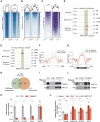
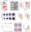
The cardiomyocyte-specific deprivation of TRIM28 led to endogenous retrovirus resurgence and heart failure. A, Immunohistochemical staining of left ventricles from human nonfailing control (CTR) and dilated cardiomyopathy (DCM) hearts. TRIM28 (tripartite motif–containing protein 28) expression is significantly reduced in human DCM hearts. Quantification of TRIM28-positive nuclei (right) was performed using ImageJ. Data represent mean±SD; n=3 hearts per group. Two-tailed Student t test. B, Western blot analysis of TRIM28 protein in CTR and DCM human left ventricles. TRIM28 signal intensity was quantified using ImageJ. The TRIM28 protein level decreased significantly in human DCM hearts. Data are mean±SD; n=3 CTR and 5 DCM hearts. Two-tailed Student t test. C, RNA sequencing analysis of endogenous retrovirus expression in Myh6-MerCreMer (CTR) and Myh6-MerCreMer TRIM28f/f (TRIM28iCKO) mouse hearts 1 week after tamoxifen induction, showing endogenous retrovirus resurgence in TRIM28iCKO hearts. RLTR6_Mm and MMVL30-int exhibited the most significant upregulation. D, Adjusted P value ranking indicated that endogenous retrovirus class 1 exhibited the most significant alterations after TRIM28 depletion. MMVL30-int, RLTR6_Mm, RLTR6-int, RLTR6C_Mm, and RLTR30D_RN of mouse endogenous retrovirus class 1, in red, ranked in the top 10 in differentially expressed RTEs. The red horizontal line indicates adjusted P=0.05. E, The structure of the MMVL30 provirus in the mouse genome. F, RNA fluorescence in situ hybridization detecting the MMVL30 transcripts (green signal) in CTR and TRIM28iCKO cardiomyocytes (red signal). The MMVL30 signal intensity (left) was quantified with ImageJ (right). Data are mean±SD; n=3 hearts per group. Two-tailed Student t test. G, The survival plot of CTR (n=18) and TRIM28iCKO (n=22) mice. Ten of 22 (46%) of TRIM28iCKO mice died within 2 weeks after tamoxifen induction. The statistical significance was calculated with a log-rank test. P<0.05 indicated significance. H through J, M-mode echocardiogram illustrating the attenuated ejection fraction (H), increased left ventricle diastolic internal diameter (I), and decreased left ventricular posterior wall thickness at diastole (J) of TRIM28CKO hearts. Data are mean±SD; n=10 mice per group. Two-tailed Student t test. K, The ratio of heart weight to tibial length in CTR (n=7) and TRIM28iCKO (n=4) mice. Data are mean±SD. Two-tailed Student t test. L, Gross anatomy showing the significant enlargement of TRIM28iCKO hearts. M and N, Wheat germ agglutinin staining of left ventricular sections, illustrating the enlarged cardiomyocytes in TRIM28iCKO hearts. The areas of cardiomyocytes (M) were calculated with ImageJ (N). N=110 to 118 cells from 3 measured hearts per group. Mean±SD, 2-tailed Student t test. O, Hematoxylin-eosin staining illustrates the dilated cardiomyopathy of TRIM28iCKO hearts. P, Hematoxylin-eosin histology showing sarcomere disarrangement, cardiomyocyte death, and inflammatory cell infiltration in TRIM28iCKO hearts. Q, Masson trichrome staining showing increased collagen deposition (blue region) in TRIM28iCKO hearts. R, The quantification of trichrome staining revealed elevated collagen deposition in TRIM28iCKO hearts. Mean±SD, n=6 mice per group. Two-tailed Student t test. EF indicates ejection fraction; ERV, endogenous retrovirus; Gag, group-specific antigen; HW, heart weight; LTR, long terminal repeat; LVIDd, left ventricle diastolic internal diameter; LVPWd, left ventricular posterior wall thickness at diastole; Pol, polyprotein; and TL, tibial length.
Figure 3.
Figure 3.
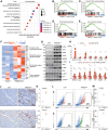
TRIM28 controlled the surveillance of endogenous retrovirus class 1 by manipulating trimethylation at lysine 9 of histone H3 and N6-methyladenosine. A, Trimethylation at lysine 9 of histone H3 (H3K9me3) chromatin immunoprecipitation sequencing signals on all targeted endogenous retroviruses (ERVs). B, H3K9me3 chromatin immunoprecipitation sequencing signals on upregulated ERVs. C, RNA sequencing heatmap of those upregulated ERVs. D, H3K9me3, RNA, and methylated RNA immunoprecipitation sequencing signals on upregulated MMVL30-int_dup726 RTE genomic loci, illustrating the declined H3K9me3 and intact N6-methyladenosine (m6A). E, H3K9me3, RNA sequencing, and methylated RNA immunoprecipitation sequencing signals on the upregulated MMVL30-int_dup810 transcript, illustrating the increased m6A and unchanged H3K9me3. F, Aggregation plot showing that the average m6A signal was lower on upregulated ERVs in TRIM28iCKO hearts. G, Aggregation plot showing that the average m6A signal was downregulated on up_MMVL30 RTE transcripts in TRIM28iCKO hearts. Shown beneath the aggregation plot is the genomic structure of MMVL30. H, Venn diagram showing the minimal overlap between m6A- and H3K9me3-modulated upregulated ERVs. Only 23% (102 of 437) of m6A-modified upregulated ERVs were also marked by H3K9me3. P=0.9161 by χ2 test. I and J, Immunoprecipitation revealed the interactions between TRIM28 (tripartite motif–containing protein 28) and METTL3. K, RNA immunoprecipitation-quantitative polymerase chain reaction revealed that the binding of METTL3 to ERVs decreased in TRIM28iCKO hearts. GAPDH served as a negative control. Mean±SD, n=4; **P<0.01, ***P<0.001; 2-tailed Student t test. L, RT-qPCR revealed that METTL3 overexpression suppressed ERV activation in TRIM28iCKO CM. Mean±SD, n=6, 2-tailed Student t test; *P<0.05, **P<0.01, ***P<0.001. ChIP-seq indicates chromatin immunoprecipitation sequencing; MeRIP-seq, methylated RNA immunoprecipitation sequencing; ns, no significance; RNA-seq, RNA sequencing; and RT-qPCR, reverse transcription-quantitative polymerase chain reaction.
Figure 4.
Figure 4.
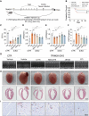
Endogenous retrovirus resurgence leads to myocarditis and heart failure through activation of TLR7/9. A, Gene Ontology analysis of upregulated differentially expressed genes. The top 10 enriched terms are shown. The color scale indicates the adjusted P value (P-adjust). B through E, Gene set enrichment analysis analyzing the gene enrichment in nucleic acid–sensing pathways of TLR (Toll-like receptor; B), cGMP-AMP (cGAS; C), NF-κB (D), and IFN (interferon; E). Significant enrichments were observed in TLR (normalized enrichment score [NES]=1.5781; P-adjust<0.05), cGAS (NES=1.5801; P-adjust<0.05), and NF-κB (NES=1.7215; P-adjust<0.01) pathways but not IFN (NES=1.2341; P-adjust=0.4838). F, Heatmap showing differentially expressed genes in the cardiac hypertrophy, nucleotide sensing, and NF-κB pathways. Genes in the IFN pathway were not significantly altered. G, Western blot illustrating the activation of the PRR (pattern recognition receptor; including cGAS, TLR7, TLR8, TLR9, MyD88, TAK1, and p-TAK1) and NF-κB (p-P65 and P65) pathways but not IFN (p-IRF7 and IRF7) in TRIM28iCKO hearts. GAPDH served as a control. H, RT-qPCR examination of TLR7/9 and cGAS activation in primary cardiomyocytes transduced with MMVL30. Data were normalized to GAPDH. Mean±SD, n=6 independent replicates, 2-tailed Student t test; *P<0.05, **P<0.01, ***P<0.001. I, RT-qPCR indicated that MMVL30 siRNA suppressed TLR7/9 and cGAS pathway activation in TRIM28 (tripartite motif–containing protein 28) knockout cardiomyocytes. Primary cardiomyocytes were treated with 4-hydroxytamoxifen to induce TRIM28 deletion and then transfected with METTL3-modified RNA for 48 hours. Mean±SD, n=6 independent replicates. One-way ANOVA followed by post hoc Tukey test. *P<0.05, **P<0.01, ***P<0.001. J and K, Immunochemistry revealing the infiltration of CD68+ macrophages (J, top) and CD3+ lymphocytes (J, bottom). Arrows indicate infiltrated immune cells. The intensity of CD68+ macrophages (K, top) and CD3+ lymphocytes (K, bottom) was calculated with ImageJ. Data are mean±SD, n=5 hearts. Two-tailed Student t test. L and M, Flow cytometry demonstrating increased CD68+ macrophages and CD3+ lymphocytes in TRIM28iCKO mice (L). The numbers of CD68+ macrophages (M, top) and CD3+ lymphocytes (M, bottom) were calculated in FlowJo. Mean±SD, n=3 mice, Two-tailed Student t test. 4-OHT indicates 4-hydroxytamoxifen; FDR, false discovery rate; GO, Gene Ontology; and RT-qPCR, reverse transcription-quantitative polymerase chain reaction.
Figure 5.
Figure 5.
Blocking the endogenous retrovirus—Toll-like receptor–NF-κB pathway ameliorated myocarditis and cardiac dysfunction of TRIM28iCKO mice. A, Dosing strategies of variable small-molecule inhibitors. NSC4375 (a TLR7/9 [Toll-like receptor 7/9] inhibitor, 20 mg/kg), JSH23 (an NF-κB inhibitor, 6 mg/kg), and C176 (a cGMP-AMP inhibitor, 8 mg/kg) as well as vehicle (5% Dimethyl sulfoxide+40% PEG300+5% Tween 80+50% double distilled H2O) were administered via intraperitoneal injection 3 times per week. The reverse transcriptase inhibitor (RTi; zidovudine, 4 mg/day) was dosed via drinking water. Mice were subsequently evaluated with echocardiography and pathology. B, The survival of TRIM28iCKO mice with or without inhibitor treatments. Treatments with NSC4375, JSH23, or RTi for 3 weeks increased the survival of TRIM28iCKO mice. The statistical significance was calculated with log-rank tests. P<0.05 indicated significance. C through E, Echocardiogram revealed that NSC4375, JSH23, and RTi significantly alleviated the pathological remodeling of TRIM28iCKO hearts. NSC4375, JSH23, and RTi rather than C176 improved the systolic function of TRIM28iCKO mice (C), decreased the left ventricle diastolic internal diameter (D) and increased the left ventricular posterior wall thickness at diastole (E). Mean±SD, n=8 to 9 mice, 1-way ANOVA followed by post hoc Tukey test; *P<0.05, **P<0.01, ***P<0.001. F, Heart weight/tibial length ratio verified that NSC4375, JSH-23, and RTi rather than C176 reduced the heart mass of TRIM28iCKO mice. Mean±SD; n=7 mice; 1-way ANOVA followed by post hoc Tukey test. **P<0.01, ***P<0.001. G, The M-mode echocardiograms of TRIM28iCKO mice 9 days after inhibitor treatments. NSC4375, JSH23, and RTi improved cardiac function. H, The gross anatomy (top) and hematoxylin-eosin staining sections (bottom) revealed that NSC4375, RTi, and JSH23 ameliorated the ventricular dilation of TRIM28iCKO hearts. I, Immunohistochemistry revealed that NSC4375, JSH23, and RTi mitigated the infiltration of CD68+ macrophages. Arrows indicate infiltrating CD68+ macrophages. CD68+ macrophages in hearts are quantified in Figure S8C. HW indicates heart weight; LVIDd, left ventricle diastolic internal diameter; LVPWd, left ventricular posterior wall thickness at diastole; ns, no significance; and TL, tibial length.
Figure 6.
Figure 6.
Curbing endogenous retrovirus resurgence mitigates heart failure. A, Heatmap of RT-qPCR showing the time-course expression profiling in the ischemia/reperfusion mouse model. TRIM28 (tripartite motif–containing protein 28) and SETDB1 were significantly downregulated, whereas endogenous retroviruses and cytokines were activated immediately after surgery, which was ahead of Nppa (natriuretic peptide A). Mean, n=4. B, Western blot showing that adeno-associated virus serotype 9 (AAV9)-TRIM28 inhibited activation of the TLR7/9 (Toll-like receptor 7/9) and NF-κB pathways in ischemia/reperfusion hearts. C, AAV9-TRIM28 suppressed endogenous retrovirus class 1 and cytokine expression in the ischemia/reperfusion hearts, as revealed by reverse transcription-quantitative polymerase chain reaction. n=6. Mean±SD, 2-way ANOVA followed by post hoc Sidak test. D, Evans blue/triphenyl-2H-tetrazolium chloride staining illustrating that AAV9-TRIM28 reduced the infarct area. Blue, nonischemic region; red, ischemic region or area at risk; white, infarct region. E and F, The calculation of the infarct area. The infarct areas (infarction/area at risk) of AAV9-TRIM28–treated hearts were smaller than those of control hearts. n=6 mice, mean±SD, 2-tailed Student t test, **P<0.01. G, Echocardiography indicating that AAV9-TRIM28 significantly improved the systolic function of ischemia/reperfusion hearts. Ejection fraction was measured for 3 consecutive weeks. n=8 mice. Mean±SD, mixed-effects ANOVA with post hoc Sidak test. *P<0.05, **P<0.01, ***P<0.001. H and I, Masson trichrome staining showing that AAV9-TRIM28 ameliorated scar formation 3 weeks after injection. The ratio of fibrotic region (blue) to total region (blue+red) was calculated by ImageJ. n=6 mice. Mean±SD, 2-way ANOVA followed by post hoc Sidak test. ***P<0.001. AAR indicates area at risk; EF, ejection fraction; IF, infarction; I/R, ischemia/reperfusion; LV, left ventricle; ns, no significance; RT-qPCR, reverse transcription-quantitative polymerase chain reaction; and TTC, triphenyl-2H-tetrazolium chloride.
Figure 7.
Figure 7.
Repressing endogenous retrovirus–mediated innate inflammation mitigates heart failure. A, NSC4375 dosing strategy: 20 mg/kg of NSC4375 injected intraperitoneally 3 times per week 2 days before ischemia/reperfusion (I/R) surgery. Serial echocardiography was performed at the relevant times, and hearts were collected for pathological analysis 3 weeks after I/R surgery. B, Western blot illustrating that NSC4375 suppressed activation of the TLR7/9 (Toll-like receptor 7/9) and NF-κB pathways in I/R hearts. C, RT-qPCR showed that NSC4375 suppressed the activation of cytokines (Il6, Il1b, and Tnfa) rather than endogenous retrovirus class 1 (MMVL30, RLTR6, and MuLV) in I/R hearts (I/R+NSC4375 vs I/R+Vehicle). Mean±SD, n=6, 1-way ANOVA followed by post hoc Tukey test. D, Evans blue/triphenyl-2H-tetrazolium chloride staining showing the reduced infarct area in NSC4375-treated I/R mice. E and F, Calculating the infarct area. The infarct areas (infarction/area at risk) were smaller in NSC4375-treated hearts than in controls (E), whereas area at risk/left ventricle remained unchanged (F). n= 6 mice, mean±SD, 2-tailed Student t test, *P<0.05. G, Echocardiography showing that NSC4375 significantly improved the systolic function of I/R hearts. Ejection fraction was measured for 3 consecutive weeks. Mean±SD, n=8 mice. Mixed-effects ANOVA with post hoc Sidak test. *P<0.05, **P<0.01, ***P<0.001. H and I, Trichrome staining revealed that NSC4375 reduced the fibrosis in I/R hearts. Fibrosis was revealed by Masson trichrome staining (H) and quantified (I) 3 weeks post I/R. Mean±SD, n=5 hearts, 2-tailed Student t test, **P<0.01. AAR indicates area at risk; EF, ejection fraction; IF, infarction; LV, left ventricle; ns, no significance; RT-qPCR, reverse transcription-quantitative polymerase chain reaction; and TTC, triphenyl-2H-tetrazolium chloride.

References

    1. Savarese G, Becher PM, Lund LH, Seferovic P, Rosano GMC, Coats AJS. Global burden of heart failure: a comprehensive and updated review of epidemiology. Cardiovasc Res. 2023;118:3272–3287. doi: 10.1093/cvr/cvac013 - PubMed
    1. Hernandez AF, Granger CB. Advancing care for acute heart failure--no time to relax. Lancet. 2009;373:1401–1402. doi: 10.1016/S0140-6736(09)60653-X - PubMed
    1. Tschope C, Ammirati E, Bozkurt B, Caforio ALP, Cooper LT, Felix SB, Hare JM, Heidecker B, Heymans S, Hubner N, et al. Myocarditis and inflammatory cardiomyopathy: current evidence and future directions. Nat Rev Cardiol. 2021;18:169–193. doi: 10.1038/s41569-020-00435-x - PMC - PubMed
    1. Hang W, Chen C, Seubert JM, Wang DW. Fulminant myocarditis: a comprehensive review from etiology to treatments and outcomes. Signal Transduct Target Ther. 2020;5:287. doi: 10.1038/s41392-020-00360-y - PMC - PubMed
    1. Lanciano S, Cristofari G. Measuring and interpreting transposable element expression. Nat Rev Genet. 2020;21:721–736. doi: 10.1038/s41576-020-0251-y - PubMed