An Adolescent Football Player With Persistent Headaches Following a Concussion: A Case of Primary Arachnoid Cyst
- PMID: 40821167
- PMCID: PMC12357769
- DOI: 10.7759/cureus.88183
An Adolescent Football Player With Persistent Headaches Following a Concussion: A Case of Primary Arachnoid Cyst
Abstract
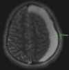
Post-concussion syndrome (PCS) is a common sequela of mild traumatic brain injury in adolescent athletes, typically resolving within weeks. However, persistent or atypical symptoms warrant further investigation to exclude structural pathology. Arachnoid cysts, though often incidental, can become symptomatic following trauma and may mimic or exacerbate PCS. A previously healthy adolescent American football player presented with persistent headaches and cognitive symptoms 14 weeks after a sports-related concussion. Despite completing a return-to-play protocol, his symptoms worsened. A brain MRI revealed a large left-sided arachnoid cyst with a 5 mm midline shift, consistent with a primary arachnoid cyst. The patient was admitted to the medical unit of the pediatric hospital for monitoring on the medical floor with 48 hours of observation and repeat imaging to verify that the MRI findings were not worsening. Serial imaging over three months demonstrated regression of the cyst and resolution of mass effect. He remained asymptomatic at follow-up but was permanently restricted from contact sports. This case underscores the importance of considering structural brain lesions in athletes with prolonged or atypical post-concussive symptoms. MRI played a critical role in identifying a potentially life-threatening condition that mimicked PCS. Conservative management may be appropriate in select cases of arachnoid cysts with favorable clinical and radiographic progression. Surgical management may be necessary in cases of increased intracranial pressure, neurologic deficit, or radiographic progression.
Keywords: brain concussion; congenital arachnoid cyst; management of arachnoid cyst; pediatric sports medicine; post-concussion headache.
Copyright © 2025, Guin et al.
Conflict of interest statement
Human subjects: Informed consent for treatment and open access publication was obtained or waived by all participants in this study. Conflicts of interest: In compliance with the ICMJE uniform disclosure form, all authors declare the following: Payment/services info: All authors have declared that no financial support was received from any organization for the submitted work. Financial relationships: All authors have declared that they have no financial relationships at present or within the previous three years with any organizations that might have an interest in the submitted work. Other relationships: All authors have declared that there are no other relationships or activities that could appear to have influenced the submitted work.
Figures


References
-
- Intracranial arachnoid cysts in children: a review of pathogenesis, clinical features, and management. Gosalakkal JA. Pediatr Neurol. 2002;26:93–98. - PubMed
-
- Washington, DC: American Psychiatric Publishing, Inc.; 1994. Diagnostic and Statistical Manual of Mental Disorders. Fourth Edition.
-
- Intracranial arachnoid cysts: current concepts and treatment alternatives. Cincu R, Agrawal A, Eiras J. Clin Neurol Neurosurg. 2007;109:837–843. - PubMed
-
- Pathogenesis of arachnoid cyst: congenital or traumatic? Choi JU, Kim DS. https://pubmed.ncbi.nlm.nih.gov/9917544/ Pediatr Neurosurg. 1998;29:260–266. - PubMed
-
- Incidental findings on pediatric MR images of the brain. Kim BS, Illes J, Kaplan RT, Reiss A, Atlas SW. https://pubmed.ncbi.nlm.nih.gov/12427622/ AJNR Am J Neuroradiol. 2002;23:1674–1677. - PMC - PubMed
Publication types
LinkOut - more resources
Full Text Sources
