

A rare presentation of peritoneal mesothelioma: Esophageal encasement and Sister Mary Joseph nodule
- PMID: 40821375
- PMCID: PMC12355503
- DOI: 10.1016/j.radcr.2025.07.033
A rare presentation of peritoneal mesothelioma: Esophageal encasement and Sister Mary Joseph nodule
Abstract
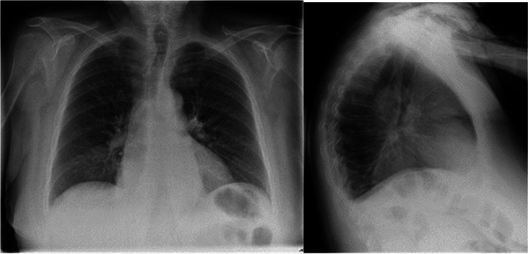
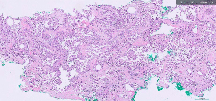
Malignant peritoneal mesothelioma (MPeM) is a rare malignancy that accounts for approximately 10%-30% of all mesothelioma cases, with the more common type being pleural mesothelioma. In this case report, we describe an atypical presentation of MPeM in a 73-year-old female presenting with periumbilical swelling and subacute chest pain. Imaging demonstrated a large periumbilical mass and a posterior mediastinal mass encasing, but not obstructing, the esophagus. A brief literature review examining the broad range of clinical and imaging manifestations of MPeM is presented.
Keywords: Malignant peritoneal mesothelioma; Mediastinal mass; Mesothelioma; Periumbilical mass; Sister Mary Joseph nodule.
Crown Copyright © 2025 Published by Elsevier Inc. on behalf of University of Washington.
Figures


References
-
- Price B. Analysis of current trends in United States mesothelioma incidence. Am J Epidemiol. 1997;145(3):211–218. - PubMed
-
- Price B., Ware A. Time trend of mesothelioma incidence in the United States and projection of future cases: an update based on SEER data for 1973 through 2005. Crit Rev Toxicol. 2009;39(7):576–588. - PubMed
-
- Bianchi C., Bianchi T. Malignant mesothelioma: global incidence and relationship with asbestos. Ind Health. 2007;45(3):379–387. - PubMed
Publication types
LinkOut - more resources
Full Text Sources
