

Difference between Okinawan and Dutch older adults in prefrontal brain activation
- PMID: 40821617
- PMCID: PMC12350274
- DOI: 10.3389/fnagi.2025.1454068
Difference between Okinawan and Dutch older adults in prefrontal brain activation
Abstract
Background: Older adults in Okinawa (Japan) are known for healthy aging and longevity. This is the first study to explore brain activation during executive functioning in Okinawan older adults in comparison to Western-European (Dutch) older adults.
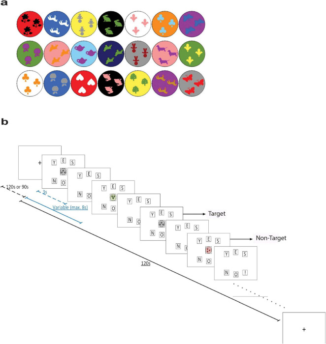
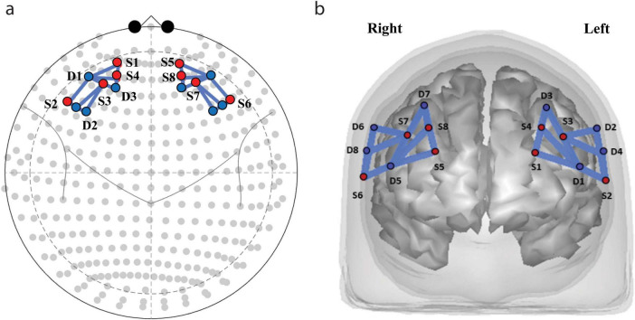
Methods: A total number of 80 participants were included in the study (41 from the Netherlands and 39 from Okinawa), with ages between 65 and 80 years). The groups did not differ for sex and handedness. Brain activation was measured during a visual working memory task and a verbal fluency task, for bilateral frontal cortex using functional near infrared spectroscopy (fNIRS). We investigated oxygenated hemoglobin (HbO) levels and laterality index.
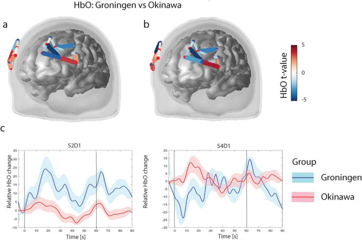
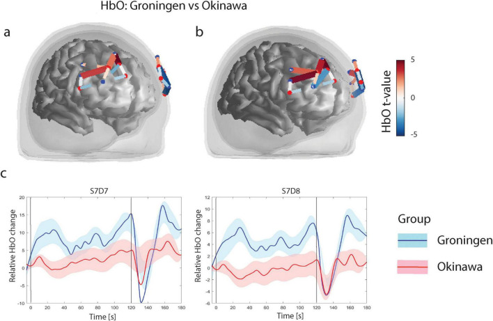
Results: Both groups performed within the normal range for their population. During verbal fluency, less activation in the left frontal gyrus was observed in Okinawa participants as compared to Dutch participants, and more activation in the anterior superior parts of the frontal gyrus. For the n-back task, the Okinawa group exhibited less activation in the right dorsolateral prefrontal cortex and more activation in the bilateral anterior frontal gyrus. Although laterality indices were similar for both tasks, Okinawa participants showed stronger left lateralization during category fluency.
Conclusion: Our results reveal less activation of the task-relevant areas in participants from Okinawa as compared to Dutch participants. It could be hypothesized, with caution, that Okinawan older adults may need less executive processing resources to perform the task. Other differences in activation may be related to different strategy use, which may be studied in more detail in future investigations.
Keywords: Okinawa; blue zones; executive function; functional near infrared spectroscopy (fNIRS); healthy aging; prefrontal brain activation.
Copyright © 2025 Ćurčić-Blake, Futenma, Willcox, Tazangi, Wardana, Ueda and Aleman.
Conflict of interest statement
The authors declare that the research was conducted in the absence of any commercial or financial relationships that could be construed as a potential conflict of interest.
Figures
Similar articles
-
Prescription of Controlled Substances: Benefits and Risks.2025 Jul 6. In: StatPearls [Internet]. Treasure Island (FL): StatPearls Publishing; 2025 Jan–. 2025 Jul 6. In: StatPearls [Internet]. Treasure Island (FL): StatPearls Publishing; 2025 Jan–. PMID: 30726003 Free Books & Documents.
-
Neural mechanisms of the relationship between aerobic fitness and working memory in older adults: An fNIRS study.Imaging Neurosci (Camb). 2024 May 10;2:imag-2-00167. doi: 10.1162/imag_a_00167. eCollection 2024. Imaging Neurosci (Camb). 2024. PMID: 40800507 Free PMC article.
-
Short-Term Memory Impairment.2024 Jun 8. In: StatPearls [Internet]. Treasure Island (FL): StatPearls Publishing; 2025 Jan–. 2024 Jun 8. In: StatPearls [Internet]. Treasure Island (FL): StatPearls Publishing; 2025 Jan–. PMID: 31424720 Free Books & Documents.
-
Technological aids for the rehabilitation of memory and executive functioning in children and adolescents with acquired brain injury.Cochrane Database Syst Rev. 2016 Jul 1;7(7):CD011020. doi: 10.1002/14651858.CD011020.pub2. Cochrane Database Syst Rev. 2016. PMID: 27364851 Free PMC article.
-
Falls prevention interventions for community-dwelling older adults: systematic review and meta-analysis of benefits, harms, and patient values and preferences.Syst Rev. 2024 Nov 26;13(1):289. doi: 10.1186/s13643-024-02681-3. Syst Rev. 2024. PMID: 39593159 Free PMC article.
References
-
- Aleman A. (2015). Our aging brain: How our mental capacities develop as we get older. Brunswick VIC: Scribe Publications Pty Ltd.
-
- Birchwood M., Smith J., Cochrane R., Wetton S., Copestake S. (1990). The social functioning scale. the development and validation of a new scale of social adjustment for use in family intervention programmes with schizophrenic patients. Br J Psychiatry 157 853–859. 10.1192/bjp.157.6.853 - DOI - PubMed
-
- Birn R. M., Kenworthy L., Case L., Caravella R., Jones T. B., Bandettini P. A., et al. (2010). Neural systems supporting lexical search guided by letter and semantic category cues: A self-paced overt response fMRI study of verbal fluency. Neuroimage 49 1099–1107. 10.1016/j.neuroimage.2009.07.036 - DOI - PMC - PubMed
LinkOut - more resources
Full Text Sources