

Biomimetic periosteum combining BMP-2-loaded M2 macrophage-derived exosomes for enhanced bone defect repair
- PMID: 40821662
- PMCID: PMC12354627
- DOI: 10.3389/fbioe.2025.1639394
Biomimetic periosteum combining BMP-2-loaded M2 macrophage-derived exosomes for enhanced bone defect repair
Abstract
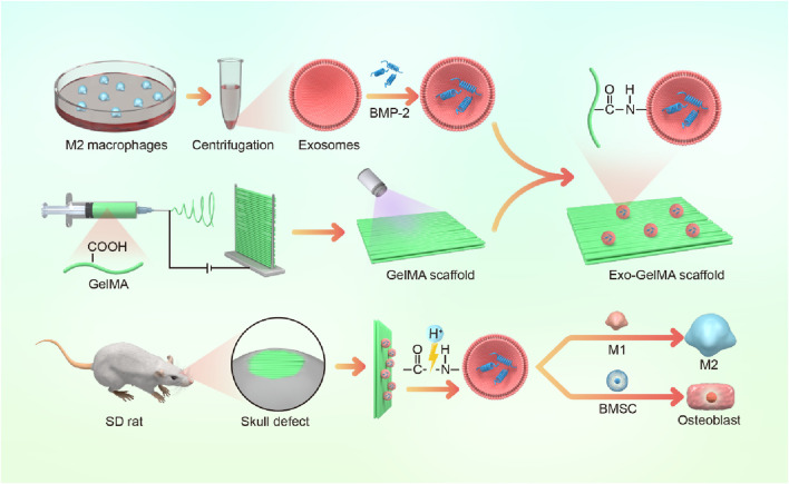
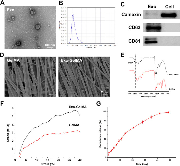
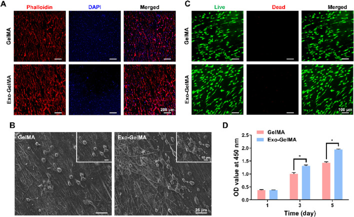
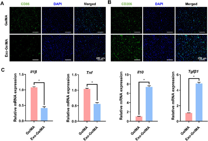
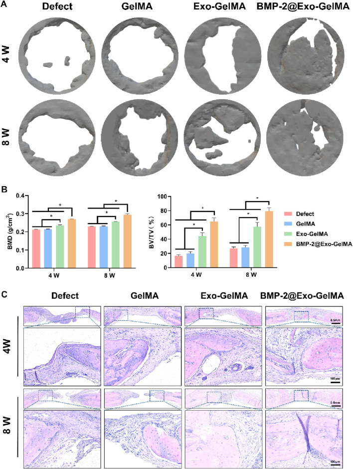
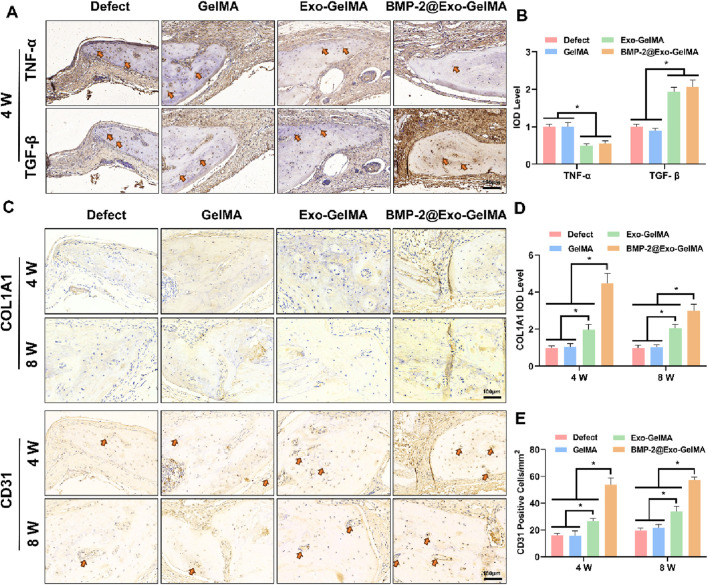
Bone defect repair continues to present a significant clinical challenge due to the limitations of traditional grafting techniques and the complexity involved in establishing a conducive regenerative microenvironment. In this study, we described the development of a multifunctional biomimetic periosteum based on electrospun gelatin methacryloyl (GelMA) membranes functionalized with bone morphogenetic protein-2 (BMP-2)-loaded M2 macrophage-derived exosomes. This engineered periosteum replicated the structural orientation and functional properties of natural periosteum, thereby providing a synergistic approach to promoting bone regeneration. Our findings indicated that the biomimetic periosteum served as a biocompatible scaffold that supported cell adhesion, proliferation, and differentiation. The incorporation of M2 macrophage-derived exosomes facilitated the creation of an anti-inflammatory immune microenvironment by polarizing macrophages towards the M2 phenotype, while the sustained release of BMP-2 enhances osteogenic differentiation and mineralization. In vivo experiments using a rat cranial defect model demonstrated that the BMP-2@Exo-GelMA membrane significantly accelerated bone defect repair, achieving superior outcomes in new bone formation and vascularization compared to control groups. This study underscored the potential of integrating immunomodulatory and osteoinductive strategies to develop next-generation biomaterials for bone tissue engineering. The biomimetic periosteum represented a promising therapeutic approach for addressing critical-sized bone defects and advancing clinical practices in bone regeneration.
Keywords: BMP; biomimetic periosteum; bone defect; exosomes; macrophage.
Copyright © 2025 Ling, Bai, Xie, Liu, Lu, Yuan, Li and Qian.
Conflict of interest statement
The authors declare that the research was conducted in the absence of any commercial or financial relationships that could be construed as a potential conflict of interest.
Figures
Similar articles
-
Polyethyleneimine-modified eggshell membrane for dual bone and periosteum regeneration: A multifunctional approach for guided bone healing.Acta Biomater. 2025 Jul 23:S1742-7061(25)00541-0. doi: 10.1016/j.actbio.2025.07.044. Online ahead of print. Acta Biomater. 2025. PMID: 40712727
-
Oncostatin-M functionalized cryogel microspheres for promoting diabetic bone defects regeneration.J Orthop Translat. 2025 Jun 20;53:138-148. doi: 10.1016/j.jot.2025.06.002. eCollection 2025 Jul. J Orthop Translat. 2025. PMID: 40606844 Free PMC article.
-
Reverse biogradient biomimetic periosteum with osteogenic and angiogenic characteristics for bone regeneration.Mater Today Bio. 2025 Jun 9;33:101967. doi: 10.1016/j.mtbio.2025.101967. eCollection 2025 Aug. Mater Today Bio. 2025. PMID: 40585030 Free PMC article.
-
Engineered M2 macrophage-derived exosomes: mechanisms and therapeutic potential in inflammation regulation and regenerative medicine.Acta Biomater. 2025 Jul 18:S1742-7061(25)00537-9. doi: 10.1016/j.actbio.2025.07.039. Online ahead of print. Acta Biomater. 2025. PMID: 40684929 Review.
-
Advanced therapeutic scaffolds of biomimetic periosteum for functional bone regeneration.J Nanobiotechnology. 2025 Jul 26;23(1):542. doi: 10.1186/s12951-025-03614-5. J Nanobiotechnology. 2025. PMID: 40713791 Free PMC article. Review.
References
-
- Jin J., Yang Y., Yang J., Sun Z., Wang D., Qin Y., et al. (2024a). Macrophage metabolic reprogramming-based diabetic infected bone defect/bone reconstruction though multi-function silk hydrogel with exosome release. Int. J. Biol. Macromol. 278 (Pt 4), 134830. 10.1016/j.ijbiomac.2024.134830 - DOI - PubMed
LinkOut - more resources
Full Text Sources