Review
. 2025 Aug 15;6(8):e70319.
doi: 10.1002/mco2.70319. eCollection 2025 Aug.

Mitochondrial Quality Control in Health and Disease

Affiliations
Review

Mitochondrial Quality Control in Health and Disease

Lin Ye et al. MedComm (2020). .

Abstract

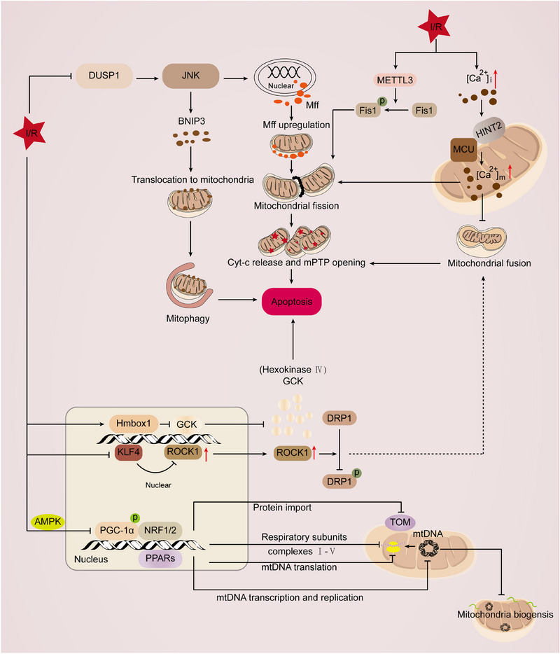
Mitochondria are central regulators of cellular energy metabolism, and their functional integrity is essential for maintaining cellular homeostasis. Mitochondrial quality control (MQC) encompasses a coordinated network of mitochondrial biogenesis, dynamics (fusion and fission), and selective autophagy (mitophagy), which together sustain mitochondrial structure and function. Under physiological conditions, MQC ensures the removal of dysfunctional mitochondria, restricts excessive reactive oxygen species production, and modulates apoptosis, thereby supporting the high energy demands of organs such as the heart and brain. Disruption of MQC contributes to the onset and progression of various diseases, including neurodegenerative disorders, cardiovascular pathologies, and metabolic syndromes, largely through accumulation of damaged mitochondria and impaired metabolic signaling. While the core components of MQC have been characterized, the mechanistic interplay among its modules and their disease-specific alterations remain incompletely defined. This review provides an integrated overview of the molecular pathways governing mitochondrial biogenesis, dynamics, and mitophagy, with a focus on their cross-talk in maintaining mitochondrial homeostasis. We further discuss how MQC dysfunction contributes to disease pathogenesis and examine emerging therapeutic approaches aimed at restoring mitochondrial quality. Understanding the regulatory logic of MQC not only elucidates fundamental principles of cellular stress adaptation but also informs novel strategies for disease intervention.

Keywords: disease intervention; mitochondria; mitochondrial quality control; therapeutic strategies.


Conflict of interest statement

The authors declare no conflicts of interest.

Figures

FIGURE 1
FIGURE 1
Mitochondrial quality control. The MQC system constitutes a multilayered regulatory network essential for preserving mitochondrial integrity and functional homeostasis. This system orchestrates a series of interconnected processes—including mitochondrial biogenesis, fission, fusion, protein degradation, and mitophagy—to maintain a healthy mitochondrial population within the cell. Over time, mitochondrial function tends to decline; under such conditions, fusion events mediated by proteins such as OPA1, MFN1, and MFN2 enable the exchange of intramitochondrial components, thereby sustaining mitochondrial performance. In contrast, persistent mitochondrial dysfunction activates fission machinery involving FIS1, DRP1, and MFF, which facilitates the segregation and elimination of damaged organelles. During this process, relatively intact mitochondria are reintegrated into the network via continued fusion, whereas impaired mitochondria are selectively removed through mitophagy, predominantly via the canonical PINK1–Parkin pathway. The dynamic interplay among fusion, fission, and mitophagy ensures optimal mitochondrial turnover and prepares the cell for the demands of mitochondrial biogenesis. Mitochondrial biogenesis, a critical regenerative mechanism, serves to replenish the mitochondrial pool by replacing senescent or damaged mitochondria. This process is tightly regulated by both mtDNA and nDNA, and is governed by transcriptional coactivators and regulators such as PGC‐1α, which drive the expression of genes involved in mitochondrial replication and oxidative metabolism. Newly synthesized mitochondria resulting from biogenesis enhance ATP production, thereby supporting cellular metabolic needs under both physiological and pathological conditions. Abbreviations: ATP, adenosine triphosphate; Drp1, dynamin‐related protein 1; FIS1, fission mitochondrial 1; MFF, mitochondrial fission factor; MFN1, mitofusin‐1; MFN2, mitofusin‐2; MQC, mitochondrial quality control; mtDNA, mitochondrial DNA; nDNA, nuclear DNA; OPA1, optic atrophy 1; PGC‐1α, peroxisome proliferators‐activated receptor γ coactivator l alpha; PINK1, PTEN‐induced putative kinase 1.
FIGURE 2
FIGURE 2
The role of mitochondrial autophagy in ischemia–hypoxia‐associated diseases. (A) In HI brain injury, PHLDA1 exacerbates brain damage by suppressing FUNDC1‐mediated mitophagy. HPC with 8% O2 alleviates hypoxic–ischemic brain injury following tGCI by activating the PINK1/Parkin pathway to promote mitophagy. In HI brain injury, activation of the BNIP3‐mediated mitophagy pathway regulates iron homeostasis and redox balance, thereby inhibiting the occurrence of ferroptosis. In HI brain injury, melatonin regulates mitophagy by upregulating the expression of NLRX1, Beclin‐1, and ATG7, and downregulating the expression of mTOR, thereby protecting neuronal cells from damage. (B) NR4A1 negatively regulates myocardial ischemia–reperfusion injury by activating Mff‐mediated mitochondrial fission and inhibiting FUNDC1‐mediated mitophagy. Additionally, NR4A1 also suppresses Parkin‐driven mitophagy, leading to impaired cardiac function. CHK1 maintains mitochondrial homeostasis and mitophagy by phosphorylating SIRT1 and inhibiting SMURF2‐mediated degradation, thereby protecting the heart from I/R injury through antioxidative stress and promoting mitochondrial biogenesis. Yiqi Huoxue alleviates I/R injury by regulating mitophagy, improving mitochondrial structure and function, and reducing oxidative stress. Gastrodin improves mitophagy by activating the PINK1/Parkin pathway, thereby reducing myocardial I/R injury. (C) In IRI, HIF‐1α CTAD transcriptionally regulates HK2 and activates PINK1/Parkin‐mediated mitophagy, thereby alleviating kidney injury. Inhibiting ACSF2 enhances IRI‐induced mitophagy, restores mitochondrial function, and further promotes mitophagy by regulating BNIP3, thereby alleviating IRI. OGG1 negatively regulates mitophagy by suppressing the PINK1/Parkin pathway in IRI, thereby exacerbating IRI. PANX1 exacerbates IRI by regulating the ATP–P2Y–mTOR signaling pathway to inhibit mitophagy. Abbreviations: HPC, hypoxic postconditioning; tGCI, transient global cerebral ischemia; PINK1, PTEN‐induced putative kinase 1; BNIP3, BCL2 interacting protein 3; NLRX1, NLR family member X1; ATG7, autophagy related 7; mTOR, mammalian target of rapamycin; NR4A1, nuclear receptor 4A1; Mff, mitochondrial fission factor; FUNDC1, FUN14 domain containing 1; CHK1, checkpoint kinase 1; SIRT1, silent information regulator 1; SMURF2, SMAD‐specific E3 ubiquitin protein ligase 2; CTAD, C‐terminal transactivation domain; HK2, hexokinase 2; ACSF2, acyl‐CoA synthetase family member 2; OGG1, 8‐oxoguanine DNA glycosylase‐1; PANX1, Pannexin1.
FIGURE 3
FIGURE 3
Molecular pathways of cell death. Apoptosis. Apoptosis involves two pathways: the intrinsic mitochondrial pathway and the extrinsic death receptor pathway. The intrinsic pathway is triggered by stress signals that activate BAX/BAK, causing MOMP and cytochrome c release, which forms the apoptosome to activate caspase‐9 and downstream caspases. The extrinsic pathway starts with death receptor activation, forming the DISC and activating caspase‐8, which cleaves caspase‐3/7. Caspase‐8 also cleaves BID to tBID, linking both pathways and amplifying apoptosis. Pyroptosis: In mice, PAMPs are recognized by PRRs, leading to activation of caspase‐1 and caspase‐11, whereas in humans, this pathway is mediated by caspase‐1, ‐4, and ‐5. Activated caspases cleave GSDMD, releasing the GSDMD‐N, which inserts into the plasma membrane to form pores, causing membrane rupture and release of proinflammatory cytokines IL‐1β and IL‐18. MOMP induces the release of mtDNA and ROS; mtDNA acts to activate the NLRP3 inflammasome, while ROS promote inflammation, establishing a positive feedback loop that exacerbates pyroptosis. Necroptosis: TNF binding induces TNFR1 trimerization and formation of Complex I, which recruits TRADD, RIPK1, and other factors to maintain cellular homeostasis. Upon dysregulation, activated RIPK1 assembles Complex IIa, composed of FADD and caspase‐8, to induce apoptosis. When caspase‐8 is inhibited, RIPK1 interacts with RIPK3 to form Complex IIb, leading to RIPK3 activation and phosphorylation of MLKL. Phosphorylated MLKL oligomerizes and translocates to the plasma membrane, disrupting membrane integrity and executing necroptosis. Ferroptosis: xCT, encoded by SLC7A11, mediates the exchange of extracellular cystine and intracellular glutamate. Once inside the cell, cystine is reduced to cysteine, which is essential for GSH synthesis. GSH acts as a cofactor for GPX4, an enzyme that prevents lipid peroxidation. Cysteine deficiency impairs GPX4 activity, enhances TCA cycle flux, and increases ROS production. Fe2⁺ drives the Fenton reaction, generating hydroxyl radicals that oxidize polyunsaturated fatty acids, leading to lipid peroxidation. The breakdown of ferritin and release of mitochondrial iron further intensify oxidative stress. Because mitochondrial membranes are close to ROS and iron sources, they are more prone to structural damage. The combination of increased membrane permeability and ROS accumulation promotes ferroptosis. Abbreviations: MOMP, mitochondrial outer membrane permeabilization; APAF‐1, apoptosis protease‐activating factor‐1; FADD, Fas‐associating protein with a novel death domain; BCL‐2, B‐cell lymphoma‐2; BIM, Bcl‐2 interacting mediator of cell death; BID, BH3 interacting domain death agonist; BAD, BCL2‐associated agonist of cell death; PUMA, p53 upregulated modulator of apoptosis; BCL‐xL, B‐cell lymphoma‐extra large; MCL1, myeloid cell leukemia‐1; SMAC, second mitochondria‐derived activator of caspases; OMI, high temperature requirement factor A2; XIAP, X‐linked inhibitor of apoptosis protein; Fas, factor‐related apoptosis; TNFR1, TNF receptor type 1; DR4, death receptor 4; DR5, death receptor 5; FasL, Fas ligand; TNF, tumor necrosis factor DED, death effector domain DISC, death‐inducing signaling complex; PAMPs, pathogen‐associated molecular patterns; PRRs, pattern recognition receptors; GSDMD, gasdermin D; DAMP, damage‐associated molecular pattern; NLRP3, NOD‐like receptor thermal protein domain‐associated protein 3; MLKL, mixed lineage kinase domain‐like protein; CYLD, cylindromatosis; SPATA2, spermatogenesis associated 2; SLC7A11, solute carrier family 7 member 11; SLC3A2, solute carrier family 3 member 2; GSH, glutathione; GPX4, glutathione peroxidase 4; TCA, tricarboxylic acid.
FIGURE 4
FIGURE 4
The role of mitochondria in the process of cell death induced by cardiac I/R. (A) PGAM5 is upregulated at both the transcriptional and expression levels during I/R. Deficiency of PGAM5 increases mitochondrial DNA copy number and transcription levels, normalizes mitochondrial respiration, inhibits mitochondrial ROS production, and prevents abnormal mPTP opening during I/R. PGAM5 deficiency disrupts I/R‐induced Drp1 dephosphorylation, partially suppressing mitochondrial fission and ultimately rescuing cardiomyocyte necrosis. Inhibiting ADK can reduce cardiomyocyte necrosis. ADK inhibition decreases MLKL and its phosphorylation, as well as the phosphorylation of CaMKII. XIAP, which is phosphorylated and stabilized through the adenosine receptors A2B and A1/Akt pathways, plays a crucial role in the effects of ADK inhibition on necrosis. RIP3‐induced CaMKII activation may trigger the opening of the mPTP through phosphorylation, oxidation, or both, leading to myocardial necrosis. (B) lncRNA SNHG1 alleviates cardiomyocyte pyroptosis during myocardial ischemia–reperfusion injury by acting as a “sponge” for miR‐137‐3p, thereby regulating the KLF4/TRPV1/AKT axis. Circ‐DGKZ regulates miR‐345‐5p and, through direct interaction, modulates TLR4 in cardiomyocytes. This, in turn, disrupts cardiomyocyte pyroptosis and induces autophagy via the TLR4/NF‐κB axis. PCSK9 can induce mtDNA damage and activate the NLRP3 inflammasome signaling pathway. (C) The Alox15‐derived intermediate metabolite 15‐HpETE promotes the binding of PGC‐1α to the ubiquitin ligase ring finger protein 34, leading to its ubiquitin‐dependent degradation, which causes impaired mitochondrial biogenesis and abnormal mitochondrial morphology, ultimately resulting in ferroptosis. In I/R, Nrf2‐mediated upregulation of Hmox1 leads to systemic accumulation of nonheme iron and induces ferroptosis. DIC and OGC play roles in GSH transport, and their inhibition exacerbates ferroptosis. Abbreviations: PGAM5, phosphoglycerate mutase family member 5; ROS, reactive oxygen species; ADK, adenosine kinase; MLKL, mixed lineage kinase domain‐like protein; XIAP, X‐linked inhibitor of apoptosis protein; RIP3, receptor‐interacting protein kinase 3; CaMKII, calcium/calmodulin‐dependent protein kinase II; TRPV1, transient receptor potential vanilloid 1; PCSK9, proprotein convertase subtilisin/kexin type 9; NLRP3, NOD‐like receptor protein 3; Alox15, arachidonate 15‐lipoxygenase; PGC‐1α, peroxisome proliferator‐activated receptor gamma coactivator 1‐alpha; Nrf2, nuclear factor erythroid 2‐related factor 2; Hmox1, heme oxygenase 1; DIC, dicarboxylate carrier; OGC, oxoglutarate carrier; GSH, glutathione.
FIGURE 5
FIGURE 5
Mitochondrial DAMP signaling mediates inflammatory responses. Its principal mechanism involves the release of mitochondrial components and byproducts arising from mitochondrial dysfunction or cell death, which accumulate intracellularly or in the extracellular space and initiate inflammation. mtDNA can exit via BAX–BAK1 pores or the PTPC, subsequently acting as a potent activator of cGAS. Once cGAS is activated, STING1 signaling is triggered, leading to the production of cytokines such as IFNβ1, IL‐6, and TNF. Furthermore, under conditions of mitochondrial impairment, mtDNA and ROS released from mitochondria can promote IL‐1β and IL‐18 secretion through inflammasome pathways. ETC function may also modulate intracellular ATP availability via PCr, thereby influencing inflammasome activation independently of ROS. Cells undergoing death release ATP through lysosomal exocytosis and PANX1 channels; this ATP subsequently binds purinergic receptors to regulate the chemotaxis and immunostimulatory activity of APCs. Abbreviations: mtDNA, mitochondrial DNA; PTPC, permeability transition pore complex; cGAS, cyclic GMP–AMP synthase; IFNβ1, interferon‐β1; TNF, tumor necrosis factor; ETC, electron transport chain; PCr, phosphocreatine; PANX1, pannexin 1; APCs, antigen‐presenting cells.
FIGURE 6
FIGURE 6
Regulation of mitochondrial biogenesis during I/R. I/R‐induced DUSP1 deficiency promotes the activation of JNK, which upregulates the expression of the Mff. Elevated levels of Mff are associated with increased mitochondrial fission and apoptosis. Additionally, the absence of DUSP1 amplifies BNIP3 phosphorylation activation via JNK, leading to enhanced mitophagy. The increased mitophagy markedly depletes mitochondrial quality, resulting in mitochondrial metabolic dysfunction. METTL3 deficiency can interfere with DNA‐PKcs phosphorylation, thereby preventing the downstream activation of Fis1 and subsequently inhibiting pathological mitochondrial fission. This ultimately improves cardiac function. Ca2+ overload is a key initiator of mitochondrial fission. HINT2 regulates the MCU complex by directly interacting with MCU in cardiac microvascular endothelial cells, thereby inhibiting Ca2+ overload. Overexpression of HINT2 suppresses the MCU complex‐induced mitochondrial calcium overload, preventing mitochondrial fission and apoptosis pathways, and thus alleviates cardiac microvascular ischemia–reperfusion injury. Under pathological conditions, the expression of Hmbox1 is upregulated. Inhibition of Hmbox1 activates the Akt/mTOR/P70S6K pathway and increases the transcription of GCK, leading to reduced cardiomyocyte apoptosis and improved mitochondrial respiration and glycolysis. This ultimately protects the heart. KLF4 deficiency can affect the expression of ROCK1 at the transcriptional level, thereby inducing DRP1‐mediated mitochondrial fission and ultimately exacerbating myocardial injury. Under pathological conditions, the process of mitochondrial biogenesis involves the inhibition of the interaction between PGC‐1α and Nrf1/2, PPARs, and suppresses the maintenance of mitochondrial oxidative phosphorylation function, inhibits the synthesis and import of nuclear gene‐encoded mitochondrial proteins, as well as the transcription, replication, and translation of mitochondrial DNA, thereby exacerbating damage. Abbreviations: DUSP1, dual specificity phosphatase 1; JNK, c‐Jun N‐terminal kinase; Mff, mitochondrial fission factor; BNIP3, BCL2 interacting protein 3; METTL3, methyltransferase like 3; DNA‐PKcs, DNA‐dependent protein kinase catalytic subunit; Fis1, fission 1; HINT2, histidine triad nucleotide binding protein 2; MCU, mitochondrial calcium uniporter; Hmbox1, heme oxygenase 1; Akt/mTOR/P70S6K: protein kinase B/mechanistic target of rapamycin/ribosomal protein S6 kinase beta‐1; GCK, glucokinase; KLF4, Krüppel‐like factor 4; ROCK1, Rho‐associated protein kinase 1; DRP1, dynamin‐related protein 1; PPARs, peroxisome proliferators‐activated receptors; TOM, translocator of the outer mitochondrial membrane; PGC1‐α, peroxisome proliferator‐activated receptor γ coactivator 1‐α.

Similar articles

References

    1. Glover H. L., Schreiner A., Dewson G., Tait S. W. G., “Mitochondria and Cell Death,” Nature Cell Biology 26, no. 9 (2024): 1434‐1446. - PubMed
    1. Wen H., Deng H., Li B., et al., “Mitochondrial Diseases: From Molecular Mechanisms to Therapeutic Advances,” Signal Transduction and Targeted Therapy 10, no. 1 (2025): 9. - PMC - PubMed
    1. Nunnari J., Suomalainen A., “Mitochondria: In Sickness and in Health,” Cell 148, no. 6 (2012): 1145‐1159. - PMC - PubMed
    1. Ashrafi G., Schwarz T. L., “The Pathways of Mitophagy for Quality Control and Clearance of Mitochondria,” Cell Death and Differentiation 20, no. 1 (2013): 31‐42. - PMC - PubMed
    1. Bock F. J., Tait S. W. G., “Mitochondria as Multifaceted Regulators of Cell Death,” Nature Reviews Molecular Cell Biology 21, no. 2 (2020): 85–100. - PubMed

LinkOut - more resources