. 2025 Aug 1:12:1595173.
doi: 10.3389/fmolb.2025.1595173. eCollection 2025.

Immunoglobulin A carries sulfated and O-acetylated N-glycans primarily at the tailpiece site - strategies for site-specific N-glycan identification

Affiliations

Immunoglobulin A carries sulfated and O-acetylated N-glycans primarily at the tailpiece site - strategies for site-specific N-glycan identification

Frania J Zuniga-Banuelos et al. Front Mol Biosci. .

Abstract

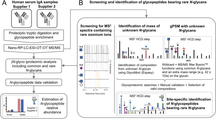
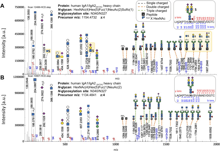
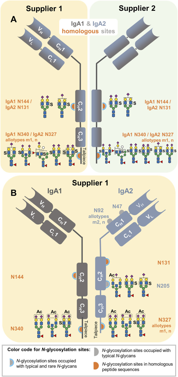
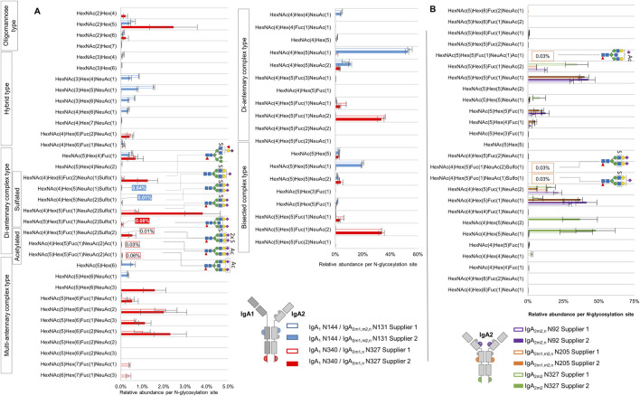
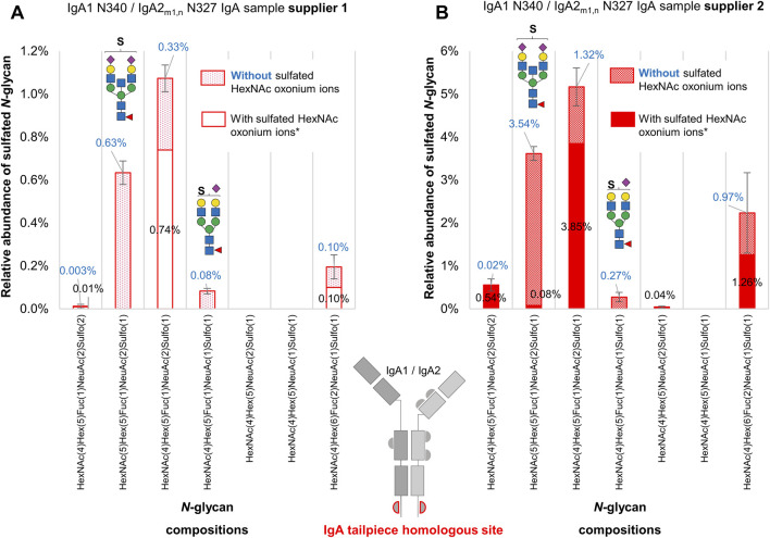
Sulfated N-glycans from human immunoglobulin A (IgA) were recently discovered via glycomic approaches. However, their site-specific description is still pending. Certain N-glycan structures at specific N-glycosylation sites in IgA are crucial for microbial neutralization and effector functions. For instance, sialylated N-glycans on the C-terminal tailpiece mediate anti-viral activity by interfering with sialic-acid-binding viruses. Sulfated N-glycan epitopes can be ligands for viral proteins and thus play a role in the immune response. In this study, we performed a site-specific screening for sulfated and other rare N-glycans in two commercially available human serum IgA samples employing an in-depth N-glycoproteomic approach, previously developed by us. We found evidence of complex-type and hybrid-type N-glycans containing sulfated N-acetylhexosamine (sulfated HexNAc) attached to the N-glycosylation sites in the tailpiece and the CH2 domain of both IgA subclasses. Also, complex-type N-glycan compositions bearing O-acetylated sialic acid were identified primarily at the tailpiece site. Surprisingly, N-glycans bearing glucuronic acid were identified in the commercial IgA samples, but from peptides of "contaminant" glycoproteins. A detailed comparison of the N-glycosylation profiles of human serum IgA samples from two suppliers showed such N-glycans with sulfated HexNAc consistently in higher abundance in the tailpiece region. These findings have not been described before for a site-specific glycopeptide analysis. Overall, our work provides strategies for performing a dedicated site-specific search for sulfated and O-acetylated N-glycans that can be easily transferred, e.g., to human IgA derived from mucosal tissues, milk, or saliva. We expect that a wider and deeper micro-heterogeneity description of clinically relevant glycoproteins, such as immunoglobulins, can expand the screening for biomarkers or treatment options.

Keywords: N-glycosylation; O-acetylated N-glycans; glycoproteomics; immunoglobulin A (IgA); mass spectrometry; oxonium marker ions; rare N-glycans; sulfated N-glycans.


Conflict of interest statement

ER is the founder and CEO of glyXera GmbH. FZ-B is an employee of glyXera GmbH and Max Planck Institute. glyXera provides high-performance glycoanalytical products and services, and holds several patents for xCGE-LIF-based glycoanalysis. UR is a shareholder of glyXera GmbH. The remaining authors declare that the research was conducted in the absence of any commercial or financial relationships that could be construed as a potential conflict of interest.

Figures

FIGURE 1
FIGURE 1
Experimental design for expanding the N-glycoproteomic analysis of IgA by applying strategies for the identification of sulfated and other rare N-glycopeptides. (A) Analysis of human serum IgA of two commercial suppliers where the identification of rare N-glycopeptides is integrated. (B) Strategies to determine the software search parameters for maximizing the identification of rare N-glycopeptides. Nano-RP-LC-ESI-OT-OT-M/MS: nano-reversed phase-liquid chromatography with electrospray-ionization coupled orbitrap tandem mass spectrometric measurement for precursor and fragment ions. HCD.step: higher-energy collisional dissociation with stepped normalized collisional energy [NCE of 20, 35, and 50]. gPSM: glycopeptide spectra match.
FIGURE 2
FIGURE 2
Comparison of HCD.step fragment ion spectra derived from two N-glycopeptides (with the same peptide backbone) - one bearing the sulfated versus the one bearing the non-sulfated N-glycan at the N-glycosylation site in the IgA tailpiece. (A) HCD.step spectrum shows B and Y ions confirming the sulfated N-glycan HexNAc4Hex5Fuc1NeuAc2Sulfo1. B and Y ions supporting structural evidence on sulfated N-glycans highlighted in yellow. (B) HCD.step spectrum shows the B and Y ions observed in the common non-sulfated N-glycan HexNAc4Hex5Fuc1NeuAc2. The annotation of the N-glycan composition enclosing the numbers in parentheses is required by the search engine.
FIGURE 3
FIGURE 3
Site-specific description of the here identified N-glycans carrying HexNAc sulfation and NeuAc O-acetylation in commercially available human serum IgA samples from two suppliers. (A) Sulfated N-glycans identified in IgA1 and IgA2 homologous sites. (B) Sulfated N-glycans identified in unique N-glycosylation sites of IgA2 and NeuAc O-acetylated N-glycans identified within the entire analysis. The sample in which each N-glycan composition was identified is also indicated in yellow and green for the IgA sample from supplier 1 and 2, respectively.
FIGURE 4
FIGURE 4
Comparison of micro-heterogeneity of N-glycosylation identified in commercially available human serum IgA samples from two suppliers. (A) Relative abundance of N-glycans at the homologous N-glycosylation sites N340-IgA1/N327-IgA2(m1/n) and N144-IgA1/N131-IgA2. (B) Relative abundance of N-glycans at the N-glycosylation sites that are exclusively present in IgA2 subclasses. The color and format of the labels with values agree with the IgA sample supplier color code indicated in the legend. Results for relative quantification are shown in the Supplementary Tables S6–S9.
FIGURE 5
FIGURE 5
A comparative analysis of sulfated N-glycans with and without HexNAc-sulfated oxonium ions attached to the IgA tailpiece site in commercially available human serum IgA samples from two suppliers. (A) The sample from supplier 1. (B) The sample from supplier 2. Sulfated N-glycopeptides were identified, manually validated, and verified for oxonium marker ions through Byonic™. Two sulfated N-glycan structures, one with bisecting HexNAc and another mono-sialylated, appear more abundant in glycopeptide spectra matches when their spectra do not present oxonium marker ions of sulfated HexNAc. Asterisk (*) indicates that oxonium-ion-based evidence refers to detection of Sulfo1HexNAc1 [M+H]+ or Sulfo1HexNAc1Hex1 [M+H]+ oxonium ions.

Similar articles

References

    1. Alagesan K., Silva D. V., Seeberger P. H., Kolarich D. (2019). A novel, ultrasensitive approach for quantitative carbohydrate composition and linkage analysis using LC-ESI ion trap tandem mass spectrometry. bioRxiv. 10.1101/853036 - DOI
    1. Alocci D., Mariethoz J., Gastaldello A., Gasteiger E., Karlsson N. G., Kolarich D., et al. (2019). GlyConnect: Glycoproteomics goes visual, interactive, and analytical. J. Proteome. Res. 18 (2), 664–677. 10.1021/acs.jproteome.8b00766 - DOI - PubMed
    1. Atkin J. D., Pleass R. J., Owens R. J., Woof J. M. (1996). Mutagenesis of the human IgA1 heavy chain tailpiece that prevents dimer assembly. J. Immunol. 157 (1), 156–159. 10.4049/jimmunol.157.1.156 - DOI - PubMed
    1. Bastian A., Kratzin H., Eckart K., Hilschmann N. (1992). Intra- and interchain disulfide bridges of the human J chain in secretory immunoglobulin A. Biol. Chem. Hoppe Seyler 373 (12), 1255–1263. 10.1515/bchm3.1992.373.2.1255 - DOI - PubMed
    1. Boisgard R., Charpigny G., Chanat E. (1999). Polymeric IgA are sulfated proteins. FEBS Lett. 463 (3), 250–254. 10.1016/s0014-5793(99)01631-2 - DOI - PubMed

LinkOut - more resources