. 2025 Aug 8:2025:4443773.
doi: 10.1155/ijin/4443773. eCollection 2025.

MLR Corresponds to the Functional Status of Monocytes in Chronic Lymphocytic Leukemia

Affiliations

MLR Corresponds to the Functional Status of Monocytes in Chronic Lymphocytic Leukemia

Wioleta Grzegorzewska et al. Int J Inflam. .

Abstract

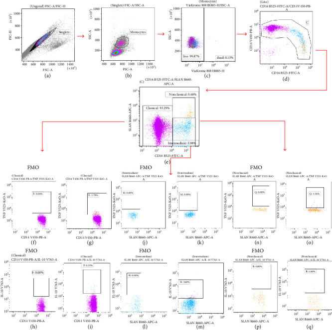
Background: The role of the inflammatory microenvironment in initiating and progressing chronic lymphocytic leukemia (CLL) is still not clarified. To date, it has been shown that the only way to reflect inflammation in the systemic circulation is to assess inflammatory markers in peripheral blood. However, in the age of modern technology, a more detailed analysis of inflammatory cells circulating in the blood of CLL patients would be useful. Objectives: The study aimed to evaluate the relationship between one of the hematological inflammatory indexes-the monocyte/lymphocyte ratio (MLR) and the risk of CLL progression associated with disease activity. In addition, we wanted to analyze whether the MLR parameter in CLL could suggest the functional immune status of circulating main monocyte subsets. Methods: The study included peripheral blood samples from 54 untreated, newly diagnosed CLL patients and 20 healthy volunteers (HVs). Immunological characterization of monocyte subpopulations included their detailed assessment by multiparametric flow cytometry, including evaluation of surface markers and intracellular expression of cytokines. In addition, the relative expression of selected microRNA (miR-21-3p, miR-150-5p, miR-106a-5p) was determined in FACS-sorted monocyte subsets. Results: In our study, CLL patients had significantly lower values of MLR parameters compared to HVs (p < 0.0001). However, the value of MLR was higher in CLL patients with negative clinical and laboratory prognostic factors, i.e., increased percentage of CD5+/CD19+ cells with ZAP-70 and CD38 expression. We noticed that the percentage of intermediate monocytes is significantly higher, but classical and nonclassical ones are significantly lower in MLR-high compared to MLR-low CLL patients. Moreover, among the monocyte subsets circulating in the blood of MLR-high, ZAP-70+, and CD38+, CLL patients' intermediate monocytes were characterised by increased intracellular expression of IL-10 and decreased miR-150-5p relative expression compared to intermediate monocytes in the MLR-low, ZAP-70-, and CD38- groups, suggesting a potential link between hematological inflammatory index and the formation of intermediate monocytes that promote CLL burden. Conclusions: The MLR index may serve not only as a marker of CLL activity, but also indirectly indicate changes in the phenotype and function of monocyte subpopulations present in the blood microenvironment. Moreover, the MLR-high parameter seems to correspond to an increase in the percentage of intermediate monocytes with anti-inflammatory properties, which may potentially promote disease progression and worsen its prognosis.


Conflict of interest statement

The authors declare no conflicts of interest.

Figures

Figure 1
Figure 1
Example illustrating the gating during the cytometric analysis of monocyte subpopulations with intracellular expression of TNF and IL-10. Dot plot of FSC-H versus FSC-A—singlets separation (a). Dot plot SSC versus FSC gating of monocytes (b). Dot plot: ViaKrome 808 versus SSC—exclusion of dead cells from further analysis. Only live cells were used for further evaluation (c). Differentiation of monocyte subpopulations by CD14 and CD16 expression (dot plot: CD16 FITC vs. CD14 V450) (d). Dot plot SLAN APC versus CD16 FITC allowed the identification of classical (CD14++/CD16−/SLAN−), intermediate (CD14+/CD16+/SLAN−), and nonclassical (CD14+/CD16++/SLAN+) monocytes (e). Dot plots Fluorescence Minus One (FMO) are control tubes containing all panel antibodies minus one for TNF BV510 (f), (j), (n) or for IL-10 BV786 (h), (l), (p). Selected classical monocytes with intracellular expression of TNF BV510 (g) or IL-10 BV786 (i). Dot plot showing the percentage of intermediate monocytes TNF-positive (k) or IL-10-positive (m). Dot plot revealing percentage of nonclassical monocytes with intracellular expression of TNF (o) or IL-10 (q).
Figure 2
Figure 2
The comparison of the MLR values in CLL patients and HVs (a). Comparison of MLR values in CLL patients in different disease clinical Rai stages. (b) The median is marked with a solid red line. The whiskers represent the IQR. Variables were compared using the Mann–Whitney U test and the Kruskal–Wallis test with Dunn correction; ∗∗∗∗p < 0.0001, ∗∗p < 0.01, MLR, monocyte-to-lymphocyte ratio, HVs, healthy volunteers.
Figure 3
Figure 3
Distribution of MLR values in CLL patients differentiated based on ZAP-70 expression in the leukemic cells into ZAP-70− and ZAP-70+ groups (a). CD38 expression on leukemic cells into CD38+ and CD38− CLL patients (b). The median is marked with a solid red line. The whiskers represent the IQR. Variables were compared using the Mann–Whitney U test. A cutoff point for ZAP-70+ in CD19+/CD5+ cells was ≥ 20% and CD38+ in CD19+/CD5+ cells was ≥ 30%. ∗∗∗p < 0.001, ∗∗∗∗p < 0.0001.
Figure 4
Figure 4
ROC curve analysis was useful for the determination of the cutoff point for MLR values differentiating ZAP-70-positive from ZAP-70-negative CLL patients. The red line is the ROC curve reference line. The blue line represents the performance of the MLR in distinguishing between ZAP-70-positive and ZAP-70-negative CLL patients. The green line marks the MLR threshold that best separates ZAP-70-positive from ZAP-70-negative CLL patients (a). Kaplan–Meier survival curves were used to assess the difference between the MLR-low and MLR-high CLL patients for time to first treatment (TTT) (b). Time-to-treatment initiation was statistically significant different between the MLR < 0.11 (MLR-low) and MLR > 0.11 (MLR-high) groups (p=0.007).
Figure 5
Figure 5
The differentiation of percentages of monocytes subpopulation of classical monocytes (a), intermediate monocytes (b), and nonclassical monocytes (c) in groups of MLR-high and MLR-low CLL patients. The median is marked with a solid red line. The whiskers represent the IQR. Variables were compared using the Mann–Whitney U test. ∗∗p < 0.01, ∗∗∗p < 0.001.
Figure 6
Figure 6
Comparison of the percentage of classical (a), (d), intermediate (b), (e), and nonclassical (c), (f) monocytes depending on ZAP-70 and CD38 expression on leukemic cells in MLR-high and MLR-low CLL patients. The median is represented by the top line of the bar. The whiskers represent the IQR. Variables were compared using the Mann–Whitney U test. A cutoff point for ZAP-70+ in CD19+/CD5+ cells was ≥ 20% and CD38+ in CD19+/CD5+ cells was ≥ 30%. p < 0.05; ∗∗p < 0.01.
Figure 7
Figure 7
Percentage distribution of classical (a), (b), intermediate (c), (d), and nonclassical (e), (f) monocytes with intracellular expression of TNF and IL-10 in MLR-high and MLR-low CLL patients. The median is marked with a solid red line and the bar plot. The whiskers represent the IQR. Variables were compared using the Mann–Whitney U test. p < 0.05.
Figure 8
Figure 8
Intracellular expression of TNF (a), (b) and IL-10 (c), (d) presented by MFI in classical, intermediate, and nonclassical monocytes in MLR-high and MLR-low CLL patients. The median is marked with a solid red line and the bar plot. The whiskers represent the IQR. Variables were compared using the Kruskal–Wallis test followed by Dunn's post hoc test. p < 0.05, ∗∗p < 0.01, ∗∗∗p < 0.001, ∗∗∗∗p < 0.0001.
Figure 9
Figure 9
The differentiation of percentage of classical (a), (d) and nonclassical (c), (f) monocytes with intracellular expression of TNF and intermediate monocytes with intracellular IL-10 (b), (e) expression in ZAP-70+, ZAP-70−, CD38+, and CD38− in MLR-high and MLR-low CLL patients. The median is marked with the bar plot. The whiskers represent the IQR. The whiskers represent the IQR. Variables were compared using the Mann–Whitney U test. A cutoff point for ZAP-70+ in CD19+/CD5+ cells was ≥ 20% and CD38+ in CD19+/CD5+ cells was ≥ 30%. p < 0.05, ∗∗p < 0.01.
Figure 10
Figure 10
The relative expression of miRNA: miR-106a-5p (a), miR-150-5p (b), and miR-21-3p (c) in purified classical, intermediate, and nonclassical monocytes from CLL patients and healthy volunteers. The median is marked with the bar plot. The whiskers represent the IQR. Variables were compared using the Mann–Whitney U test. CLL, chronic lymphocytic leukemia, HV, healthy volunteers, p < 0.05.
Figure 11
Figure 11
miR-106a-5p expression in classical monocytes (a), miR-150-5p expression in intermediate (b), and miR-21-3p expression in nonclassical monocytes (c) in MLR-high and MLR-low CLL patients. The median is marked with the bar plot. The whiskers represent the IQR. Variables were compared using the Mann–Whitney U test. p < 0.05, ∗∗p < 0.01.
Figure 12
Figure 12
miR-106a-5p expression in classical monocytes (a), miR-150-5p expression in intermediate (b), and miR-21-3p expression in nonclassical monocytes (c) in ZAP-70-positive, ZAP-70-negative, CD38-positive, and CD38-negative CLL patients. The median is marked with the bar plot. The whiskers represent the IQR. Variables were compared using the Mann−Whitney U test. A cutoff point for ZAP-70 + in CD19+/CD5+ cells was ≥ 20% and CD38+ in CD19+/CD5+ cells was ≥ 30%. p < 0.05.

Similar articles

References

    1. Vilchis-Ordoñez A., Ramírez-Ramírez D., Pelayo R. The Triad Inflammation-Microenvironment-Tumor Initiating Cells in Leukemia Progression. Current Opinion in Physiology . 2021;19:211–218. doi: 10.1016/j.cophys.2020.10.010. - DOI
    1. Saluja S., Bansal I., Bhardwaj R., Beg M. S., Palanichamy J. K. Inflammation as a Driver of Hematological Malignancies. Frontiers in Oncology . 2024;14:p. e1347402. doi: 10.3389/fonc.2024.1347402. - DOI - PMC - PubMed
    1. Mukkamalla S. K. R., Taneja A., Malipeddi D., et al. Statpearls . StatPearls Publishing; 2023. Chronic Lymphocytic Leukemia. https://europepmc.org/books/NBK470433 . - PubMed
    1. Marionneaux S. M., Keohane E. M., Lamanna N., King T. C., Mehta S. R. Smudge Cells in Chronic Lymphocytic Leukemia: Pathophysiology, Laboratory Considerations, and Clinical Significance. Laboratory Medicine . 2021;52(5):426–438. doi: 10.1093/labmed/lmaa119. - DOI - PubMed
    1. Hallek M., Cheson B. D., Catovsky D., et al. Iwcll Guidelines for Diagnosis, Indications for Treatment, Response Assessment, and Supportive Management of CLL. Blood . 2018;131(25):2745–2760. doi: 10.1182/blood-2017-09-806398. - DOI - PubMed

LinkOut - more resources