. 2025 May 20;14(7):103560.
doi: 10.1016/j.eats.2025.103560. eCollection 2025 Jul.

Metal-Free Cerclage Method of Arthroscopic Latarjet Using a Printed Poly Lactic Acid Coracoid Protector to Prevent Coracoid Stress Riser While Tensioning

Affiliations

Metal-Free Cerclage Method of Arthroscopic Latarjet Using a Printed Poly Lactic Acid Coracoid Protector to Prevent Coracoid Stress Riser While Tensioning

Sujit Jos et al. Arthrosc Tech. .

Abstract

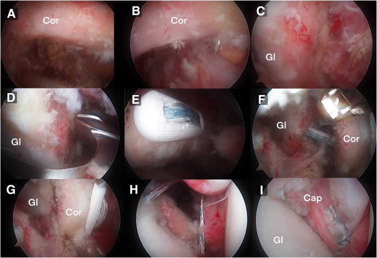
Although the arthroscopic Latarjet procedure has evolved as the most reliable bony sling procedure for shoulder instability with bone loss, the soft cancellous nature of the coracoid makes the FiberTape option for metal-free fixation difficult because of cheese wiring and cutting into the coracoid when the tape is tensioned. Hence, long titanium screws or titanium buttons are used to fix the coracoid in all existing methods for the Latarjet procedure. Rigid and metal-based methods can cause abrasion of the bony moving surfaces coated with cartilage and can cause wear to contact areas, especially when osteolysis occurs during remodeling of the coracoid bone. We describe an absorbable poly lactic acid 3-dimensional printed coracoid protector (which we call the SJ Buckler) to protect the coracoid process resting on its anterior surface and prevent cut through, with laboratory-verified strength to withstand greater than 120 N. This allows us to perform metal-free fixation of the coracoid in the Latarjet procedure, which can be performed arthroscopically or open, where the coracoid tensioning can be done without fear of cut-through. The poly lactic acid material is absorbable and will integrate to bone or disintegrate after the purpose of bony union to glenoid has been achieved.


Conflict of interest statement

The authors declare the following financial interests/personal relationships which may be considered as potential competing interests: S.J. reports consulting or advisory with Malankara Orthodox Syrian Church Medical College and Hospital. P.P. reports employment with Malankara Orthodox Syrian Church Medical College Hospital. S.S. reports employment with Malankara Orthodox Syrian Church Medical College Hospital. K.B. reports employment with Malankara Orthodox Syrian Church Medical College Hospital. R.J. reports employment with Malankara Orthodox Syrian Church Medical College Hospital. V.V. reports employment with Malankara Orthodox Syrian Church Medical College Hospital.

Figures

Fig 1
Fig 1
Illustration of the Latarjet procedure. (A) The coracoid bone is passed through the split in the subscapularis muscle and seated over the anteroinferior glenoid. (B) View from the front of the final construct after Latarjet procedure, with the coracoid fixed to the ideal position using the poly lactic acid coracoid protector and cerclage tapes. (C) Sagittal section showing the split subscapularis and cerclage tapes passed through the glenoid and coracoid with the Buckler looped in the anterior segment of the tapes so as to place it on the anterior coracoid during tensioning of the tapes. (D) View from the side of the final construct with the coracoid graft placed flush to the anteroinferior glenoid through the split subscapularis and the Buckler protecting the anterior glenoid.
Fig 2
Fig 2
Poly lactic acid coracoid protector (which we call the SJ Buckler). (A) Dimensions of the product: length 18 mm and width 7 mm, which is smaller than the size of the smallest coracoid. The thickness is 2.5 mm and the 3-mm holes are placed 2.5 mm from the ends, with a distance of 10 mm between the midpoints of the holes. There is a groove connecting the holes for proper sliding and seating of the cerclage tapes during tensioning. (B-C) Superior and side views of the 3-dimensional rendering of the Buckler. (D) Cerclage tapes (Arthrex), the FiberLink sutures, drill bits, and passing instruments.
Fig 3
Fig 3
(A) In vitro strength testing of the poly lactic acid coracoid protector, which revealed a breaking strength above 190 N, which is almost double the maximum tension applied for seating the coracoid bone during the Latarjet procedure (100 N). (B) The device is loaded on a polyvinylchloride circular cross section to emulate the drill holes through the glenoid, showing survival of compression forces above the breaking strength of the cerclage tapes.
Fig 4
Fig 4
Pictures from the operating theater showing the patient placed in the beach-chair position using a Spider II (Smith & Nephew) limb positioner (A), arthroscopy cannula inserted through the P (posterior) portal of the right shoulder (B), portals marked on the anterior aspect (C), glenoid jig for cerclage tape glenoid drilling (Arthrex) (D), glenoid jig being inserted from the P portal, with arthroscope in the AS portal and feeding tube in the H portal to retract the upper subscapularis superiorly (E), and cerclage tape end being passed through the poly lactic acid coracoid protector in the C portal (F). (AS, anterosuperior portal; C, central portal; H, high portal; I, inferior portal.)
Fig 5
Fig 5
Arthroscopic images of the prepared under surface of the glenoid of right shoulder joint (A), guidewires drilled through the coracoid, placed 10 mm apart (B), prepared anteroinferior surface of the glenoid with the capsule elevated and cancellous bone exposed (C), the hook of the glenoid drill jig placed over the anteroinferior glenoid, centralized over the area of maximum bone loss (D), the coracoid Buckler seated over the coracoid with the cerclage tapes being tensioned (E), coracoid being guided by the coracoid grasper (F), final position of the coracoid over the anteroinferior aspect of the glenoid, well compressed by the cerclage tapes, with the poly lactic acid coracoid protector seated over the anterior surface of the graft (G), all-suture anchors placed at the glenoid edge, for capsulolabral repair (H), and capsulolabral repair completed, exteriorizing the coracoid (I). (Cap, capsule; Cor, coracoid; Gl, glenoid.)
Fig 6
Fig 6
Postoperative imaging of the right glenoid (A-B) radiographs at 6 months, true anteroposterior and Bernageau showing the coracoid graft well seated on the anteroinferior glenoid. (C) Computed tomography scan showing the coracoid positioned in line with the glenoid face in axial section.

References

    1. Lafosse L., Lejeune E., Bouchard A., Kakuda C., Gobezie R., Kochhar T. The arthroscopic Latarjet procedure for the treatment of anterior shoulder instability. Arthroscopy. 2007;23:1242.e1–1242.e5. - PubMed
    1. Latarjet M. Treatment of recurrent dislocation of the shoulder. Lyon Chir. 1954;49:994–997. - PubMed
    1. Chaudhary D., Goyal A., Joshi D., Jain V., Mohindra M., Mehta N. Clinical and radiological outcome after mini-open Latarjet technique with fixation of coracoid with Arthrex wedge mini-plate. J Clin Orthop Trauma. 2016;7:23–29. - PMC - PubMed
    1. Frank R.M., Roth M., Wijdicks C.A., et al. Biomechanical analysis of plate fixation compared with various screw configurations for use in the Latarjet procedure. Orthop J Sports Med. 2020;8 - PMC - PubMed
    1. Dolan C.M., Hariri S., Hart N.D., McAdams T.R. An anatomic study of the coracoid process as it relates to bone transfer procedures. J Shoulder Elbow Surg. 2011;20:497–501. - PubMed

LinkOut - more resources