. 2025 Jun 3;14(7):103620.
doi: 10.1016/j.eats.2025.103620. eCollection 2025 Jul.

Superior Capsular Reconstruction Combined With Latissimus Dorsi Transfer for Massive Irreparable Rotator Cuff Tears With Shoulder Hyperlaxity

Affiliations

Superior Capsular Reconstruction Combined With Latissimus Dorsi Transfer for Massive Irreparable Rotator Cuff Tears With Shoulder Hyperlaxity

Gianezio Paribelli et al. Arthrosc Tech. .

Abstract

Massive rotator cuff injuries are a frequent and disabling shoulder disorder. Over the years, many treatment options have been proposed, including superior capsular reconstruction and latissimus dorsi transfer. However, in our experience, when these 2 approaches are performed individually, they have achieved suboptimal results in patients with massive cuff injury and concomitant shoulder hyperlaxity, as is most often the case in female patients with Beighton scores ≥4. Therefore, we considered the possibility of combining the 2 treatments, taking advantage of the dual stabilizing action of both approaches in cases of hyperlaxed shoulders with massive and irreparable rotator cuff lesions.


Figures

Fig 1
Fig 1
Lateral decubitus on the left side with the right shoulder at 70° abduction for the arthroscopic phase of the procedure.
Fig 2
Fig 2
View from the posterior portal of the subscapularis tendon (S) reinserted on the lesser tuberosity of the humerus and humeral head (H).
Fig 3
Fig 3
Glenoid (G) seen from the posterior portal during cleaning of the superior glenoid rim (red arrow).
Fig 4
Fig 4
View from the posterior portal of the glenoid (G), humeral head (H), and its anchors inserted on the upper edge of the glenoid. Anterior (red arrow) and posterior (blue arrow) anchorage.
Fig 5
Fig 5
View from the posterior portal of shaving and cleaning of the greater tuberosity of the humerus (T).
Fig 6
Fig 6
View from the posterior portal of the SwiveLock anchor insertion (red asterisk) in anterior position on the greater tuberosity of the humerus (T). The glena (G) appears in the background.
Fig 7
Fig 7
View from the posterior portal of the SwiveLock (Arthrex) anchor insertion (blue asterisk) in a posterior position on the greater tuberosity of the humerus (T). The glena (G) appears in the background.
Fig 8
Fig 8
Measurement of the length of the anterior margin of the patch with the dedicated Arthrex instrument (SCR Guide; Arthrex). View from the posterior portal, the glenoid (G) and humeral head (H) are appreciated.
Fig 9
Fig 9
Preparation of the patch and its doubling by suturing the 2 membranes together.
Fig 10
Fig 10
Full doubled patch. The upper side and the lateral and medial portions should be marked.
Fig 11
Fig 11
Detail of the passage of anchor wires over the patch using Scorpion suture passers (Arthrex).
Fig 12
Fig 12
Portals configuration. The PassPort cannula (Arthrex) (red arrow) is placed in posterolateral portal, arthroscope is placed in posterior portal (patient's right shoulder).
Fig 13
Fig 13
Arthroscopic detail of the patch passage through the PassPort cannula (Arthrex) as seen from the posterior portal.
Fig 14
Fig 14
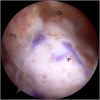
View from the posterolateral portal of the patch (P) fixed to the glenoid rim (Gr).
Fig 15
Fig 15
Insertion of the second SwiveLock anchor (Arthrex) (red asterisk) on the distal part of the greater tuberosity of the humerus to complete the double-row suture for lateral patch fixation (P). View from the posterior portal.
Fig 16
Fig 16
Detail of the double-row suture as seen from the posterolateral portal.
Fig 17
Fig 17
View from the posterolateral portal of the suture between the patch (P) and the posterior cuff remnant (Pc) with a 2 mm TigerTape wire (Arthrex).
Fig 18
Fig 18
View from the posterolateral portal of the final patch configuration (P), attached to the glenoid rim (Gr) and sutured to the posterior cuff remnant (Pc).
Fig 19
Fig 19
Example of the positioning of the patient's arm with the shoulder at 90° of abduction and the elbow at 90° of flexion, during the withdrawal phase of the latissimus dorsi transfer (patient's right shoulder).
Fig 20
Fig 20
Latissimus dorsi tendon basted with different colors FiberWire threads (patient's right shoulder).
Fig 21
Fig 21
Surgical forceps indicate the space behind the long head tendon of the triceps (red asterisk) where the latissimus dorsi transfer will pass. Tendon of latissimus dorsi (LD) and teres major (TM) (patient's right shoulder).
Fig 22
Fig 22
Detail of the meniscus clamp passage (red arrow) with the 2 silicone tubes loaded for intra-articular transport. Teres major (TM), Latissimus dorsi (LD), and the long head of the triceps brachii (red asterisk) are shown. (patient's right shoulder)
Fig 23
Fig 23
Detail of the passage of latissimus dorsi tendon wires through silicone tubes using a wire loop (patient's right shoulder).
Fig 24
Fig 24
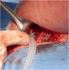
Detail of the passage of the latissimus dorsi (LD) transfer through the space posterior to the long head tendon of the triceps brachii by means of 2 transparent silicone suction tubes (patient's right shoulder).
Fig 25
Fig 25
View from the posterolateral portal of the latissimus dorsi (LD) transfer passing into the space between posterior cuff remnant and deltoid, and above the SCR membrane.
Fig 26
Fig 26
View from the anterolateral portal of the latissimus dorsi (LD) transfer passing over the SCR membrane, final surgery configuration.
Fig 27
Fig 27
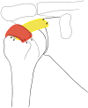
Representation of SCR (in yellow) and LD transfer (in red) final configuration.

References

    1. Kim J.Y., Park J.S., Rhee Y.G. Can preoperative magnetic resonance imaging predict the reparability of massive rotator cuff tears? Am J Sports Med. 2017;45:1654–1663. - PubMed
    1. Kanatlı U., Özer M., Ataoğlu M.B., et al. Arthroscopic-assisted latissimus dorsi tendon transfer for massive, irreparable rotator cuff tears: Technique and short-term follow-up of patients with pseudoparalysis. Arthroscopy. 2017;33:929–937. - PubMed
    1. Ozturk B.Y., Ak S., Gultekin O., Baykus A., Kulduk A. Prospective, randomized evaluation of latissimus dorsi transfer and superior capsular reconstruction in massive, irreparable rotator cuff tears. J Shoulder Elbow Surg. 2021;30:1561–1571. - PubMed
    1. Cofield R.H., Parvizi J., Hoffmeyer P.J., Lanzer W.L., Ilstrup D.M., Rowland C.M. Surgical repair of chronic rotator cuff tears. J Bone Joint Surg Am. 2001;83:71–77. - PubMed
    1. Zumstein M.A., Jost B., Hempel J., Hodler J., Gerber C. The clinical and structural long-term results of open repair of massive tears of the rotator cuff. J Bone Joint Surg Am. 2008;90(11):2423–2431. - PubMed

LinkOut - more resources