. 2025 Jul 31:16:1613560.
doi: 10.3389/fphar.2025.1613560. eCollection 2025.

The functional roles of deoxyelephantopin potential target circTNPO3 in regulating pancreatic cancer malignant phenotype and gemcitabine chemoresistance via miR-188-5p/CDCA3/TRAF2-mediated remodeling of NF-κB signaling pathway

Affiliations

The functional roles of deoxyelephantopin potential target circTNPO3 in regulating pancreatic cancer malignant phenotype and gemcitabine chemoresistance via miR-188-5p/CDCA3/TRAF2-mediated remodeling of NF-κB signaling pathway

Daolin Ji et al. Front Pharmacol. .

Abstract

Background: Pancreatic cancer (PC) has been one of the most severe digestive system malignant tumor with poor prognosis that threatens human health. Chemotherapy is essential for patients with advanced PC, but unfortunately the curative effect is limited by chemoresistance. CircTNPO3, a recently discovered circular RNA (circRNA), has been indicated to be associated with multi-types of tumors. However, the function and mechanism of circTNPO3 in regulating PC malignant phenotype and chemoresistance still remain obscure.

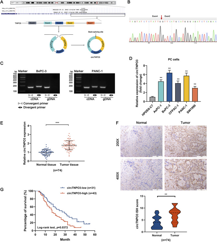
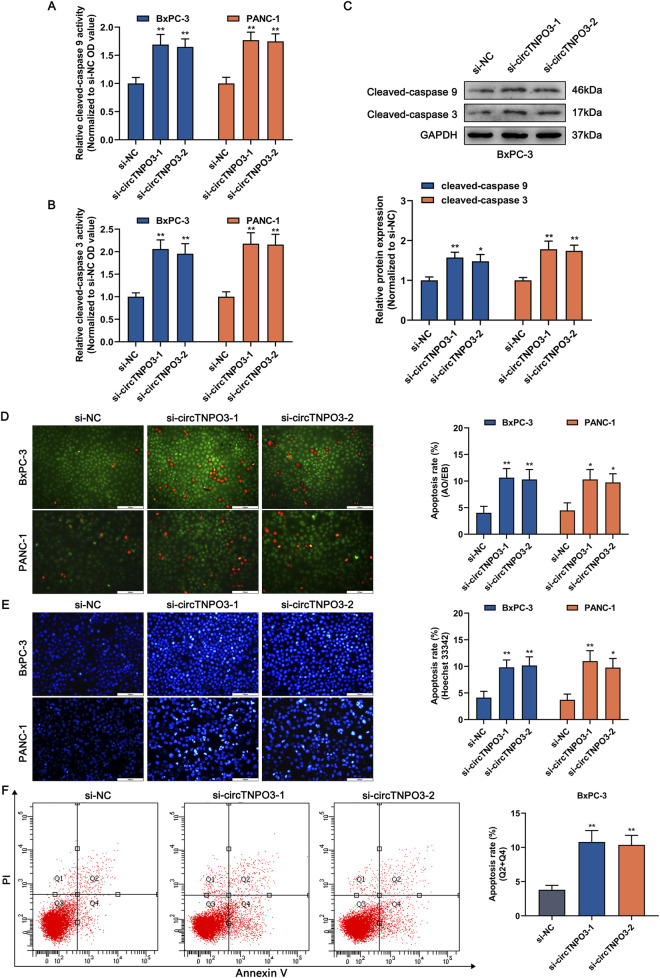
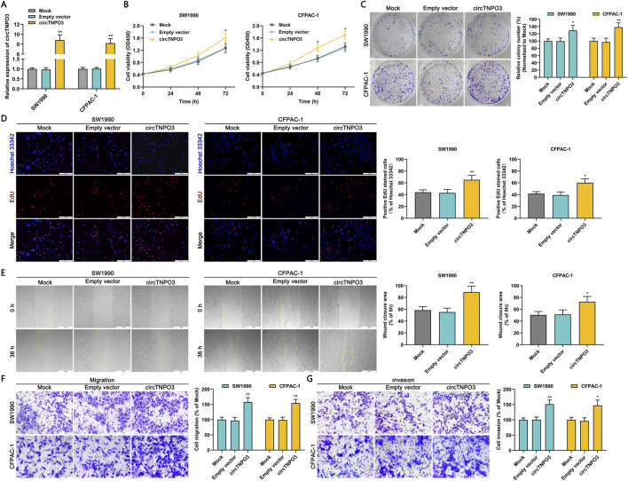
Methods: qRT-PCR and ISH were used to analyze circTNPO3 expression in PC cells and pathological specimens. The subcellular localization of circTNPO3 was visualized through nucleoplasmic RNA separation and FISH assays. The effect of cicTNPO3 on PC cell proliferation, migration and invasion was assessed using EdU, colony formation, wound healing and Transwell assays respectively. Cell apoptosis was detected using ELISA, AO/EB, Hoechst 33342 and flow cytometry assays. The binding potential between circTNPO3, miR-188-5p and CDCA3 was verified by Ago2-RIP, RNA pull down and dual-luciferase reporter assays. The relationship between CDCA3, TRAF2 and NF-κB-p65 was analyzed using Pearson correlation, and the expression was detected using immunoblotting. The nucleus translocation of p65 was evaluated using IF assay. The effect of circTNPO3 on PC growth and metastasis was analyzed using subcutaneous and lung metastatic tumor models in vitro. Deoxyelephantopin, a small molecule extract from traditional Chinese medicine, was applied to evaluate the potential of circTNPO3 as therapeutic target.

Results: CircTNPO3 was aberrantly highly expressed in PC cells and tissues, and negatively associated with patient prognosis and gemcitabine chemotherapy sensitivity. Functionally, silencing circTNPO3 attenuated the malignant phenotypes and chemoresistance of PC in vitro and in vivo, conversely, facilitated by circTNPO3 overexpression. Mechanically, cytoplasmic circTNPO3 functioned as a sponge of miR-188-5p, and partially alleviated the effect of miR-188-5p on downstream molecules, which further upregulate the CDCA3 and TRAF2 expression and NF-κB activity, finally promoted PC progression and chemoresistance. More innovatively, the potential of circTNPO3 as a novel diagnostic biomarker and therapeutic target for PC was primarily validated in present study.

Conclusion: CircTNPO3 acted as an oncogenic and chemoresistant gene in PC, mechanically through targeting miR-188-5p and regulating CDCA3, TRAF2 and NF-κB signaling pathway.

Keywords: NF-κB signaling pathway; chemoresistance; circTNPO3; deoxyelephantopin; gemcitabine; pancreatic cancer; stemness.


Conflict of interest statement

The authors declare that the research was conducted in the absence of any commercial or financial relationships that could be construed as a potential conflict of interest.

Figures

FIGURE 1
FIGURE 1
The heterogeneity of circTNPO3 in PC cells and tissues, and association with clinicopathological characteristics. (A) The chromosome localization and splicing schematic of has_circ_0001741 (circTNPO3) were presented. (B) The specific primers designed for circTNPO3 were validated by qRT-PCR and the PCR product was verified through Sanger sequencing. The red arrow represented the back-splicing site. (C) The cyclization characteristic of circTNPO3 was confirmed through agarose gel electrophoresis using divergent and convergent primers in BxPC-3 and PANC-1 cells. (D) The differential expression of circTNPO3 between human normal pancreatic ductal epithelial (HPDE6-C7) and PC (AsPC-1, BxPC-3, CFPAC-1, PANC-1 and SW1990) cell lines was detected by qRT-PCR. (E) qRT-PCR analysis of circTNPO3 expression between 74 pairs of PC tissues and adjacent peritumoral tissues. (F) The expression of circTNPO3 in PC tissues and adjacent peritumoral tissues was detected by ISH assay. (n = 74). (G) The overall survival of PC patients with lower (n = 31) and higher (n = 43) circTNPO3 expression was compared using Kaplan–Meier curve. ∗∗P < 0.01, ∗∗∗P < 0.001. Magnification, ×200 (first row of F), × 400 (second row of F). Scale bar, 100 μm (first row of F), 50 μm (second row of F). PC, pancreatic cancer. ISH, in situ hybridization.
FIGURE 2
FIGURE 2
Silencing circTNPO3 suppressed the proliferation, migration and invasion of PC cells in vitro. (A) The expression of circTNPO3 in BxPC-3 and PANC-1 cells after transfection with si-NC or interference sequences targeting circTNPO3 (si-circTNPO3-1/2) was detected by qRT-PCR. (B) The viability of BxPC-3 and PANC-1 cells after transfection with siRNAs was assessed by CCK-8 assay. (C) The proliferation of BxPC-3 and PANC-1 cells was analyzed by colony forming assay. (D) The EdU positive staining representing proliferation of BxPC-3 and PANC-1 cells was quantified by EdU assay. (E) Wound healing assay was performed to detect the locomotive ability of BxPC-3 and PANC-1 cells after circTNPO3 silencing. (F,G) Transwell assay was conducted to assess the migration and invasion of BxPC-3 and PANC-1 cells. *P < 0.05, **P < 0.01. Magnification, ×40 (E), × 200 (D,F,G). Scale bar, 500 μm (E), 100 μm (D,F,G). EdU, 5-Ethynyl-2′-deoxyuridine.
FIGURE 3
FIGURE 3
Silencing circTNPO3 induced the apoptosis of PC cells. (A,B) The activation of apoptosis-associated markers including upstream caspase 9 and downstream caspase 3 was determined using Caspase 9 and Caspase 3 Activity Assay Kits. (C) The expression level of cleaved-caspase 9 and cleaved-caspase 3 after circTNPO3 silencing was measured using immunoblotting in BxPC-3 cells. (D,E) The apoptosis rate of BxPC-3 and CFPAC-1 cells after circTNPO3 silencing was detected by AO/EB and Hoechst 33342 fluorescent staining assays. (F) Flow cytometry analysis was applied to analyze the apoptosis of BxPC-3 and CFPAC-1 cells after circTNPO3 silencing. Q1 quadrant, mechanically damaged cells. Q2 quadrant, non-viable apoptotic cells. Q3 quadrant, normal viable cells. Q4 quadrant, viable apoptotic cells. *P < 0.05, **P < 0.01. Magnification, ×200 (D,E). Scale bar, 100 μm (D,E). AO, Acridine Orange. EB, Ethidium Bromide.
FIGURE 4
FIGURE 4
Silencing circTNPO3 attenuated PC growth and metastasis in vivo. (A) BxPC-3 cells after transfection with sh-NC or sh-circTNPO3-1/2 were inoculated into the left axilla of nude mice to construct subcutaneous xenograft model, and the mice were divided to three groups. (n = 5). (B) Tumor growth curves were plotted according to the tumor volume recorded every 3 days. (C) The final weight of the subcutaneous tumor. (D) The functional protein associated with proliferation (Ki-67, PCNA) and EMT (E-cadherin) in tumor samples were detected by IHC assay. (E) The lung metastatic model was established by nude mice tail vein injection of transfected BxPC-3 cells, and the alveolar structure was evaluated by H&E staining. (n = 5). *P < 0.05, **P < 0.01. Magnification, ×100 (E), × 200 (D). Scale bar, 200 μm (E), 100 μm (D). H&E, hematoxylin-eosin. EMT, epithelial to mesenchymal transition.
FIGURE 5
FIGURE 5
Overexpression circTNPO3 promoted the proliferation, migration and invasion of PC cells in vitro. (A) The expression of circTNPO3 in SW1990 and CFPAC-1 cells after transfection with empty vector or overexpression vector was detected by qRT-PCR. (B) The viability of SW1990 and CFPAC-1 cells after circTNPO3 overexpression was analyzed by CCK-8 assay. (C) The proliferation of SW1990 and CFPAC-1 cells after circTNPO3 overexpression was assessed by colony forming assay. (D) The EdU positive staining representing proliferation of SW1990 and CFPAC-1 cells after circTNPO3 overexpression was quantified by EdU assay. (E) The locomotive ability of SW1990 and CFPAC-1 cells after circTNPO3 overexpression was tested by wound healing assay. (F,G) The migration and invasion of SW1990 and CFPAC-1 cells after circTNPO3 overexpression was analyzed by Transwell assay. *P < 0.05, **P < 0.01. Magnification, ×40 (E), × 200 (D,F,G). Scale bar, 500 μm (E), 100 μm (D,F,G).
FIGURE 6
FIGURE 6
CircTNPO3 was involved in the regulation of GEM chemoresistance in PC. (A) The differential expression of circTNPO3 between chemoresistant PC cells and non-chemoresistant parental PC cells was analyzed using qRT-PCR. **P < 0.01 versus HPDE6-C7 group. #P < 0.05 versus respective parental non-chemoresistant control (BxPC-3, CFPAC-1) group. (B) The expression of circTNPO3 after transfection with sh-circTNPO3-1/-2 was detected using qRT-PCR. (C) The expression of circTNPO3 after transfection with overexpression vector was monitored by qRT-PCR. (D) Stem cell self-renewal associated with chemoresistance was tested using spheroid formation assay. (E) The viability of BxPC-3/GR and CFPAC-1/GR cells after circTNPO3 silencing combined with GEM treatment was tested by CCK-8 assay. (F) The viability of BxPC-3 and CFPAC-1 cells after circTNPO3 overexpression combined with GEM treatment was tested by CCK-8 assay. *P < 0.05, **P < 0.01. Magnification, ×100 (D). Scale bar, 200 μm (D). (G) The apoptosis rate of BxPC-3/GR and CFPAC-1/GR cells after circTNPO3 silencing combined with GEM treatment was detected by AO/EB fluorescent staining assay. (H) The apoptosis rate of BxPC-3 and CFPAC-1 cells after circTNPO3 overexpression combined with GEM treatment was detected by AO/EB fluorescent staining assay. *P < 0.05, **P < 0.01 versus sh-NC group. #P < 0.05, ##P < 0.01 versus sh-NC combined GEM or empty vector combined GEM group. GEM, gemcitabine. BxPC-3/GR, BxPC-3 gemcitabine resistant cell line. CFPAC-1/GR, CFPAC-1 gemcitabine resistant cell line.
FIGURE 7
FIGURE 7
The subcellular localization of circTNPO3 and the identification of interacting miRNAs. (A) The localization of circTNPO3 in BxPC-3/GR and CFPAC-1/GR cells was visualized using FISH assay. (B) The cellular distribution of circTNPO3 in BxPC-3/GR and CFPAC-1/GR cells was analyzed using Cytoplasmic and Nuclear RNA Purification Kit and qRT-PCR assay. (C) The expression of circTNPO3 and corresponding linear TNPO3 mRNA after RNase R treatment in BxPC-3/GR and CFPAC-1/GR cells was tested using qRT-PCR. (D) The half-life of circTNPO3 and linear TNPO3 mRNA after actinomycin D treatment in BxPC-3/GR and CFPAC-1/GR cells was assessed using qRT-PCR. (E) The binding capacity between circTNPO3 and miRNAs was investigated using Ago2-RIP assay. (F) The intersection of the target miRNAs of circTNPO3 was intersectively identified using CircBank, ENCORI and TargetScan databases, and displayed through Venn diagram. (G) The binding ability between circTNPO3 and four predicted miRNAs including miR-188-5p, miR-199a-5p, miR-199b-5p and miR-552-3p was verified using RNA pulldown assay. (H) The binding sequence between circTNPO3 and miR-188-5p was showed, and the direct binding potential was determined using dual-luciferase reporter assay. *P < 0.05, **P < 0.01. (I) The differential expression of miR-188-5p between chemoresistant PC cells and non-chemoresistant parental PC cells was analyzed using qRT-PCR. *P < 0.05, **P < 0.01 versus HPDE-C7 group. #P < 0.05 versus respective parental non-chemoresistant control (BxPC-3, CFPAC-1) group. (J) The expression of miR-188-5p after circTNPO3 silencing in BxPC-3/GR and CFPAC-1/GR cells was detected using qRT-PCR. (K) The viability of BxPC-3/GR and CFPAC-1/GR cells after transfection with sh-circTNPO3-1 and miR-188-5p inhibitor was measured using CCK-8 assay. (L) The migration ability of BxPC-3/GR and CFPAC-1/GR cells after transfection with sh-circTNPO3-1 and miR-188-5p inhibitor was determined using Transwell assay. (M) The apoptosis of BxPC-3/GR and CFPAC-1/GR cells after transfection with sh-circTNPO3-1 and miR-188-5p inhibitor was evaluated using Hoechest 33342 fluorescence staining assay. *P < 0.05, **P < 0.01 versus sh-NC group. #P < 0.05, ##P < 0.01 versus sh-circTNPO3-1 group.
FIGURE 8
FIGURE 8
circTNPO3 facilitated GEM chemoresistance through competitively binding miR-188-5p, upregulating CDCA3 and activing NF-κB nuclear translocation. (A) The binding sequence between miR-188-5p and CDCA3 mRNA was listed, and the direct binding potential was confirmed using dual-luciferase reporter assay. (B) The binding ability between miR-188-5p and CDCA3 mRNA was determined using miR-188-5p probe RNA pulldown assay. (C) The differential expression of CDCA3 between chemoresistant PC cells and parental non-chemoresistant PC cells was analyzed using qRT-PCR and immunoblotting assays. *P < 0.05, **P < 0.01 versus HPDE6-C7 group. #P < 0.05, ##P < 0.01 versus respective parental non-chemoresistant control (BxPC-3, CFPAC-1) group. (D) Pan-cancer atlas of CDCA3 in various human malignant tumors, and the differential expression of CDCA3 in PC was labeled by a red arrow. (E,F) The expression level of CDCA3 in PC was assessed by TCGA, GTEx and TNMplot databases. (G) Kaplan-Meier curve according to GEPIA database was used to analyze the overall survival of PC patients with lower and higher CDCA3 expression level. (H) The expression level of CDCA3 between PC tumor tissues and adjacent normal tissues was compared using IHC assay. (I) The correlation between miR-188-5p and CDCA3 in PC was analyzed through Pearson correlation coefficient. (J,K) The expression level of CDCA3 in BxPC-3/GR and CFPAC-1/GR cells after transfection with sh-circTNPO3-1 and miR-188-5p inhibitor was detected using qRT-PCR and immunoblotting assays. *P < 0.05, **P < 0.01 versus sh-NC group. #P < 0.05, ##P < 0.01 versus sh-circTNPO3-1 group. (L) The correlation between CDCA3 and TRAF2 in PC was analyzed through Pearson correlation coefficient from TNMplot database. (M,N) The expression level of CDCA3 in BxPC-3/GR and CFPAC-1/GR cells after transfection with sh-CDCA3-1/-2 was examined using immunoblotting assay. (O) The expression level of TRAF2 and the NF-κB-p65 nuclear translocation in BxPC-3 and BxPC-3/GR cells after CDCA3 knockdown were determined using immunoblotting assay. *P < 0.05 versus sh-NC group. ##P < 0.01 versus sh-CDCA3-2 group. (P) The NF-κB-p65 nuclear translocation in BxPC-3 and BxPC-3/GR cells after CDCA3 knockdown was evaluated using IF assay. IF, immunofluorescence.
FIGURE 9
FIGURE 9
The mechanism of circTNPO3 regulating the malignant phenotype of PC.

Similar articles

References

    1. Beutel A. K., Halbrook C. J. (2023). Barriers and opportunities for gemcitabine in pancreatic cancer therapy. Am. J. Physiol. Cell Physiol. 324 (2), C540–c552. 10.1152/ajpcell.00331.2022 - DOI - PMC - PubMed
    1. Bray F., Laversanne M., Sung H., Ferlay J., Siegel R. L., Soerjomataram I., et al. (2024). Global cancer statistics 2022: GLOBOCAN estimates of incidence and mortality worldwide for 36 cancers in 185 countries. CA Cancer J. Clin. 74 (3), 229–263. 10.3322/caac.21834 - DOI - PubMed
    1. Chen X., Zhou M., Yant L., Huang C. (2022). Circular RNA in disease: basic properties and biomedical relevance. Wiley Interdiscip. Rev. RNA 13 (6), e1723. 10.1002/wrna.1723 - DOI - PubMed
    1. Conn V. M., Chinnaiyan A. M., Conn S. J. (2024). Circular RNA in cancer. Nat. Rev. Cancer 24 (9), 597–613. 10.1038/s41568-024-00721-7 - DOI - PubMed
    1. Danpanichkul P., Suparan K., Tothanarungroj P., Dejvajara D., Rakwong K., Pang Y., et al. (2024). Epidemiology of gastrointestinal cancers: a systematic analysis from the global burden of disease study 2021. Gut 74 (1), 26–34. 10.1136/gutjnl-2024-333227 - DOI - PubMed

LinkOut - more resources